m
or phogenes i s and t he m
ai nt enanc e of
s per m
at ogenes i s i n adul t m
i c e
著者
Shaw
ki H
os s am
H
. , O
i s hi H
i s as hi , U
s ui
Tos hi aki , Ki t adat e Yu, Bas ha W
al aa A. ,
Abdel l at i f Ahm
ed M
. , H
as egaw
a Kaz unor i , O
kada
Ri s a, M
oc hi da Kei j i , El - Shem
y H
any A. ,
M
ur at ani M
as af um
i , O
gur a At s uo, Yos hi da
Shos ei , Takahas hi Sat or u
j our nal or
publ i c at i on t i t l e
PLO
S O
N
E
vol um
e
13
num
ber
1
page r ange
e0190800
year
2018- 01
権利
( C) 2018 Shaw
ki et al . Thi s i s an open ac c es s
ar t i c l e di s t r i but ed under t he t er m
s of t he
Cr eat i ve Com
m
ons At t r i but i on Li c ens e, w
hi c h
per m
i t s unr es t r i c t e d us e, di s t r i bu t i on, and
r epr oduc t i on i n any m
edi um
, pr ovi ded t he
or i gi nal aut hor and s our c e ar e c r edi t ed.
U
RL
ht t p: / / hdl . handl e. net / 2241/ 00150896
MAFB is dispensable for the fetal testis
morphogenesis and the maintenance of
spermatogenesis in adult mice
Hossam H. Shawki1,2*, Hisashi Oishi1,3*, Toshiaki Usui1, Yu Kitadate4, Walaa A. Basha1, Ahmed M. Abdellatif1, Kazunori Hasegawa5, Risa Okada1, Keiji Mochida6, Hany A. El-Shemy7, Masafumi Muratani8, Atsuo Ogura6, Shosei Yoshida4, Satoru Takahashi1
1Department of Anatomy and Embryology, Graduate School of Comprehensive Human Science, University of Tsukuba, Tsukuba, Japan,2Department of Animal Genetic Resources, National Gene Bank, Giza, Egypt,
3Department of Comparative and Experimental Medicine, Graduate School of Medical Sciences, Nagoya City University, Nagoya, Japan,4Division of Germ Cell Biology, National Institute for Basic Biology, Okazaki, Japan,5Department of Medicine, Stanford University School of Medicine, Stanford, California, United States of America,6BioResource Center, RIKEN, Tsukuba, Japan,7Cairo University Research Park, Cairo University, Giza, Egypt,8Department of Genome Biology, Faculty of Medicine, University of Tsukuba, Tsukuba, Japan
*[email protected](HHS);[email protected](HO)
Abstract
The transcription factor MAFB is an important regulator of the development and differentia-tion of various organs and tissues. Previous studies have shown that MAFB is expressed in embryonic and adult mouse testes and is expected to act as the downstream target of reti-noic acid (RA) to initiate spermatogenesis. However, its exact localization and function remain unclear. Here, we localized MAFB expression in embryonic and adult testes and analyzed its gene function usingMafb-deficient mice. We found that MAFB and c-MAF are the only large MAF transcription factors expressed in testes, while MAFA and NRL are not. MAFB was localized in Leydig and Sertoli cells at embryonic day (E) 18.5 but in Leydig cells, Sertoli cells, and pachytene spermatocytes in adults.Mafb-deficient testes at E18.5 showed fully formed seminiferous tubules with no abnormal structure or differences in testic-ular somatic cell numbers compared with those of control wild-type mice. Additionally, the expression levels of genes related to development and function of testicular cells were unchanged between genotypes. In adults, the expression of MAFB in Sertoli cells was shown to be stage specific and induced by RA. By generatingMafbfl/flCAG-CreER™(Mafb -cKO) mice, in which Cre recombinase was activated upon tamoxifen treatment, we found that the neonatal cKO mice died shortly uponMafbdeletion, but adult cKO mice were alive upon deletion. Adult cKO mice were fertile, and spermatogenesis maintenance was normal, as indicated by histological analysis, hormone levels, and germ cell stage-specific markers. Moreover, there were no differences in the proportion of seminiferous stages between cKO mice and controls. However, RNA-Seq analysis of cKO Sertoli cells revealed that the down-regulated genes were related to immune function and phagocytosis activity but not spermatogenesis. In conclusion, we found that MAFB is dispensable for fetal testis morpho-genesis and spermatomorpho-genesis maintenance in adult mice, despite the significant gene
a1111111111 a1111111111 a1111111111 a1111111111 a1111111111 OPEN ACCESS
Citation:Shawki HH, Oishi H, Usui T, Kitadate Y, Basha WA, Abdellatif AM, et al. (2018) MAFB is dispensable for the fetal testis morphogenesis and the maintenance of spermatogenesis in adult mice. PLoS ONE 13(1): e0190800.https://doi.org/ 10.1371/journal.pone.0190800
Editor:Jean-Marc A Lobaccaro, Universite Clermont Auvergne, FRANCE
Received:March 9, 2017
Accepted:December 20, 2017
Published:January 11, 2018
Copyright:©2018 Shawki et al. This is an open access article distributed under the terms of the
Creative Commons Attribution License, which permits unrestricted use, distribution, and reproduction in any medium, provided the original author and source are credited.
Data Availability Statement:All relevant data are available at the following URL:https://www.ncbi. nlm.nih.gov/geo/query/acc.cgi?acc=GSE94297.
Funding:Dr. Shosei Yoshida received a Grant-in-Aid for Scientific Research on Innovative Areas #25114004 "Mechanisms regulating gamete formation in animals". Dr. Shosei Yoshida also received a Grant-in-Aid for Scientific Research(A) #16H02507.
i-expression in different cell types, but MAFB might be critical for phagocytosis activity of Ser-toli cells.
Introduction
The testes are divided into several tubules known as seminiferous tubules, which are the houses of sperm production. Each tubule comprises multiple germinal cell types and only one somatic cell type, Sertoli cells, which support sperm development. Leydig cells, another type of somatic cell, are located outside the tubules and produce androgens required for the maturation of sex-ual organs and sexsex-ual characteristics as well as sperm development. The testes produce sperm through a process known as spermatogenesis. Spermatogenesis is a complex process of cellular transformation that depends on numerous factors for successful production of haploid sperm from diploid spermatogonial stem cells [1].
Spermatogenesis comprises three main phases (mitosis, meiosis, and post-meiosis). matogonia are diploid and divide by mitosis into several other types of spermatogonia. Sper-matogonia are present as undifferentiated type A sperSper-matogonia (A single, A paired, A aligned), which all retain stem cell properties; differentiated type A spermatogonia (A1, A2, A3, A4); intermediate spermatogonia; and type B spermatogonia. Type B spermatogonia are then divided by mitosis to form preleptotene, leptotene and zygotene spermatocytes, which subsequently undergo meiosis I to form secondary spermatocytes and meiosis II to form hap-loid round spermatids. Spermiogenesis is the post-meiosis process that transforms spherical, haploid spermatids into elongated spermatid and mature sperm that are released into the lumen of the seminiferous tubules.
MAF family of proteins is a subgroup of basic region-leucine zipper (bZIP) transcription factors that recognize a long palindromic DNA sequence [TGCTGAC(G)TCAGCA] known as MAF recognition element (MARE) [2]. The MAF family is subdivided into two groups; large MAFs and small MAFs. Large MAFs contain an acidic domain that promotes transcriptional activation. In contrast, small MAFs lack the acidic domain and act as transcriptional repressors unless they form heterodimers with other proteins that harbor transcriptional activation domains [3–6]. In mice, large MAFs include MAFA, MAFB, c-MAF and Neural Retina Leu-cine (NRL), while small MAFs include MAFF, MAFG, and MAFK. InDrosophila melanoga-ster, there is only one large MAF transcription factor calledTraffic Jam(TJ) and one small MAF called MAF-S. Several studies have shown that each protein plays a key role in cellular differentiation and regulation of tissue-specific gene expression [7,8].
Inactivation ofTJ, the large MAF factor inDrosophila, revealed that it plays an essential role in gonad morphogenesis and that its expression in somatic gonadal cells in direct contact with germline cells is required for female and male fertility [9]. InTJmutant gonads, somatic cells fail to intermingle and properly envelop germline cells, causing an early block in germ cell dif-ferentiation.TJencodes an orthologue of the typical bZIP transcription factors MAFB and c-MAF in vertebrates.
In particular, the large MAF transcription factor in vertebrates, MAFB, is first expressed in mouse embryonic gonads along the gonad-mesonephros border in both sexes as early as embryonic day (E) 11.5. Between E12.0 and E14.5, MAFB expression expands in the interstitial compartment and then becomes restricted to Leydig cells in XY gonads, but the expression pattern does not change significantly in XX gonads [10]. On the other hand, MAFB in post-natal mouse testes has been detected in Sertoli cells within the seminiferous tubules [11] and in testicular macrophages outside the tubules [12].
The active metabolite of vitamin A, retinoic acid (RA), is essential for the initial differentia-tion and meiotic entry of spermatogonia. Vitamin A-deficient (VAD) mice result in blockage of A to A1 spermatogonia transition, and only undifferentiated type A spermatogonia and Ser-toli cells remain within the seminiferous tubules in the testes [13]. This indicates that removing RA inhibits the ability of undifferentiated spermatogonia to differentiate in adult mouse testes. Treating VAD mice with retinol or RA results in the complete recovery of spermatogenesis [13]. Two models have been suggested for how RA drives male germ cell development [11,14– 19]. First, RA that generated by Sertoli cells in the neonatal testes acts in an autocrine manner to induce the first wave of A1 spermatogonia differentiation. Second, after the first wave, the transition of A to A1 spermatogonia appears to be generated by the activity of RA in either pachytene spermatocytes or preleptotene spermatocytes. However, the target genes that are regulated by RA and that control spermatogonia differentiation remain to be discovered.
A previous report revealed that MAFB is one of the RA target genes that induces spermato-gonia differentiation [11]. The authors specifically knocked out RA-synthesizing enzymes (RALDH1 to RALDH3 encoded byAldh1a1toAldh1a3genes) in Sertoli cells and found a blockage of spermatogonial differentiation, similar to VAD mice. After treatment with a RARA agonist, differentiated spermatogonia were detected with highly increased MAFB expression in Sertoli cells [11]. In addition, they detected a robust RAR-binding site at the end of theMafbcoding region, indicating that this gene is a direct target of RA.
However, the complete role of MAFB in spermatogonia differentiation remains unclear becauseMafbknockout mice die by birth, and conditional alleles are not generated by this time. In the current study, we generated conditional alleles ofMafband investigated the role of Mafbin male gonads.
Materials and methods
Animals
All mice used in this study were of the C57BL/6 background. Animal experiments were approved by the Animal Experiment Committee of the University of Tsukuba (Permit Num-ber: 14–049) and performed in accordance with the Guide for the Care and Use of Laboratory Animals of the National Institutes of Health. All the mice received humane care, were main-tained in specific pathogen-free conditions and were euthanized with carbon dioxide gas in the Laboratory Animal Resource Center at the University of Tsukuba to minimize suffering. Animals were checked daily and the specific criteria for animal euthanasia when they displayed early markers associated with death or specific signs of severe suffering or distress, including slow or no mobility or an absence of heartbeat or respiratory movement.
Control mice lacked the Cre transgene (Mafbfl/fl). To induce gene deletion, mice were injected with tamoxifen. The primer sequences used for genotyping ofMafb−/−,MafbloxP, andMafb excision (MafbΔ) are listed inTable 1.
Cre driver mice efficiency
R26GRR reporter mice, as previously described [21,22], were mated with the CAG-CreER™ driver mouse strain obtained from Jackson laboratories. The progeny were genotyped using the Cre and GFP primers listed inTable 1. Frozen sections from the mice carrying double heterozygous mice carrying GRR and Cre recombinase were then analyzed with or without tamoxifen injection.
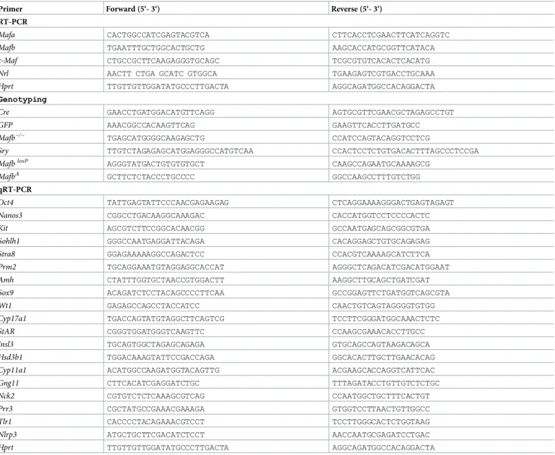
Table 1. Complete list of primers used in this study.
Primer Forward (5‘- 3‘) Reverse (5‘- 3‘)
RT-PCR
Mafa CACTGGCCATCGAGTACGTCA CTTCACCTCGAACTTCATCAGGTC
Mafb TGAATTTGCTGGCACTGCTG AAGCACCATGCGGTTCATACA
c-Maf CTGCCGCTTCAAGAGGGTGCAGC TCGCGTGTCACACTCACATG
Nrl AACTT CTGA GCATC GTGGCA TGAAGAGTCGTGACCTGCAAA
Hprt TTGTTGTTGGATATGCCCTTGACTA AGGCAGATGGCCACAGGACTA
Genotyping
Cre GAACCTGATGGACATGTTCAGG AGTGCGTTCGAACGCTAGAGCCTGT
GFP AAACGGCCACAAGTTCAG GAAGTTCACCTTGATGCC
Mafb−/− TGAGCATGGGGCAAGAGCTG CCATCCAGTACAGGTCCTCG
Sry TTGTCTAGAGAGCATGGAGGGCCATGTCAA CCACTCCTCTGTGACACTTTAGCCCTCCGA
MafbloxP AGGGTATGACTGTGTGTGCT CAAGCCAGAATGCAAAAGCG
MafbΔ GCTTCTCTACCCTGCCCC GGCCAAGCCTTTGTCTGG
qRT-PCR
Oct4 TATTGAGTATTCCCAACGAGAAGAG CTCAGGAAAAGGGACTGAGTAGAGT Nanos3 CGGCCTGACAAGGCAAAGAC CACCATGGTCCTCCCCACTC
Kit AGCGTCTTCCGGCACAACGG GCCAATGAGCAGCGGCGTGA
Sohlh1 GGGCCAATGAGGATTACAGA CACAGGAGCTGTGCAGAGAG Stra8 GGAGAAAAAGGCCAGACTCC CCACGTCAAAAGCATCTTCA
Prm2 TGCAGGAAATGTAGGAGGCACCAT AGGGCTCAGACATCGACATGGAAT
Amh CTATTTGGTGCTAACCGTGGACTT AAGGCTTGCAGCTGATCGAT
Sox9 ACAGATCTCCTACAGCCCCTTCAA GCCGGAGTTCTGATGGTCAGCGTA
Wt1 GAGAGCCAGCCTACCATCC CAACTGTCAGTAGGGGTGTGG
Cyp17a1 TGACCAGTATGTAGGCTTCAGTCG TCCTTCGGGATGGCAAACTCTC
StAR CGGGTGGATGGGTCAAGTTC CCAAGCGAAACACCTTGCC
Insl3 TGCAGTGGCTAGAGCAGAGA GTGCAGCCAGTAAGACAGCA Hsd3b1 TGGACAAAGTATTCCGACCAGA GGCACACTTGCTTGAACACAG Cyp11a1 ACATGGCCAAGATGGTACAGTTG ACGAAGCACCAGGTCATTCAC Gng11 CTTCACATCGAGGATCTGC TTTAGATACCTGTTGTCTCTGC
Nck2 CGTGTCTCTCAAAGCGTCAG CCAATGGCTGCTTTCACTGT
Prr3 CGCTATGCCGAAACGAAAGA GTGGTCCTTAACTGTTGGCC
Tlr1 CACCCCTACAGAAACGTCCT TCCTTGGGCACTCTGGTAAG
Nlrp3 ATGCTGCTTCGACATCTCCT AACCAATGCGAGATCCTGAC
Hprt TTGTTGTTGGATATGCCCTTGACTA AGGCAGATGGCCACAGGACTA
Tamoxifen administration
Tamoxifen (Sigma; T5648) was dissolved in corn oil. The tamoxifen-oil mixture was stored at
−20˚C until used. Neonatal mice received 50μg tamoxifen/day intragastrically from P1 to P3
[23]. Starting 6 weeks after birth, mice received 75 mg tamoxifen/kg body weight intraperito-neally once daily for 5 consecutive days [24]. All littermates were injected with the same dose to generate the necessary controls.
Reverse transcription PCR (RT-PCR)
Total RNA was extracted from various tissues with a Nucleospin RNA II kit (TaKaRa 740955), and cDNA was synthesized with a QuantiTect Reverse Transcription kit (QIAGEN; 205313), each according to the manufacturer’s instructions. PCR was performed forMafa,Mafb,c-Maf, andNrlusing TaKaRa Ex Taq (RR001).Hprtwas used as an internal control for cDNA quality and PCR efficiency. The sequences of the primers used for RT-PCR are shown inTable 1.
Quantitative RT-PCR (qRT-PCR)
Total RNA was extracted from the testes with a Nucleospin RNA II kit (TaKaRa; 740955), and cDNA was synthesized with a QuantiTect Reverse Transcription kit (QIAGEN 205313), each according to the manufacturer’s instructions. Quantitative PCR reactions were run on a Ther-mal Cycler Dice Real-Time System Single (Takara) with SYBR Green PCR master mix (Takara; RR820). All qPCR analyses were performed in duplicate. Amplification products were quanti-fied by the standard curve method. The mRNA levels of each gene were normalized to that of Hprt. The primers used for qPCR are listed inTable 1.
Tissue collection and histological analysis
Tissues were dissected from mice immediately after euthanasia, fixed in 4% (mass/vol) para-formaldehyde for up to 24 h (5 h in case of embryos and neonatal mice), stored in 70% (vol/vol) ethanol, and embedded in paraffin. Sections of 5-μm thickness were prepared and mounted on glass slides. After deparaffinization, slides were used either for immunohisto-chemical analyses or stained with periodic acid–Schiff (PAS) and haematoxylin/eosin (HE).
Immunohistochemical analysis
Immunocytochemical analysis
Isolated Sertoli cells were plated onto a silanized glass slide using a Shandon Cytospin™3 cen-trifuge. Cells were then fixed with 4% paraformaldehyde in PBS for 15 min at room tempera-ture. Cells were washed with PBS and blocked with 3% skim milk plus 0.1% Triton X-100 in PBS. Cells were then incubated with a 1:500 dilution of guinea pig polyclonal anti-vimentin antibody (Progen Biotechnik; GP53) overnight at 4˚C. After washing, cells were incubated with a 1:1000 dilution of Alexa Fluor 594-conjugated secondary antibody (Life Technologies, Gaithersburg, MD, USA) for an hour at room temperature. Cells were counterstained with DAPI.
Counting of Leydig and Sertoli cells
Leydig and Sertoli cells were identified in 18.5-dpc mouse testes. Testes from WT andMafb KO embryos (n= 3) were sectioned, and 4 sections for each gonad were randomly selected and stained with an antibody against GATA4 or STAR. The number of STAR-positive cells outside seminiferous tubules (Leydig cells) and GATA4-positive cells inside the tubules (Ser-toli cells) were counted, and the whole gonadal areas were measured.
Isolation of stage-specific seminiferous tubules
The specific stages of seminiferous tubules were isolated for qPCR analysis. Briefly, mature WT male mice were sacrificed, and the testes were dissected and decapsulated in a Petri dish containing PBS. The tubules were viewed on a trans-illuminating dissection microscope. The light absorption pattern was used to identify the different stages as previously described [25].
Primary Sertoli cells culture
Primary Sertoli cells were isolated and cultured as previously described [26,27] with slight modifications. Briefly, three-week-old testes without tunica albuginea were sequentially treated with 0.5 mg/ml collagenase (Wako, 034–22363), 1 mg/ml hyaluronidase (Sigma, H3506) plus 1 mg/ml collagenase, and 1 mg/ml hyaluronidase in Dulbecco’s modified Eagle’s medium (DMEM) containing DNase I. Small pieces of seminiferous tubules were removed via filtering through a 100μm-pore-size filter. The purity of the isolated cells was confirmed by immunocy-tochemical staining with an anti-vimentin antibody (Progen Biotechnik; GP53). Isolated Ser-toli cells were cultured with F12-DMEM (Invitrogen, 10565–018) mixed with 10μg/ml insulin (Nacalai Tesque, 19251–24), 5μg/ml transferrin (Sigma, T1147), and 5 ng/ml epidermal growth factor (BD Bioscience, 40010) at 34˚C. The culture medium was changed at days 2 and 4, and Sertoli cells were stimulated with 1μM RA (Sigma) at day 5 for 24 hours. Then, qPCR analyses were performed.
Protein extraction and western blotting
hour at room temperature. As a loading control, we used an HRP-conjugated anti-β-actin antibody (1:3000; MBL PM057-7). Signals were detected via western blotting with luminol reagents (Nacalai). The protein marker used was WIDE-VIEW prestained protein size marker III (Wako, Osaka, Japan).
Testosterone measurement
Male mice of each genotype (n= 4) were analyzed three times at 3 and 8 months of age. Blood was collected by cardiac puncture post-euthanasia, allowed to clot for 30 min, and centrifuged at 6,000 rpm for 10 min at 4˚C to separate the plasma. Plasma testosterone concentration was determined using a testosterone ELISA kit from Enzo Life Sciences (Sapphire Bioscience; ADI-900-065) according to the manufacturer’s recommendations.
Breeding test
Four adult males, 3 and 8 months old, of each genotype were mated with WT females (male/1 female). Females were subsequently observed for parturition 3 weeks (the gestation period of mice) after exposure to the male for a duration of 2 weeks. After the female mice gave birth, the number of pups per litter was recorded.
Sperm analysis
Four adult male mice, 3 and 8 months old, of each genotype were evaluated. Total sperm obtained from the cauda epididymis were counted using a haemocytometer. Sperm motility parameters in samples containing>300 sperm were evaluated by loading the sample onto a
microslide (0.1×2.0 mm; HTR 1099; VitroCom) and were measured using a TOX IVOS auto-mated system (Hamilton Thorne).
Proportion of seminiferous stages
Three male mice from each genotype were scarified; testes were dissected, fixed in 4%PFA, embedded in paraffin, and sectioned. The sections were then stained using periodic acid– Schiff (PAS) stain. In total, 100 seminiferous tubules were scored per animal. The tubules were staged as previously described [29].
Flow cytometry
ForMafbexpression analysis, adultMafb-GFP knock-in mice in which the GFP gene was inserted into one allele of theMafblocus [20] were used for the flow cytometric analysis as pre-viously described [30]. Briefly, testes were treated twice with 1 mg/ml collagenase in DMEM containing 25 U/ml DNaseI twice at room temperature for 10 min and then with 2 mg/ml hyaluronidase and 1 mg/ml collagenase in DMEM containing 25 U/ml DNaseI at 32˚C for 30 min with agitation. After filtration using a 35-μm filter, cells were suspended in PBS containing 2% fetal bovine serum (FBS) and 100μg/ml propidium iodide (PI). PI-negative cells were sorted against GFP with a Beckman Coulter Gallios (Beckman Coulter), and Sertoli cells were distinguished from germ cells according to the parameters explained previously [30]. WT mice were used as a control. The data were analyzed using FlowJo software (Tree Star, Inc).
RNA sequencing and bioinformatic analysis
Total RNA was extracted using a Nucleospin RNA XS Kit (TaKaRa; 740902) from the isolated Sertoli cells of cKO and control mice at three months of age (n= 3, each group). The concen-tration and purity of the RNA were determined by automated optical density evaluation (OD 260/OD 2801.8 and OD 260/OD 2301.8) using a Nanodrop system. RNA sequencing libraries were prepared with a NEBNext rRNA Depletion Kit and an ENBNext Ultra Direc-tional RNA Library Prep Kit (New England Biolabs) according to the manufacturer’s instruc-tions using 9 to 70 ng of total RNA samples. Then, 2x36 base paired-end sequencing was performed with a NextSeq500 sequencer (Illumina) by Tsukuba i-Laboratory LLP (Tsukuba, Japan). FASTQ files were imported to the CLC Genomics Workbench (Version 7.5.1), mapped to the mm10 mouse genome and quantified for annotated genes in the ENSEMBLE database. FASTQ files containing the unmapped reads were deposited into NCBI GEO (accession no. GSE94297). Up-regulation and down-regulation were defined as relative transcription levels above Log2-fold change (FC)|±5|. Genes that were up-regulated or down-regulated by more than two folds were selected and filtrated by scatter plot. A heatmap was generated by R 3.2.1 GUI 1.66 Snow Leopard build (6956) software. The Database for Annotation, Visualiza-tion and Integrated Discovery (DAVID) was used for KEGG pathway analyses of the identified differentially expressed genes (DEGs) with thresholds ofP-value<0.05 and enrichment gene
count3.
Statistical analysis
Values were recorded as the mean±standard error (SEM) or±standard deviation (S.D.) of the mean. The statistical significance of the differences among the means of several groups was determined using Student’st-tests.P<0.05 was considered significant.
Results
MAF family of transcription factors in mouse testes
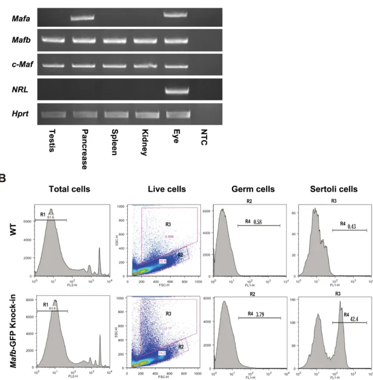
The functions of genes are determined in part by their expression patterns. The expression of large MAF transcription factors in mouse testes has not been fully elucidated. To identify which MAF factor is expressed in testes, MAF mRNAs were analyzed by RT-PCR in adult mouse tissues. We extracted the total RNA from various mouse tissues, including the testes, pancreas, spleen, kidney, and eye. After synthesizing the cDNAs, PCR was performed for Mafa,Mafb,c-Maf,Nrl, and the internal controlHprt. The results showed thatMafband c-Mafare the only large MAF transcription factors expressed in testes, in addition to their expression in the pancreas, spleen, kidney, and eye.Mafawas expressed in the pancreas and eye, whileNrlwas only detectable in the eye (Fig 1A). Next, we focused on the transcription factorMafb. To determine whetherMafbexpression in testes was from somatic cells or germ cells within the seminiferous tubules, we used adultMafb-GFP knock-in mice in which the GFP gene was inserted into one allele of theMafblocus, and WT mice were used as the con-trol. Testicular cells were analyzed by flow cytometry as explained previously in the Materials and Methods. Testicular live cells (R1 population) had two subpopulations: R2, which repre-sented germ cells, and R3, which reprerepre-sented Sertoli cells (Fig 1Band SupplementalS1 Fig). The proportion of GFP-positive cells (R4) from both germ cells and Sertoli cells revealed that a small population (3.79%) of germ cells expressedMafb, while 42.4% of the Sertoli cells expressedMafb(Fig 1B).
Fig 1. Expression of MAFB in mouse testes.(A) Reverse transcription-PCR (RT-PCR) of the large MAF transcription factorsMafa,Mafb,c-MafandNrlin various mouse tissues. Total RNA isolated from mouse testis, pancreas, spleen, kidney, and eye was used for RT-PCR analysis as described in the Materials and Methods section. RT-PCR ofHprtwas carried out as a positive control. NTC was a non-template control. The RT-PCR analysis shown is representative of two independent experiments. (B) Detection of MAFB expression at Sertoli and germ cells in adult mouse testes. Testicular cells obtained from WT (upper panel) orMafb-GFP knock-in mice (lower panel) were analyzed by FACS. Propidium iodide (PI)-negative cells (R1) were selected for flow sorting, while PI-positive dead cells were removed. The R1 fractions from WT orMafb-GFP knock-in testicular cells were classified into two fractions, R2 representing germ cells and R3 representing Sertoli cells. GFP-positive cells are represented by the R4 fraction. The proportion of each fraction is shown above the bar.
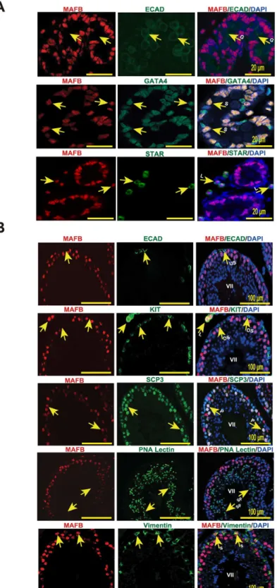
was detected in Leydig cells, as indicated by MAFB co-localization with STAR+cells outside seminiferous tubules, and in Sertoli cells, as indicated by MAFB co-localization with GATA4+ cells inside the tubules. In contrast, germ cells (E-Cadherin+) were negative for MAFB at the E18.5 stage (Fig 2A).
At the adult stage, Mafb was localized by staining of anti-MAFB together with markers for undifferentiated spermatogonia (E-cadherin), differentiated spermatogonia (KIT+inside the tubule), spermatocytes (SCP3), spermatids (PNA-Lectin), Sertoli cells (vimentin), and Leydig cells (KIT+outside the tubule) (Fig 2B). The results revealed that MAFB was localized at Leydig cells and Sertoli cells as indicated by MAFB co-localization with KIT+cells outside the tubule and with vimentin, respectively. In addition, pachytene spermatocytes were found to be the only germ cells that expressed MAFB, as determined by MAFB co-localization with SCP3 cells and the morphology of their DNA. However, neither spermatogonia (ECAD+or KIT+) nor other advanced germ cells, including spermatids (PNA-Lectin+) and testicular sperm expressed MAFB protein.
Testis morphogenesis developed normally in
Mafb
KO mice
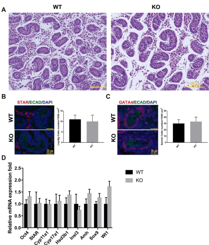
As described above,Mafbis expressed in embryonic testes. The effects ofMafbdisruption on fetal testis morphogenesis were examined at the histological level and molecular level. We first analyzed the histology of prenatal mouse testes just before birth at E18.5 by staining with HE staining. The results showed that in for theMafbKO mice, the seminiferous tubules were fully formed, and no abnormal structure was found in the testis sections (Fig 3A). Furthermore, Leydig and Sertoli cell numbers, the two types of cells that expressMafbin embryonic testes, were counted and shown as the number of cells per unit area (Fig 3B and 3C). Immunostain-ing for STAR (a Leydig marker) and GATA4 (a Sertoli marker within seminiferous tubules) were used as indicators. The average number of Leydig cells was 11±2 for WTvs. 10±3 in KO mice per 10000μm2area. The average number of Sertoli cells was 30±6 for WTvs. 33±7 in KO mice per 10000μm2area. In total, Leydig and Sertoli cell numbers did not differ significantly between KO and age-matched WT testes. In addition, no abnormal distribution or clustering of the Leydig cells was observed in the KO testes. To confirm and extend the histological data, the expression levels of cell type-specific genes involved in normal development and function of the testes were examined at E18.5 using qRT-PCR (Fig 3D). The expression level ofOct4 (also known asPou5f1), which is expressed exclusively in embryonic gonads by early pluripo-tent germ cells, was not altered between WT and KO testes. On the other hand, the expression levels of genes involved in steroid hormone synthesis as indictors of fetal Leydig cell develop-ment and function were examined in the KO testes. The steroidogenic protein levels ofStAR, which facilitates the rapid transport of cholesterol into mitochondria,Cyp11a1, which catalyzes the conversion of cholesterol to pregnenolone,Cyp17a1, which catalyzes the conversion of pregnenolone into hydroxyprogesterone, andHsd3b1, which converts pregnenolone to pro-gesterone did not differ significantly between WT and KO testes, indicating that steroidogenic output was not compromised in the KO testes. Moreover, the expression ofInsl3, which is pro-duced by Leydig cells and is important for fetal testis descent, was unaltered. Similarly, the expression levels of the Sertoli cell markersAmh,Sox9, andWt1were statistically unchanged between KO and WT mice. These data indicated that the development of the germ cell and somatic cell lineages in fetal testis are not disrupted in the absence ofMafb.
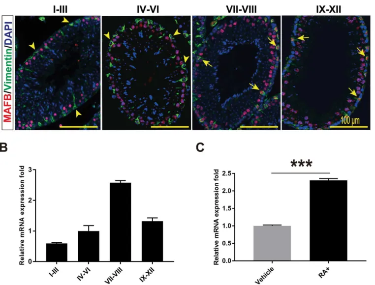
MAFB in Sertoli cells is a stage-specific and induced by RA
of Sertoli cells marker vimentin together with MAFB for the different seminiferous stages: I-III, IV-VI, VII-VIII and IX-XII. We found that the expression of MAFB in Sertoli cells was not observed in stages I-VI but was highly detectable in stages VII-VIII followed by stages IX-XII (Fig 4A). This observation was further confirmed by isolating the different seminifer-ous stages from mature WT mice testis followed by RNA extraction and qRT-PCR. Similarly, the mRNA level ofMafbwas dramatically increased in stages VII-VIII (2.58±0.07-fold) fol-lowed by stages IX-XII (1.32±0.11-fold) (Fig 4B).Mafbin stages I-III and IV-VI was 0.59
±0.03-fold and 1.00±0.18-fold, respectively. These data indicate thatMafbexpression in Sertoli
cells is stage specific.
Next, because it is that stages VII-VIII are the stages in which A aligned spermatogonia transform into A1 under the control of RA [31–36], we examined whetherMafbis induced by RA synthesized in Sertoli cells. For this purpose, primary Sertoli cells were isolated and cultured in serum-free medium for 4 days followed by stimulation with RA at day 5. The expression changes inMafbwere quantified by qRT-PCR. The data revealed that theMafb expression level was increased significantly by RA compared to that in control cells (Fig 4C). Altogether,Mafbshowed the highest expression during stages VII-VIII, and the expression was increased by RA treatment in cultured Sertoli cells.
Generation of
Mafb
conditional KO mice
We generated a tamoxifen-induced time-dependent deletion of conditionalMafballeles. First, mice carrying floxed alleles ofMafb(Mafbfl/fl) were generated and then crossed with mice expressing Cre-ER™recombinase (CAG-CreER™), which was driven by the chickenβ-actin promoter and ubiquitously expresses the inducible Cre recombinase gene in all cell types [37].
After two generations, we obtained mice carrying bothMafbfloxed alleles and one copy of the Cre-ER™transgene (Mafbfl/fl;CAG-CreER™). Mice of theMafbfl/fl;CAG-CreER™genotype are herein designatedMafb-cKO, and age-matchedMafbfl/flmice were considered controls (Fig 5A). To determine the efficiency of the Cre deleter line used, we crossed CAG-CreER™ driver mice with double-fluorescent Cre reporter mice R26GRR (ROSA26CAG-GFP/tdsRed) that express GFP fluorescence constitutively in all cell types prior to Cre-mediated excision and tdsRED after excision [21,22]. The resulting male offspring were genotyped, and double het-erozygous mice carrying GRR and Cre recombinase were then injected with tamoxifen at 6 weeks age. Two weeks later, the expression of green or red fluorescence in the testes was ana-lyzed under a fluorescence microscope. Sections of the testes switched from green to red fluo-rescence, indicating that Cre-ER™was efficient (Fig 5B). Next, to check the ablation ofMafbin the cKO mice, six-week-old mice were intraperitoneally injected with tamoxifen, and time-courses of Mafb excision from tail DNA were examined at post tamoxifen injection days 1, 5, 10 and 15 usingMafbexcision primer (MafbΔ).Mafbwas found to be excised efficiently in adult mice starting from day 10 post-tamoxifen injection (Fig 5C). To further confirm MAFB deletion in the testes, two weeks following tamoxifen injection, mice were sacrificed, and MAFB expression in the testes was analyzed by immunohistochemistry (IHC) and western blotting. IHC results showed that no fluorescence signals were detected in cKO testes com-pared with those in the controls (Fig 5D). In addition, western blotting confirmed the absence of MAFB protein in the cKO mice testes (Fig 5E). These results indicated thatMafb condi-tional alleles were successfully generated and tamoxifen induces MAFB deletion in cKO mice. represent stage VII. US; undifferentiated spermatogonia. DS; differentiated spermatogonia. P; pachytene
spermatocytes. Sp; spermatids. S; Sertoli cells. L; Leydig cells. MAFB was specifically detected in Leydig cells, Sertoli cells, and pachytene spermatocytes.
Fig 3. Testis morphogenesis ofMafbKO embryos developed normally.(A) Histological section of WT and KO E18.5 testes stained with HE. No morphological alteration or distribution was detectable. (B and C) Counts of Leydig and Sertoli cells from E18.5 WT and KO testes. Embryonic testes (n= 3 per genotype) were sectioned, and 4 sections for each gonad were randomly selected and stained with the germ cell marker E-cadherin (green) together with either STAR (red) or GATA4 (red). Numbers of STAR-positive cells outside seminiferous tubules (Leydig cells) or GATA4-positive cells inside the tubules (Sertoli cells) were counted per unit area. The values are the mean±S.D.P<0.05. There was no significant difference between WT and
MAFB is dispensable for spermatogenesis maintenance in adult mouse
testes
At first, we aimed to investigate whether MAFB is required for initiation of spermatogenesis in neonatal mice. For this purpose, we induced MAFB deletion before spermatogonia differentia-tion begins by tamoxifen injecdifferentia-tion on postnatal day 1 to 3. Unexpectedly, cKO mice died soon after MAFB deletion at postnatal days 5–10 (unpublished data). In contrast, at postnatal day 45 (Oct4), Leydig cells (Cyp17a1,StAR,Insl3,Hsd3b1, andCyp11a1), and Sertoli cells (Amh,Sox9, andWT1) was determined by qRT-PCR in E18.5 WT and MafbKO testes. Gene expression levels were normalized toHprt. The bars represent the mean±SEM of five individuals.
P<0.05.
https://doi.org/10.1371/journal.pone.0190800.g003
Fig 4. MAFB expression in seminiferous is stage specific and induced by RA.(A) Seminiferous tubules at stages I-III, IV-VI, VII-VIII and IX-XII were double immunostained with MAFB (red) and vimentin (green). Nuclei were counterstained with DAPI (blue). Arrowhead indicates MAFB-negative Sertoli cells, while Arrows indicated MAFB-positive Sertoli cells. MAFB in Sertoli cells was specifically detected in stages VII to XII.(B)Seminiferous tubules at stages I-III, IV-VI, VII-VIII and IX-XII were isolated from mature mouse testis, mRNAs were extracted, and the expression levels ofMafbwere then compared by qRT-PCR. The proportion of stage-dependentMafbexpression is shown as columns.Hprtwas used as an internal control. Bars represent the mean±S.D. (C) Cultured primary Sertoli cells were treated with 1μM RA for 24 hours and expression changes inMafbwere quantified by qRT-PCR (n= 3).Hprtwas used as an internal control. The bars represent the mean±S.D.
P<0.001.
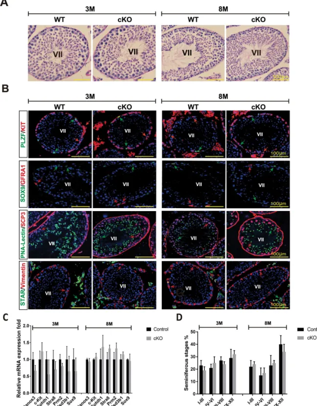
(adult stage), all mice were alive upon MAFB deletion, indicating that MAFB deletion led to neonatal death. Therefore, we analyzed whether MAFB deficiency would result in a disruption of spermatogenesis of adult mice. For this purpose, cKO and control mice (n= 4, per group) were injected with tamoxifen at 6 weeks of age and analyzed at 3 and 8 months of age. Each single animal was sacrificed, testes were dissected and histological sections were prepared and analyzed by PAS staining. The results showed that the seminiferous tubules had no abnormal morphological structure and exhibited complete spermatogenesis at both time points exam-ined (Fig 6A). We then examexam-ined the absence of any testicular cell types by immunostaining with various cell type-specific markers. The results showed that the undifferentiated spermato-gonia (GFRA1+or PLZF+), differentiated spermatogonia (KIT+inside the tubule), spermato-cytes (SCP3+), spermatids (PNA-Lectin+), Sertoli cells (SOX9+or vimentin+), and Leydig cells Fig 5. Generation ofMafb-cKO and efficiency evaluation.(A) Targeting strategy to generateMafbfl/flCAG CreER™(Mafb-cKO) mouse. The genomic structure of the mouseMafbgene (top line) and illustrations of the targeting vector (second line) are shown. TheMafbsingle exon flanked by loxP sites (green triangles), and a neomycin (Neo) resistance cassette was inserted downstream flanked by FRT sites (orange triangles). The resultant targeted allele is shown (third line). The neomycin cassette was excisedin vivoby crossing with the general FLP deleter strain, and the resultant floxed allele (fourth line) is shown. The floxed allele was crossed with the desired CAG-CreER™(fifth line). The deleted allele after induction by tamoxifen is presented (bottom line). Restriction enzyme sites are shown in blue. PCR primers sites are indicated (blue triangles). (B) Efficiency of the Cre driver mice. The R26GRR reporter mouse, containing two cassettes EGFP flanked by loxP sites and tdsRED, was mated with the CAG-CreER™driver mouse strain that ubiquitously expresses CreER™in all cell types. The testes from a resultant male CAG-CreER™;R26GRR mouse were sectioned and analyzed pre- (upper panel) and post-tamoxifen injection (lower panel). The ability of the reporter mice to express tdsRED exclusively in testes was confirmed by microscopic examination of unstained sections. The mice crossing strategy is shown on the right. (C) Time-course ofMafbexcision in the cKO mice. Six-week-old controls as well as cKO mice were intraperitoneally injected with tamoxifen. gDNA was extracted from mouse tails at post-tamoxifen injection days 1, 5, 10 and 15, and PCR was performed using aMafbΔprimer: 400 bp represents the deletedMafbexon. D; Day. M; Marker. bp; base pair. (D) IHC was performed to verify MAFB deletion from cKO mouse testes after tamoxifen injection. Immunostaining for MAFB (red); nuclei were counterstained with DAPI (blue) as shown. The upper panel shows the control, while the lower panel shows cKO mouse testes. (E) Western blot confirming MAFB deletion in cKO mouse testes after tamoxifen injection. Total testis proteins were isolated, ~80μg was loaded per well, and then the blot was probed with anti-MAFB or anti-β-actin antibody (internal control) for both the control and cKO mice, as shown. kDa; kilodaltons.
(KIT+outside the tubule or STAR) were all present in the testicular sections from 3 and
8-month-old mice (Fig 6B). To confirm the histological data, the expression levels of cell type-specific genes involved in spermatogenesis development and testis functions were examined in 3- and 8-month-old cKO mice compared with age-matched control mice using qRT-PCR (Fig 6C). The expression levels of the following germ cell marker genes were not significantly dif-ferent between cKO and control mice:Nanos3, expressed by undifferentiated spermatogonia; c-Kit, expressed by differentiated spermatogonia;Sohlh1, expressed by undifferentiated and differentiated spermatogonia;Stra8, expressed by differentiated spermatogonia and prelepto-tene spermatocytes with the onset of mitotic/meiotic switching; andPrm2, expressed at post-meiotic by haploid spermatids. Similarly, the expression level of the Leydig cell protein Hsd3b1involved in steroidogenic synthesis and the Sertoli cell proteinSox9were statistically unchanged between cKO and control mice. Furthermore, we examined whether there were any differences in the proportion of seminiferous stages in 3- and 8-month-old cKO mice compared with to age-matched controls (Fig 6D). Testis sections were stained by PAS staining and scored as explained in Materials and Methods. The data showed that the percentage of seminiferous stages at the examined ages were not significantly different. Collectively, we con-cluded that testicular function and spermatogenesis of theMafb-cKO mice were apparently normal.
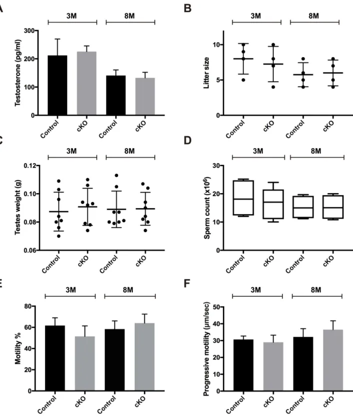
As MAFB is expressed in Leydig cells, we then examined if the lack of MAFB would affect the testosterone levels produced by Leydig cells, which is a mandatory hormone for spermato-genesis (Fig 7A). Therefore, we collected blood samples and checked plasma testosterone levels from 3- and 8-month-old male cKOversusage-matched control mice (n= 4, each group). There were no changes in plasma testosterone levels in either the 3-month-old mice (cKO = 225.56±20.07vs. control = 212.23±57.83 pg/ml (mean±SEM);P= 0.8382) or the 8-month-old mice (cKO = 132.6±19.75vs. control = 141±19.73 pg/ml (mean±SEM);
P= 0.7802). Each single animal was then tested for fertility by mating with one WT C57BL/6J female. The cKO mice were shown to be fertile, with a litter size comparable to the age-matched control (Fig 7B). The mean pup number for the 3-month-old group was cKO = 7.25
±1.25vs. control = 8±1.8 pups (mean±SEM;P= 0.0.6658), and the 8-month-old group was
cKO = 6±0.91vs. control = 5.75±0.85 pups (mean±SEM;P= 0.8481). The mice from 3- and 8-month-old groups were then sacrificed, testis weight was measured, and the cauda epididy-mis was dissected for sperm analyses. The results showed that testis weight, sperm count, sperm motility, and progressive motility were not statistically different between cKO and age-matched control mice (Fig 7C–7F). Together these data illustrate that MAFB is not required for spermatogenesis maintenance in adult mice.
Changes in the transcriptome profile of
Mafb
-cKO Sertoli cells
Since MAFB is a transcription factor, we were interested in examining whether the expres-sion of the transcriptome of Sertoli cells was affected by the absence of MAFB. Sertoli cells were sorted from 3-month-old cKO as well as age-matched control mice (n= 3, each group). Total RNA was extracted from the isolated cells, and RNA samples (pooling for WT or cKO,n= 3 each) were analyzed by RNA-Seq. The purity of the isolated cells was The marker genes representing undifferentiated spermatogonia (Nanos3), differentiated spermatogonia (c-Kit), undifferentiated and differentiated spermatogonia (Sohlh1), differentiated spermatogonia and preleptotene spermatocytes (Stra8), spermatids (Prm2), Leydig cells (Hsd3b1), and Sertoli cells (Sox9) were analyzed by qRT-PCR. Each reaction was performed in duplicate for each gene. The data represent the means±SEM and are shown as relative mRNA expression after normalization toHprt.P<0.05. (D) The proportion of
seminiferous stages: I-III, IV-VI, VII-VIII, and IX-XII from each genotype.P<0.05. M; Month.
Fig 7. Examination of the testosterone levels and male fertility of adult cKO mice.Six-week-old cKO mice were injected with tamoxifen and examined at 3- and 8-months of age compared with age-matched controls (n= 4 for each group).(A)Blood plasma testosterone levels. (B) Litter size. (C) Testis weights. (D) Epididymal sperm counts. (E) Sperm motility %. (F) Progressive motility. M; Month. The data represent the mean±SEM.
P<0.05.
confirmed by immunostaining using a Sertoli cell-specific marker (S2A Fig). The peaks rep-resenting RNA-Seq tag counts of theMafbsingle exon confirmed the reduction ofMafbin cKO Sertoli cells (S2B Fig). RNA-Seq analysis showed that 1937 transcripts with a fold change2 in cKO Sertoli cells were differentially regulated, and among them, 1103 were down-regulated and 834 were up-regulated. Detailed information regarding the down-regu-lated and up-regudown-regu-lated transcripts is listed inS1andS2Tables, respectively.Gng11,Nck2, Prr3,Tlr1, andNlrp3were randomly selected and examined by qRT-PCR which validated the RNA-Seq data (S2C Fig).
DAVID Database was used for KEGG pathway analyses of the identified DEGs with fold change2 inMafb-cKO Sertoli cells as indicated inTable 2. The pathways withP-value<0.05
and enrichment gene count3 of the down- and up-regulated genes were not related to repro-duction. Notably, the identified down regulated genes were related to immunodeficiency and phagocytosis activity of Sertoli cells, as revealed inTable 2.
Discussion
The present study was undertaken in an effort to define the physiological role of theMafbgene in mammalian testes. MAFB is a transcription factor that plays an important role in regulating the development and differentiation of various tissues. A previous study of embryonic mouse gonads showed that MAFB is expressed in the XY gonads along the gonad-mesonephros bor-der as early as E11.5 and is then restricted to Leydig cells by E14.5 [10]. Subsequent studies have usedMafb-GFP knock-in as an early marker of the Leydig cell lineage in mice [38–40]. Whether MAFB is necessary for embryonic testis morphogenesis or not is unknown. Just before birth at E18.5, we localized MAFB expression and found that MAFB is expressed in Ser-toli cells in addition to being expressed in Leydig cells. However, examination of E18.5Mafb KO embryos via somatic cell counts, histological analysis, and molecular analysis indicated that testis morphogenesis was normal. At the adult stage, it has been reported that the active metabolite of vitamin A (RA) is essential for initial differentiation and meiotic entry of mouse spermatogonia [11]. Experiments using vitamin A-deficient mice resulted in a blockade of A to A1 spermatogonia transition, and spermatogenesis could be recovered upon RA treatment [13]. More precisely, RA drives spermatogonia differentiation in two ways: the first spermato-genic wave due to RA from Sertoli cells and subsequent waves due to RA from spermatocytes [11,14–19]. However, the downstream targets of RA are still unknown. As the evidence that the large MAF transcription factor inDrosophila melanogaster,TJ, which encodes an ortholo-gue of the typical bZIP transcription factors MAFB and c-MAF in vertebrates, was shown to be mandatory for germ cell differentiation in Drosophila [9]. In addition, RA was reported to induce MAFB expression and there is a robust RAR-binding site at the end of theMafbcoding region [11,41]. Therefore, MAFB might have a conserved function between species to induce spermatogonial differentiation under the control of RA. In this regard, we found that MAFB is localized in Sertoli cells and pachytene spermatocytes in postnatal mouse seminiferous tubules, as confirmed by immunohistochemical staining and flow cytometric analyses. The expression of MAFB protein in Sertoli cells was specific to stages VII-XII but was not observed in stages I-VI, which can be explained by the low mRNA expression level ofMafbat stages I-VI. Along the same line, it is well known that stages VII-VIII, in whichMafbwas highly expressed, are the stages of A aligned spermatogonia transformation into A1 under the control of RA [31– 36]. Our observations were consistent with the hypothesis that the transcription factor MAFB may take part in the regulation of the onset of the spermatogenic cycle.
Surprisingly, our data revealed that adult cKO mice were fertile and did not exhibit any abnor-mality in testicular histology, proportion of seminiferous stages, expression levels of germ cell markers, or plasma testosterone levels, indicating that MAFB is dispensable for spermatogene-sis. Similarly, GATA-1 expression in Sertoli cells, which is induced in response to the cyclical changes of seminiferous stages, specifically during stages VII-IX, was found to be dispensable Table 2. KEGG pathway analysis of the DEGs inMafb-cKO Sertoli cells.
KEGG_id Description Count List Total
Genes P-value
Down-regulated
mmu04620 Toll-like receptor signaling pathway
21 303 PIK3CG,CCL3,IL6,LY96,TLR1,CXCL9,TLR2,NFKBIA,FADD,CD40,TLR6,CCL4, TLR7,CXCL10,CD86,RIPK1,MAP3K8,PIK3R5,CD14,AKT3,SPP1
1.40E-07
mmu04060 Cytokine-cytokine receptor interaction
33 303 CCL3,CCL2,TNFRSF25,TNFRSF12A,CSF2RB2,CCR1,IL18,IL21R,CXCL2,CCL9, CXCL9,PF4,CCL4,CCL7,IL10,CXCL10,CCL6,CCL24,TNFRSF1B,TNFRSF11A, IL10RA,CSF3R,PDGFC,IL13RA1,IL1A,IL6,BMP2,TNFRSF17,CD40,GM614,OSM,
ACVR2B,CX3CR1
1.10E-06
mmu03010 Ribosome 18 303 GM7290,GM7808,GM12174,GM3362,RPL36A,GM12034,GM5786,GM8730,GM5621, GM14173,GM6030,GM10036,GM4149,GM14323,RPL21,GM15772,GM14217,
GM10132,RPS27A,GM15500,GM11942,GM10269
2.80E-06
mmu04062 Chemokine signaling pathway 25 303 CCL3,CCL2,PREX1,CCR1,CXCL2,CCL9,CXCL9,NFKBIA,PF4,CCL4,CCL7,PRKX, CCL6,CXCL10,CCL24,PTK2,RAC2,PIK3R5,AKT3,PIK3CG,LYN,HCK,WAS,
CX3CR1,JAK2
2.40E-05
mmu04210 Apoptosis 13 303 PIK3CG,CFLAR,CSF2RB2,RIPK1,NTRK1,CASP12,EXOG,NFKBIA,FADD,PIK3R5, PRKX,IL1A,AKT3
1.80E-03
mmu05322 Systemic lupus erythematosus 14 303 C3,GM14474,HIST1H2BG,HIST1H2BH,SNRPD1,FCGR4,SSB,CD40,IL10,TRIM21, C8G,CD86,FCGR2B,GM14484
2.70E-03
mmu04621 NOD-like receptor signaling pathway
10 303 IL6,CARD9,CCL2,NAIP2,IL18,CXCL2,NFKBIA,NLRP3,CASP1,CCL7 4.70E-03
mmu04662 B cell receptor signaling pathway
11 303 PIK3CG,CD19,LYN,RAC2,FCGR2B,NFKBIE,NFKBIA,PIK3R5,AKT3,BLNK, NFATC1
8.70E-03
mmu05340 Primary immunodeficiency 7 303 GM614,DCLRE1C,PTPRC,CD19,CD4,CD40,BLNK 1.00E-02
mmu04370 VEGF signaling pathway 10 303 PIK3CG,PTK2,PTGS2,RAC2,JMJD7,MAPKAPK3,PIK3R5,PLA2G2D,AKT3,NFATC1 1.80E-02
mmu00520 Amino sugar and nucleotide sugar metabolism
7 303 RENBP,UAP1,HK3,GFPT2,PGM1,UGDH,UXS1 2.60E-02
mmu04666 Fc gamma R-mediated phagocytosis
11 303 PIK3CG,PTPRC,PLD1,LYN,RAC2,FCGR2B,HCK,RPS6KB2,PIK3R5,WAS,AKT3 3.20E-02
mmu00500 Starch and sucrose metabolism 6 303 GANC,UGT1A6B,HK3,PGM1,UGDH,UXS1 3.90E-02
mmu04610 Complement and coagulation cascades
9 303 PLAT,C3AR1,C5AR1,C3,F13A1,SERPINE1,CFH,PLAUR,C8G 4.20E-02
mmu04660 T cell receptor signaling pathway
12 303 PIK3CG,PTPRC,NFKBIE,NCK1,MAP3K8,NFKBIA,CD4,PIK3R5,IL10,AKT3,TEC, NFATC1
4.50E-02
Up-regulated
mmu05016 Huntington’s disease 12 178 DNAH7B,NDUFB3,DNAH10,NDUFA5,NDUFB4,COX8C,CYCT,DNAH17,COX7C, DNAH1,DNAIC1,NDUFB2
5.70E-03
mmu01100 Metabolic pathways 42 178 INPP1,NDUFB3,NDUFB4,SEC1,GBGT1,COX7C,AASS,TKTL2,GPAT2,NDUFB2, PLCH2,PEMT,FUT4,ENO3,B3GNT3,CSL,PCYT2,B4GALT7,ACSL6,DCTPP1, NDUFA5,COX8C,CYCT,MCAT,FBP1,DGKH,POLR1C,CDO1,LPCAT4,APRT, NME5,PLCG1,PTGDS,PYGM,RRM2,DPM2,MTAP,PLA2G2C,SMPD4,SMPD3,
NMNAT1,DUT
1.03E-02
mmu03460 Fanconi anemia pathway 5 178 SLX4,SLX1B,FANCG,UBE2T,ERCC1 2.90E-02
mmu03013 RNA transport 9 178 AAAS,NUP62,RASL2-9,PABPC2,THOC6,THOC7,NUP188,NUPL2,POM121L2 4.10E-02
Count, number of the down-regulated genes involved in a pathway; KEGG, Kyoto Encyclopedia of Genes and Genomes.
Statistically significant,P<0.05.
for spermatogenesis as well [43,44]. Many other examples of genes expressed in testes with dispensable function have been reported [44–54]. Several possible explanations may account for these phenomena. First, spermatogenesis is a complex process that involves many genes, and the inactivation of one gene may not be sufficient to induce a detectable phenotype [55]. Consistently, we found that bothMafbandc-Mafare expressed in postnatal testes and that c-MAF protein may compensate for the function of c-MAFB, as in macrophages [56,57]. Thus, double knockout mice should be further evaluated. Second, the defects inMafb-cKO mouse testes may be too small to be detected using the techniques employed here. A detailed ultra-structural examination of testicular tissues may be needed to confirm any minor changes in cKO mice. Third, the up-regulation of theMafbexpression started at stage VII is a dramatic response to the RA signaling that is periodically activated during stages VII-XII, and this acti-vation alters the expression of various Sertoli cell’s genes [13,30].
Next, we aimed to investigate whether MAFB is required to establish the first spermato-genic wave in neonatal mice testis. Before the spermatogonia differentiation started, we induced MAFB deletion by tamoxifen injection on postnatal days 1 to 3. Spermatogonia are known to begin to differentiate between postnatal days 6–10 [58–60]. Unexpectedly, the cKO mice died shortly after MAFB deletion between postnatal days 5–10 and therefore could not be reliably analyzed. However, as Sertoli cells could not initiate the first spermatogenic wave in adult VAD mice until RA recovered, we hypothesized that if MAFB in Sertoli cells acts down-stream of RA to initiate the first spermatogenic wave, the transcriptome profile ofMafb-cKO Sertoli cells would be affected. Thus, we analyzed the transcriptome profile of Sertoli cells by RNA sequencing. Signaling pathway analyses of the DEGs were not related to spermatogene-sis. However, the down-regulated transcripts were related to immunodeficiency and phagocy-tosis pathways. These data could be the fourth explanation for the possible function of MAFB as a phagocytic modulator in Sertoli cells during the identified positive seminiferous stages. In this connection, it is known that during spermatogenesis half of differentiating germ cells undergo apoptosis before maturing into spermatozoa [61–63]. Since macrophages, the integral compartment of the immune system, are absent inside the seminiferous tubules, Sertoli cells that are endowed with the phagocytic properties respond to the inflammatory signals by rapid engulfment of the apoptotic germ cells within the seminiferous epithelium [63–67]. Further studies should clarify if MAFB, an important modulator of macrophage phagocytosis, is involved in such process in Sertoli cells [68–70]. Moreover, the possibility that MAFB might be required for initiation of the first spermatogenic wave in neonatal testis should be clarified using a Sertoli cell-specific Cre driver mouse.
Conclusions
The transcription factors MAFB and c-MAF are expressed in mouse testes. MAFB localized in Leydig and Sertoli cells in testes at E18.5 while it localized in Leydig cells, Sertoli cells, and pachytene spermatocytes in adult testes. Our current examination technique revealed that MAFB-deficient testes developed normally by E18.5, and spermatogenesis was not disrupted in adult mice. The transcriptome profile of adult cKO Sertoli cells showed the down-regulation of genes related to the immune function and phagocytosis activity of Sertoli cells.
Supporting information
S1 Table. List of the down-regulated transcripts ofMafb-cKO Sertoli cells.
S2 Table. List of the up-regulated transcripts ofMafb-cKO Sertoli cells.
(XLSX)
S1 Fig. Sertoli and germ cell separation by FACS.Immunostaining with the Sertoli cell marker vimentin (red) before and after cell sorting. Upper panels (R1): before sorting. Middle (R2) and Lower (R3) panels: after sorting. The R2 population represents germ cells as vimentin negative, while the R2 population represents Sertoli cells as vimentin positive. Nuclei were counterstained with DAPI (blue). The proportion of vimentin-positive cells is shown in the R3 merged photo. The value is the mean±S.D.
(TIF)
S2 Fig. RNA-Seq validation for the transcriptome profile ofMafb-cKO Sertoli cells.Sertoli cells were sorted from three-month-oldMafb-cKO or control mice and analyzed by RNA-Seq. (A) The purity of the isolated cells was confirmed by IHC staining with the Sertoli cell-specific marker anti-vimentin antibody (red) and nuclei were counterstained with DAPI (blue). The proportion of vimentin-positive cells is shown in the merged photo. (B) UCSC genome browser screenshot showing the RNA-Seq tag counts. Reads from both genotypes were mapped to the single exon of theMafbgene and visualized to confirm the reduction in the cKO reads. The full genome sequence for Mus musculus provided by UCSC (mm10, Dec. 2011) was used for mapping by HISAT. The arrow under the panel shows the exon and indi-cates the direction of transcription. Each panel is labeled with the genotype. The read histo-grams show the number of reads at each nucleotide. (C) qRT-PCR validation for the RNA-Seq data analyses. Expression of five differentially expressed genes (up and down) that were ran-domly selected are shown.
(TIF)
Acknowledgments
The authors express deep gratitude to Akihiro Kuno, Tony De Falco, and Yumiko Saga for valuable discussion. The authors would also like to thank Mohamed Samy, Masami Ojima, and Hyojung Jeon for technical assistance.
Author Contributions
Conceptualization:Hossam H. Shawki, Hisashi Oishi, Kazunori Hasegawa, Hany A. El-Shemy, Shosei Yoshida, Satoru Takahashi.
Data curation:Hossam H. Shawki, Yu Kitadate, Risa Okada, Masafumi Muratani, Satoru Takahashi.
Formal analysis:Hossam H. Shawki, Risa Okada, Masafumi Muratani.
Funding acquisition:Shosei Yoshida.
Investigation:Hossam H. Shawki, Yu Kitadate, Kazunori Hasegawa, Keiji Mochida, Atsuo Ogura, Satoru Takahashi.
Methodology:Hossam H. Shawki, Hisashi Oishi, Toshiaki Usui, Yu Kitadate, Walaa A. Basha, Ahmed M. Abdellatif, Kazunori Hasegawa, Keiji Mochida, Masafumi Muratani, Atsuo Ogura, Satoru Takahashi.
Project administration:Hossam H. Shawki, Hisashi Oishi.
Supervision:Hisashi Oishi, Shosei Yoshida, Satoru Takahashi.
Validation:Hossam H. Shawki, Hisashi Oishi, Risa Okada, Hany A. El-Shemy, Masafumi Muratani.
Visualization:Hossam H. Shawki, Toshiaki Usui, Yu Kitadate, Walaa A. Basha, Ahmed M. Abdellatif, Kazunori Hasegawa, Risa Okada, Keiji Mochida, Masafumi Muratani, Atsuo Ogura, Satoru Takahashi.
Writing – original draft:Hossam H. Shawki, Hisashi Oishi.
Writing – review & editing:Shosei Yoshida.
References
1. Hess RA, Renato de Franca L. Spermatogenesis and cycle of the seminiferous epithelium. Adv Exp Med Biol. 2008; 636:1–15. Epub 2009/10/27.https://doi.org/10.1007/978-0-387-09597-4_1PMID: 19856159.
2. Yoshida T, Ohkumo T, Ishibashi S, Yasuda K. The 5’-AT-rich half-site of Maf recognition element: a functional target for bZIP transcription factor Maf. Nucleic Acids Res. 2005; 33(11):3465–78. Epub 2005/06/24.https://doi.org/10.1093/nar/gki653PMID:15972792.
3. Yang Y, Cvekl A. Large Maf Transcription Factors: Cousins of AP-1 Proteins and Important Regulators of Cellular Differentiation. Einstein J Biol Med. 2007; 23(1):2–11. Epub 2007/12/27. PMID:18159220.
4. Blank V, Kim MJ, Andrews NC. Human MafG is a functional partner for p45 NF-E2 in activating globin gene expression. Blood. 1997; 89(11):3925–35. Epub 1997/06/01. PMID:9166829.
5. Fujiwara KT, Kataoka K, Nishizawa M. Two new members of the maf oncogene family, mafK and mafF, encode nuclear b-Zip proteins lacking putative trans-activator domain. Oncogene. 1993; 8(9):2371–80. Epub 1993/09/01. PMID:8361754.
6. Katsuoka F, Yamamoto M. Small Maf proteins (MafF, MafG, MafK): History, structure and function. Gene. 2016; 586(2):197–205. Epub 2016/04/09.https://doi.org/10.1016/j.gene.2016.03.058PMID: 27058431.
7. Eychene A, Rocques N, Pouponnot C. A new MAFia in cancer. Nat Rev Cancer. 2008; 8(9):683–93. Epub 2009/01/15.https://doi.org/10.1038/nrc2460PMID:19143053.
8. Kataoka K. Multiple mechanisms and functions of maf transcription factors in the regulation of tissue-specific genes. J Biochem. 2007; 141(6):775–81. Epub 2007/06/16.https://doi.org/10.1093/jb/mvm105 PMID:17569705.
9. Li MA, Alls JD, Avancini RM, Koo K, Godt D. The large Maf factor Traffic Jam controls gonad morpho-genesis in Drosophila. Nat Cell Biol. 2003; 5(11):994–1000. Epub 2003/10/28.https://doi.org/10.1038/ ncb1058PMID:14578908.
10. DeFalco T, Takahashi S, Capel B. Two distinct origins for Leydig cell progenitors in the fetal testis. Dev Biol. 2011; 352(1):14–26. Epub 2011/01/25.https://doi.org/10.1016/j.ydbio.2011.01.011PMID: 21255566.
11. Raverdeau M, Gely-Pernot A, Feret B, Dennefeld C, Benoit G, Davidson I, et al. Retinoic acid induces Sertoli cell paracrine signals for spermatogonia differentiation but cell autonomously drives spermato-cyte meiosis. Proc Natl Acad Sci U S A. 2012; 109(41):16582–7. Epub 2012/09/27.https://doi.org/10. 1073/pnas.1214936109PMID:23012458.
12. DeFalco T, Potter SJ, Williams AV, Waller B, Kan MJ, Capel B. Macrophages Contribute to the Sper-matogonial Niche in the Adult Testis. Cell Rep. 2015; 12(7):1107–19. Epub 2015/08/11.https://doi.org/ 10.1016/j.celrep.2015.07.015PMID:26257171.
13. Hogarth CA, Griswold MD. The key role of vitamin A in spermatogenesis. J Clin Invest. 2010; 120 (4):956–62. Epub 2010/04/07.https://doi.org/10.1172/JCI41303PMID:20364093.
14. Hogarth CA, Griswold MD. Retinoic acid regulation of male meiosis. Curr Opin Endocrinol Diabetes Obes. 2013; 20(3):217–23. Epub 2013/03/21.https://doi.org/10.1097/MED.0b013e32836067cfPMID: 23511242.
16. Snyder EM, Small C, Griswold MD. Retinoic acid availability drives the asynchronous initiation of sper-matogonial differentiation in the mouse. Biol Reprod. 2010; 83(5):783–90. Epub 2010/07/24.https://doi. org/10.1095/biolreprod.110.085811PMID:20650878.
17. Vernet N, Dennefeld C, Rochette-Egly C, Oulad-Abdelghani M, Chambon P, Ghyselinck NB, et al. Reti-noic acid metabolism and signaling pathways in the adult and developing mouse testis. Endocrinology. 2006; 147(1):96–110. Epub 2005/10/08.https://doi.org/10.1210/en.2005-0953PMID:16210368.
18. Sugimoto R, Nabeshima Y, Yoshida S. Retinoic acid metabolism links the periodical differentiation of germ cells with the cycle of Sertoli cells in mouse seminiferous epithelium. Mech Dev. 2012; 128(11– 12):610–24. Epub 2011/12/28.https://doi.org/10.1016/j.mod.2011.12.003PMID:22200512.
19. Gely-Pernot A, Raverdeau M, Celebi C, Dennefeld C, Feret B, Klopfenstein M, et al. Spermatogonia dif-ferentiation requires retinoic acid receptor gamma. Endocrinology. 2012; 153(1):438–49. Epub 2011/ 11/03.https://doi.org/10.1210/en.2011-1102PMID:22045663.
20. Moriguchi T, Hamada M, Morito N, Terunuma T, Hasegawa K, Zhang C, et al. MafB is essential for renal development and F4/80 expression in macrophages. Mol Cell Biol. 2006; 26(15):5715–27. Epub 2006/07/19.https://doi.org/10.1128/MCB.00001-06PMID:16847325.
21. Al-Soudy AS, Nakanishi T, Mizuno S, Hasegawa Y, Shawki HH, Katoh MC, et al. Germline recombina-tion in a novel Cre transgenic line, Prl3b1-Cre mouse. Genesis. 2016; 54(7):389–97. Epub 2016/04/29. https://doi.org/10.1002/dvg.22944PMID:27124574.
22. Hasegawa Y, Daitoku Y, Sekiguchi K, Tanimoto Y, Mizuno-Iijima S, Mizuno S, et al. Novel ROSA26 reporter knock-in C57BL/6N mice exhibiting green emission before and red emission after Cre-mediated recombination. Exp Anim. 2013; 62(4):295–304. Epub 2013/11/01.https://doi.org/10.1538/ expanim.62.295PMID:24172193.
23. Pitulescu ME, Schmidt I, Benedito R, Adams RH. Inducible gene targeting in the neonatal vasculature and analysis of retinal angiogenesis in mice. Nat Protoc. 2010; 5(9):1518–34. Epub 2010/08/21.https:// doi.org/10.1038/nprot.2010.113PMID:20725067.
24. Nagao M, Cheong CW, Olsen BR. Col2-Cre and tamoxifen-inducible Col2-CreER target different cell populations in the knee joint. Osteoarthritis Cartilage. 2015; 24(1):188–91. Epub 2015/08/11.https:// doi.org/10.1016/j.joca.2015.07.025PMID:26256767.
25. Kotaja N, Kimmins S, Brancorsini S, Hentsch D, Vonesch JL, Davidson I, et al. Preparation, isolation and characterization of stage-specific spermatogenic cells for cellular and molecular analysis. Nat Meth-ods. 2004; 1(3):249–54. Epub 2005/09/08.https://doi.org/10.1038/nmeth1204-249PMID:16144087.
26. Hasegawa K, Namekawa SH, Saga Y. MEK/ERK signaling directly and indirectly contributes to the cyclical self-renewal of spermatogonial stem cells. Stem Cells. 2013; 31(11):2517–27. Epub 2013/07/ 31.https://doi.org/10.1002/stem.1486PMID:23897718.
27. Hasegawa K, Okamura Y, Saga Y. Notch signaling in Sertoli cells regulates cyclical gene expression of Hes1 but is dispensable for mouse spermatogenesis. Mol Cell Biol. 2012; 32(1):206–15. Epub 2011/11/ 01.https://doi.org/10.1128/MCB.06063-11PMID:22037762.
28. Shawki HH, Kigoshi T, Katoh Y, Matsuda M, Ugboma CM, Takahashi S, et al. Identification, localization, and functional analysis of the homologues of mouse CABS1 protein in porcine testis. Exp Anim. 2016; 65(3):253–65. Epub 2016/03/11.https://doi.org/10.1538/expanim.15-0104PMID:26960363.
29. Meistrich ML, Hess RA. Assessment of spermatogenesis through staging of seminiferous tubules. Methods Mol Biol. 2013; 927:299–307. Epub 2012/09/21.https://doi.org/10.1007/978-1-62703-038-0_ 27PMID:22992924.
30. Hasegawa K, Saga Y. Retinoic acid signaling in Sertoli cells regulates organization of the blood-testis barrier through cyclical changes in gene expression. Development. 2012; 139(23):4347–55. Epub 2012/10/26.https://doi.org/10.1242/dev.080119PMID:23095883.
31. de Rooij DG, Russell LD. All you wanted to know about spermatogonia but were afraid to ask. J Androl. 2000; 21(6):776–98. Epub 2000/12/06. PMID:11105904.
32. de Rooij DG. Proliferation and differentiation of spermatogonial stem cells. Reproduction. 2001; 121 (3):347–54. Epub 2001/02/28. PMID:11226060.
33. Wakayama T, Nakata H, Kumchantuek T, Gewaily MS, Iseki S. Identification of 5-bromo-2’-deoxyuri-dine-labeled cells during mouse spermatogenesis by heat-induced antigen retrieval in lectin staining and immunohistochemistry. J Histochem Cytochem. 2015; 63(3):190–205. Epub 2014/12/07.https:// doi.org/10.1369/0022155414564870PMID:25479790.
34. Oakberg EF. Spermatogonial stem-cell renewal in the mouse. Anat Rec. 1971; 169(3):515–31. Epub 1971/03/01.https://doi.org/10.1002/ar.1091690305PMID:5550531.
36. Nakagawa T, Sharma M, Nabeshima Y, Braun RE, Yoshida S. Functional hierarchy and reversibility within the murine spermatogenic stem cell compartment. Science. 2010; 328(5974):62–7. Epub 2010/ 03/20.https://doi.org/10.1126/science.1182868PMID:20299552.
37. Hayashi S, McMahon AP. Efficient recombination in diverse tissues by a tamoxifen-inducible form of Cre: a tool for temporally regulated gene activation/inactivation in the mouse. Dev Biol. 2002; 244 (2):305–18. Epub 2002/04/12.https://doi.org/10.1006/dbio.2002.0597PMID:11944939.
38. Defalco T, Saraswathula A, Briot A, Iruela-Arispe ML, Capel B. Testosterone levels influence mouse fetal Leydig cell progenitors through notch signaling. Biol Reprod. 2013; 88(4):91. Epub 2013/03/08. https://doi.org/10.1095/biolreprod.112.106138PMID:23467742.
39. Jameson SA, Natarajan A, Cool J, DeFalco T, Maatouk DM, Mork L, et al. Temporal transcriptional pro-filing of somatic and germ cells reveals biased lineage priming of sexual fate in the fetal mouse gonad. PLoS Genet. 2012; 8(3):e1002575. Epub 2012/03/23.https://doi.org/10.1371/journal.pgen.1002575 PMID:22438826.
40. Liu C, Rodriguez K, Yao HH. Mapping lineage progression of somatic progenitor cells in the mouse fetal testis. Development. 2016; 143(20):3700–10. Epub 2016/11/02.https://doi.org/10.1242/dev.135756 PMID:27621062.
41. Zhang Y, Chen QY, Ross AC. Transcription factor MafB is a mediator of retinoic acid-induced monocytic cell differentiation. Faseb Journal. 2010; 24.
42. Blanchi B, Kelly LM, Viemari JC, Lafon I, Burnet H, Bevengut M, et al. MafB deficiency causes defective respiratory rhythmogenesis and fatal central apnea at birth. Nature Neuroscience. 2003; 6(10):1091–9. https://doi.org/10.1038/nn1129PMID:14513037
43. Yomogida K, Ohtani H, Harigae H, Ito E, Nishimune Y, Engel JD, et al. Developmental stage- and sper-matogenic cycle-specific expression of transcription factor GATA-1 in mouse Sertoli cells. Develop-ment. 1994; 120(7):1759–66. Epub 1994/07/01. PMID:7924983.
44. Lindeboom F, Gillemans N, Karis A, Jaegle M, Meijer D, Grosveld F, et al. A tissue-specific knockout reveals that Gata1 is not essential for Sertoli cell function in the mouse. Nucleic Acids Res. 2003; 31 (18):5405–12. Epub 2003/09/05.https://doi.org/10.1093/nar/gkg723PMID:12954777.
45. Bedard N, Hingamp P, Pang Z, Karaplis A, Morales C, Trasler J, et al. Mice lacking the UBC4-testis gene have a delay in postnatal testis development but normal spermatogenesis and fertility. Mol Cell Biol. 2005; 25(15):6346–54. Epub 2005/07/19.https://doi.org/10.1128/MCB.25.15.6346-6354.2005 PMID:16024774.
46. Fantz DA, Hatfield WR, Horvath G, Kistler MK, Kistler WS. Mice with a targeted disruption of the H1t gene are fertile and undergo normal changes in structural chromosomal proteins during spermiogene-sis. Biol Reprod. 2001; 64(2):425–31. Epub 2001/02/13. PMID:11159343.
47. Godmann M, Gashaw I, Katz JP, Nagy A, Kaestner KH, Behr R. Kruppel-like factor 4, a "pluripotency transcription factor" highly expressed in male postmeiotic germ cells, is dispensable for spermatogene-sis in the mouse. Mechanisms of Development. 2009; 126(8–9):650–64.https://doi.org/10.1016/j.mod. 2009.06.1081PMID:19539755
48. Iwamori N, Zhao M, Meistrich ML, Matzuk MM. The testis-enriched histone demethylase, KDM4D, regu-lates methylation of histone H3 lysine 9 during spermatogenesis in the mouse but is dispensable for fer-tility. Biol Reprod. 2011; 84(6):1225–34. Epub 2011/02/05.https://doi.org/10.1095/biolreprod.110. 088955PMID:21293030.
49. Liu YQ, Tao DC, Lu YJ, Yang Y, Ma YX, Zhang SZ. Targeted disruption of the mouse testis-enriched gene Znf230 does not affect spermatogenesis or fertility. Genetics and Molecular Biology. 2014; 37 (4):708–15.https://doi.org/10.1590/S1415-47572014005000013PMID:25505846
50. Mannan AU, Nayernia K, Mueller C, Burfeind P, Adham IM, Engel W. Male mice lacking the Theg (tes-ticular haploid expressed gene) protein undergo normal spermatogenesis and are fertile. Biol Reprod. 2003; 69(3):788–96. Epub 2003/05/16.https://doi.org/10.1095/biolreprod.103.017400PMID: 12748127.
51. Marschang P, Brich J, Weeber EJ, Sweatt JD, Shelton JM, Richardson JA, et al. Normal development and fertility of knockout mice lacking the tumor suppressor gene LRP1b suggest functional compensa-tion by LRP1. Mol Cell Biol. 2004; 24(9):3782–93. Epub 2004/04/15.https://doi.org/10.1128/MCB.24.9. 3782-3793.2004PMID:15082773.
52. Nayernia K, Drabent B, Adham IM, Moschner M, Wolf S, Meinhardt A, et al. Male mice lacking three germ cell expressed genes are fertile. Biol Reprod. 2003; 69(6):1973–8. Epub 2003/08/22.https://doi. org/10.1095/biolreprod.103.018564PMID:12930723.