Inorganic-Biochemical Perspectives
of Sporadic Prion Diseases
Yuzo Nishida
Chemical Institute for Neurodegeneration, Yamagata University
Preface
Between 1980 and roughly 1996, about 750,000 cattle infected with BSE (bovine spongiform encephalopathy) were slaughtered for human consumption in Great Britain, and it is now clear that BSE, also known as “mad cow disease”
is not merely a UK phenomenon, nor is it merely an economic nuisance. The sudden and explosive increase of BSE in recent Europe (1990-2000) may have been spread among cattle by the feeding of infected offal, but the majority of cases of naturally occurring prion diseases arise sporadically with no known cause. Thus, the most important problem to be solved is to elucidate the intrinsic chemical mechanism of the prion diseases which arise sporadically, i.e., we must answer the questions:
What induces the conversion of normal prion protein into an abnormal isoform, and how the abnormal isofom forms without the infected offals ?
Many years ago ALS (amyotrophic lateral sclerosis) patients were collectively found in the New Guinea and Papua islands, and its origin has been attributed to the subterranean water, which contains much Al 3+ and Mn 2+ ions. In Alzheimer’s disease specific region such as the hippocampus and the motor cortex contain elevated iron levels relative to normal, and abnormalities in brain iron metabolism have been described for several neurodegenerative disorders, including Alzheimer’s diseases, Parkinson’s disease, Huntington’s, and prion diseases. Investigations of scrapie, CJD, and chronic wasting disease clusters in Iceland, Slovakia and Colorado, respectively have indicated that the soil in these regions is low in copper and higher in manganese.
Above facts suggest that the sporadic prion and other neurodegenerative diseases are closely related with the function of several transition-metal ions, and thus inorganic-biochemical perspectives are necessary in order to elucidate the chemical mechanisms of pathogenesis of these diseases.
October 20 th , 2006 Yuzo Nishida
Chemical Institute for Neurodegeneration, Yamagata University
Contents
Chapter I Introduction ∙∙∙∙∙∙∙∙∙∙∙ 4 Chapter II Parkinson’s Disease: Deficiency of Neurotransmitters and Neural Cell Death due to Oxidative stress ∙∙∙∙∙∙∙∙ 9 Chapter III Nishida’s Concept on Activation of Oxygen Molecule ∙∙ 14 Chapter IV Copper(II)-hydroperoxide Adduct in Amyotrophic Lateral
Sclerosis (ALS) and Sporadic Prion Diseases ∙∙∙∙∙∙∙∙∙ 23 Chapter V Oxygen Activation in Tyrosine Hydroxylase and its
Derivatives: Factors to prevent the Formation of Neurotransmitters ∙∙∙∙∙∙∙∙∙∙∙ 36 Chapter VI Non-specific Iron Ion and Abnormalities of Brain Iron
Metabolism ∙∙∙∙∙∙∙∙∙∙∙ 48
Chapter VII Summary ∙∙∙∙∙∙∙∙∙∙∙ 55
Chapter I. Introduction
Between 1980 and roughly 1996, about 750,000 cattle infected with BSE (bovine spongiform encephalopathy) were slaughtered for human consumption in Great Britain, and it is now clear that BSE, also known as “mad cow disease” is not merely a UK phenomenon, nor is it merely an economic nuisance. In fact, it may be an impending world-wide health crisis, and in recent months several other European countries have found BSE in their cattle herds, and over the past few years about 100 mostly young individuals have fallen victim to a fatal condition known as new variant Creutzfeld-Jacob disease (vJCD). [1-3] BSE and vJCD are one of the transmissible spongiform encephalopathies (TSEs, or prion disease) which are group of fatal neurodegenerative disorders that include BSE, vJCD, scrapie of sheep, chronic wasting diseases (CWD) of mule deer and elk, as well as Gestmann-Straussler-Scheinker disease (GSS) and fatal familial insomnia (FFI) of humans. [4,5] At present it is generally recognized that BSE may have originated from a scrapie agent infecting small ruminants, which have been recycled through cattle and disseminated through the use of contaminated meat and bonemeal. Compelling evidence links vCJD to exposure to beef infected with BSE prions, and recent studies have suggested that blood-borne transmission of CJD is highly possible.
Some 250 years ago, a sheep disease that presented with excitability,
itching, ataxia and finally paralysis and death was recognized and this is known
today as scrapie in English-speaking countries, “the trembles” in France, “trotting
disease” in Germany and “itching disease” in Japan, reflecting the gamut of its
symptoms. The first major advance in scrapie research took place in 1936 when
Cuille and Chelle succeeded in transmitting the disease to sheep and goats by
inoculating them with lumbar cord of diseased animals. Subsequently,
transmission to mice and hamsters provided more-convenient experimental
models. It was soon recognized that the transmissible agent had quite
extraordinary properties, such as unusually long incubation periods, measured in
months to years, and uncommon resistance to high temperature, formaldehyde
treatment and UV irradiation. Enriching fractions from Syrian hamster (SHa)
brain for scrapie infectivity led to the discovery of the prion protein (PrP), and at
post-translational conversion of the normal cellular prion protein (PrP C ) into an abnormal isoform of called scrapie PrP (PrP Sc ) that has a high- b -sheet content and is associated with transmissible disease. (see Figure 1) [6] These misfodled prions (PrP Sc ) ultimately kills neurons and leaves the brain riddled with holes, like a sponge, and the 1997 Nobel Prize in Physiology and Medicine was awarded to Professor S. Prusiner of the University of California, San Francisco, for his contributions towards the identification of the infectious agent that causes TSEs.
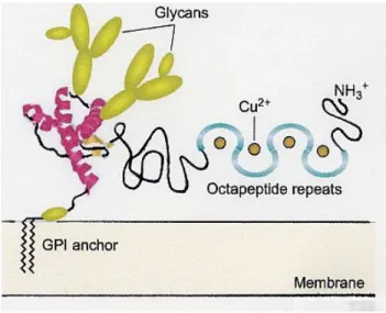
PrP C is a glycoprotein expressed on the surface of many cell types (see Figure 2) and the fact that the protein is expressed in neurons at higher levels than in any other cell types suggests that PrP C has special importance for neurons.
Additionally, PrP C is highly concentrated at the synapse and there is evidence for intense localization not only as central nerves synapse but also at endplates. PrP C is linked to the cell membrane by glycosylphosphatidylinositol (GPI) anchor. (see Fig. 2) [7] It has one or two sugar chains that are closely linked to the C-terminus and also exists in a non-glycosylated form. PrP Sc is extracted from affected brains as highly aggregated, detergent-insoluble materials that is not amenable to high-resolution
structural technique. PrP Sc is covalently indistinguishable from PrP C . During infection, theunderlying molecular events that lead to the conversion of PrP C to the scrapie agent remain ill defined.
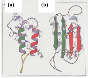
Figure 1. Plausible models for the tertiary structures of PrP Sc and PrP C . (a) The proposed three-dimensional structure of PrP C.
It has been believed that helices 1 and 2 are converted into a b-sheet structure during the formation of PrP Sc . (b) The proposed three-dimensionalstructure of PrP Sc .
(S. B. Prusiner, Trends Biochemical Sciences, 1996, 21, 482)
(a) (b)
Figure 2. Model of PrP C structural domains.
The folded C-terminal portion of PrP C that contains the short b-sheet strands and the a-helix is based on a model derived from NMR-based coordinates of residues of hamster PrP. (B. Caughey, Trends Biochemical Sciences, 2001, 25, 235)
Recent studies have showed that PrP C not only binds copper (Cu) within the octarepeat region located in the unstructured N-terminus, but under certain specific circumstances may bind along the C-terminal structured domain of protein fragments. Furthermore, recombinant PrP C can also bind other metals such as manganese at both the octarepeats and the C-terminal sites. [8] Indeed, accumulating evidence suggests that metallochemical alterations may play a role in the pathogenesis of prion diseases and other neurodegenerative diseases. It has been demonstrated that both recombinant and brain-derived PrP have superoxide dismutase (SOD)-like activity when Cu is bound to the octarepeat region resulting in conformational changes to the protein. [8]
The sudden and explosive increase of BSE in recent Europe may have
been spread among cattle by the feeding of infected offal [9-12] but the majority
cause. Thus, the most important problem to be solved is to elucidate the intrinsic chemical mechanism of the prion diseases which arise sporadically, i.e., we must answer the questions:
What induces the conversion of PrP C to PrP Sc and how the PrP Sc forms in the life process without the infected offals ?
The sporadic neurodegenerative diseases are in general endemic; many years ago ALS (amyotrophic lateral sclerosis) patients were collectively found in the New Guinea and Papua islands, and its origin has been attributed to the drinking subterranean water, which contains much Al 3+ and Mn 2+ ions, and in these regions many patients of Alzheimer’s and Parkinson’s diseases were found, [13]
and increased aluminum levels were reported in the hippocampus of patients with Alzheimer’s disease. [14] In Alzheimer’s disease specific region such as the hippocampus and the motor cortex contain elevated iron levels relative to normal, whereas the occipital cortex contains decreased levels, and abnormalities in brain iron metabolism have been described for several neurodegenerative disorders, including Alzheimer’s diseases, Parkinson’s disease, Huntington’s, and prion diseases. [14-17] Investigations of scrapie, CJD, and chronic wasting disease clusters in Iceland, Slovakia and Colorado, respectively have indicated that the soil in these regions is low in copper and higher in manganese, and Brown et al.
observed striking elevation of manganese ion accompanied by significant reduction of copper ion bound to purified PrP in all sCJD (sCJD = sporadic CJD) variants. [18] Brown et al. have reported that it loses the SOD-like activity when Cu is replaced with Mn in recombinant PrP, and also that Cu binding to PrP purified from sporadic CJD was significantly decreased while the binding of Mn and Zn was markedly increased. [18] These results suggest that altered metal-ion occupancy of PrP plays a pivotal role in the pathogenesis of prion diseases.
In this book we will show the new concept on the “oxidative stress”
induced by the metal ions such as copper, manganese, and iron, etc, [19] to
lead to the sporadic prion diseases and other neurodegenerative diseases which
include ALS, Alzheimer’s and Parkinson’s, and will postulate the comprehensive
chemical mechanism of the sporadic prion diseases..
References
[1] A. C. Chani, N. M. Ferguson, C. A. Donnell, and R. M. Anderson, Nature, 2000, 406, 583.
[2] A. J. Beale, J. Roy. Soc. Med., 2001, 94, 207.
[3] F. Houston, J. D. Foster, A. Chong, N. Hunter, and C. J. Bostock, The Lancet, 2000, 356, 955.
[4] F. E. Cohen and S. B. Prusiner, Annu. Rev. Biochem., 1998, 67, 793.
[5] J. Collinge, Annu. Rev. Neurosci., 2001, 24, 519.
[6] S. B. Prusiner, Trends Biochem. Sciences, 1996, 21, 482.
[7] B. Caughey, Trends Biochem. Sciences, 2001, 25, 235.
[8] D. Brown, Trends Neurosciences, 2001, 24, 85.
[9] B. Chesebro, Science, 2004, 305, 1918.
[10] A. Almond and J. Pattison, Nature, 1997, 389, 437.
[11] G. Legname, I. V. Baskakov, H.-O. B. Hguyen, D. Rieser, F. E. Cohen, S. J.
DeArmond, and S. B. Prusiner, Science, 2004, 305, 673.
[12] P. G. Smith and R. Bradley, British Med. Bull. 2003, 66, 185.
[13] H. Shiraki and Y. Yase, “Handbook of Clinical Neurology”, ed. By P. I.
Vinken, W. Bruyn, H. L. Klawans, Vol. 15, 1991, pp.273-300.
[14] M. Gerlach, D. B.-Schachar, P. Riederer, and M. B. H. Youdim, J.
Neurochemistry, 1994, 63, 793.
[15] M. B. H. Youdim and P. Riederer, Scientific American, 1997, 52.
[16] S. Fernaeus, K. Reis, K. Bedecs, and T. Land, Neuroscience Lett., 2005, 389, 133.
[17] S. Fernaeus, J. Halldin, K. Bedecs, and T. Land, Mol. Brain Research, 2005, 133, 266.
[18] B.-S. Wong, S. G. Chen, M. Colucci, Z. Xie, T. Pan, T. Liu, R. Li, P.
Gambetti, M.-S. Sy, and D. R. Brown, J. Neurochemistry, 2001, 78, 1400.
[19] Y. Nishida, Med. Hypothesis Res. 2004, 1, 227. http://
www.journal-mhr.com/PDF_Files/vol_1_4/1_4_PDFs/1_4_2.pdf.
Chapter II Parkinson’s Disease: Deficiency of Neurotransmitters and Neural Cell Death
due to Oxidative stress
In October 2000, the Nobel Assembly awarded the Nobel Prize in Physiology and Medicine to Carlsson of the University of Gothernburg in Sweden and to two pioneers in the study of nerve cell communications; Paul Greengard of Rockefeller University in New York City, who figured out how dopamine and other neurotransmitters trigger target neurons when they bind at the synapse, the junction between two nerve cells; and Eric Kandel of New York’s Columbia University, who built on these insights to demystify some aspects of learning and memory. Carlsson overturned conventional wisdom by proving that dopamine, once thought to be merely a precursor in the synthesis of the neurotransmitter norepinephrine (see Figure 3), is an important nervous system messenger in its own right. They gave rabbits a drug that depletes norepinephrine in the brain, putting the animals into a temporary stupor.
Carlsson found that the rabbits could be roused with injections of L-dopa, which the brain converts to dopamine. Later they discovered that Parkinson’s diseases results from degeneration of dopamine-producing neurons in the brain involved movement control. That finding led to the use of L-dopa as a therapy for Parkinson’s patients.
Greengard et al. have found that in most neurons the neurotransmitters exert their effects by triggering a so-called second messenger inside the target cells. This in turn activates an enzyme that adds phosphate groups to cellular proteins, setting off a chain of events that alter nerve cell properties. To date, they have identified more than 100 brain proteins phosphorated as a result of neurotransmitter activity, including that serves as a kind of master control switch for dopamine. The link between phosphorylation and nerve cell signaling inspired the research of Kandel into how the brain learns and remembers. They demonstrated that the responses of Aplysia’s nerve cells to various stimuli were amplified according to the strength and duration of the stimuli.
Parkinson’s disease (PD) is a common neurodegenerative disorder that is
clinically characterized by tremor, bradykinesia, rigidity, and loss of postural reflexes. It is generally believed that the major symptoms of PD are caused by a striatal dopamine (DA) deficiency, secondary to degeneration of nigrostriatal dopaminergic neurons and possibly a decreased DA-biosynthetic capacity in the surviving cells. [1,2] Although the DA loss is most pronounced, norepinephrine, serotonin, and melanin pigments are also decreased, whereas cholinergic activity seems to be increased. The selective loss of specific neurons in the central nervous system (CNS) is a characteristic feature for PD and other common neurodegenerative disorders, such as Alzheimer’s disease, Huntington’s chorea, amyotrophic lateral sclerosis (ALS), and also “mad cow disease”. Although originally discounted, hereditary factors have emerged as the focus of research in PD; recent studies suggest that hereditary factors play an important role in sporadic PD, and two genes are clearly associated with the diseases; a-synuclein and parkin, and as a third, gene ubiquitin C-terminal hydroxylase L1. [3]
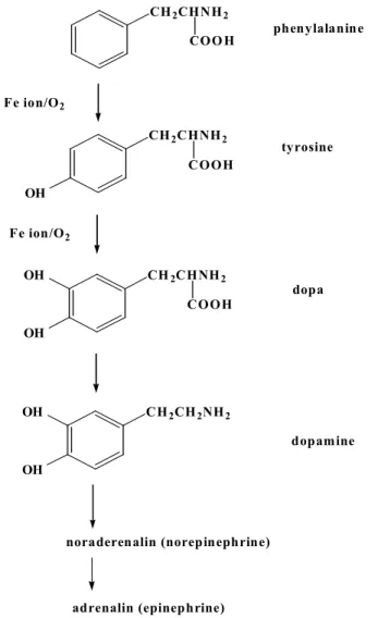
Let us at first consider the biochemical synthesis of dopamine. The chemical mechanism of dopamine synthesis has been elucidated by the biochemists, and the result is illustrated in Figure 3. Dopamine is synthesized from phenylalanine and tyrosine, one of the 20 essential amino-acids through the oxygenation reaction at the benzene ring by the enzymes, phenylalanine hydroxylase (PAH) or tyrosine hydroxylase (TH) (see Figure 4). It should be noted here that the oxygenation at the benzene ring does not occur in the air without the catalyst, and thus it is necessary for us to know the detail chemical mechanism of the enzymes, TH or PAH, and or tryptophan hydroxylase, which catalyzes the formation of serotonin from tryptophan; the deficiency of serotonin has been proposed to induce several metal diseases, such as depression etc.
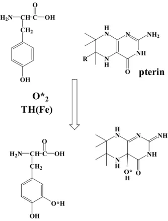
TH is a non-heme iron protein that uses one molecule of dioxygen to
hydroxylate its amino acid and tetrahydropterin substrates to hydroxy-amino
acids and 4a-hydroxytetrahydropterins, respectively. (see Figure 4) [4,5] As the
4a-hydroxytetrahydropterins subsequently dehydrates and is regenerated by the
NADH-dependent enzyme dihydropteridine reductase, it is frequently termed a
cofactor for the peteridine-dependent hydroxylases. The cofactor (BH 4 ) is the
most abundant of the unconjugated tetrahydropterins in mammalian tissues and is
considered to be the naturally tetrahydropterin substrate for these enzymes. The
hydroxylases have been investigated by kinetic and spectroscopic techniques, as well as by site-directed mutagenesis. The iron is necessary for catalytic turnover, (see chapter V) and the tetrahydropterin and amino acid substrates bind close to the iron(II) center, but probably without a direct coordination to the metal center. Thus, it is clear that iron-deficiency should lead to deficiency of neurotransmitters, such as dopamine, serotonin, etc.
Figure 3. Synthesis scheme of neurotransmitters
CH
2CHNH
2COO H phenylalanine
CH
2CHNH
2COOH OH
tyrosine
CH
2CHNH
2COOH OH
OH
dopa
CH
2CH
2NH
2OH OH
dopamine Fe ion/O
2Fe ion/O
2noraderenalin (norepinephrine)
adrenalin (epinephrine)
Figure 4. Formation of dopa from tyrosine catalyzed by TH
As indicated in Chapter I, increased brain iron concentrations at some special regions have been described in Parkinson’s disease, [1] but these iron ions do not contribute to the formation of dopamine; the reason will be developed in Chapter VI. The cause of nigral cell death in the Parkinson’s disease remains unsolved, but many authors have pointed out the hypothesis that the cellular degeneration observed results from oxidative stress. [2,6-8] Oxidative stress manifests itself as an increased oxidation of cellular constituents (lipids and proteins) and DNA damage. Lipid peroxidation and protein damage have been observed in the SN of PD patients, which suggests that oxidative stress is involved in the pathogenesis of this disease.
N N
NH N NH 2
O R
H
H
O* 2
N N
NH
N NH
O H
H O*
pterin
H 2 N CH C CH 2
OH O
H 2 N CH C CH 2
OH O
OH
H OH
TH(Fe)
O*H
The increased iron ions observed in these Parkinson’s and Alzheimer’s disorders has been frequently suggested to play a role in catalyzing the production of the so-called oxygen free radicals via the metal dependent reduction of hydrogen peroxide. This reaction, sometimes referred to as the Fenton reaction, may involve the reaction of hydrogen peroxide with ferrous ion to produce the potentially damaging hydroxyl radical (OH•) (see below), and many authors have insisted that this OH• should be a main active oxygen species in the oxidative stress,
Fe 2+ + H 2 O 2 Fe 3+ + OH - + OH•
but it should be noted that free intracellular ferrous iron concentration have been calculated to be very low, below 10 -8 M, [9] and nobody has succeeded in confirming the OH• formation by the reliable chemical methods.
I would like to show that OH• does not exert the oxidative stress in the living cell, and to propose the new concept for oxidative stress in this monograph (see chapter III) [10] and also show that deficiency of neurotransmitters due to the abnormal iron metabolism in brain leads to the neural cell death.
References
[1] M. B. H. Youdim and P. Riederer, Scientific American, 1997, 52.
[2] J. Haavik and K. Toska, Molecular Neurobiology, 1998, 16, 285.
[3] R. Kruger, O. Eberhardt, O. Riess, and J. B. Schulz, Trends Molcular Medicine, 2002, 8, 236.
[4] P. F. Fitzpatrick, Annu. Rev. Biochem., 1999, 68, 355.
[5] T. J. Kappock and J. P. Caradonna, Chem. Rev., 1996, 96, 2659.
[6] E. C. Hirsch, Molecular Neurobiology, 1994, 9, 135.
[7] J. Haavik, B. Almas, and T. Flatmark, J. Neurochemistry, 1997, 68, 328.
[8] T. Finkel and N. J. Holbrook, Nature, 2000, 408, 239.
[9] P. M. Harrison and P. Arosio, Biochim. Biophys. Acta, 1996, 1275, 161.
[10] Y. Nishida, Med. Hypothesis Res. 2004, 1, 227. http://
www.journal-mhr.com/ PDF_Files/vol_1_4/1_4_PDFs/1_4_2.pdf.
Chapter III Nishida’s Concept on Activation of Oxygen Molecule
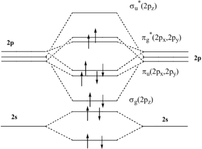
The electronic configuration of the oxygen atom is 1s 2 2s 2 2p 4 . When two O atoms combine to form O 2 , the same orbital types combine if they are of equal or approximately equal energy. Thus, the 1s, 2s, 2p x , 2p y , and 2p z on one oxygen combine with the similar orbitals on the other oxygen to give, in each case, two MO’s. The five AO’s on each atom give rise to ten MO’s in the molecule. [1]
Since the electrons will occupy the orbitals in the order of increasing energy, we must arrange our MO’s in an energy sequence so that we can place our sixteen electrons properly. One of the most instructive ways to do this is by means of the molecular orbital energy diagram method. In the oxygen molecule we have the combination of both 1s and 2s orbitals to give four s type orbitals, [1] two bonding and two antibonding, each of them
occupied by two electrons. Because the 1s electrons are not valence electrons, we usually pay little heed to them. The combination of the 2s orbitals does not result in any net bonding. (see Figure 5)
Figure 5. MO scheme for O 2 .
2s 2s
2p
2p
s g (2p z ) s u * (2p z )
p u (2p x ,2p y )
p g * (2p x ,2p y )
The three atomic p orbital levels in the isolated atom are of equal energy (degenerate); but when we bring one atom into the field of the other, the p z
orbitals pointing toward the other atom start to interact to form a s bond between the two atoms. the corresponding s* orbital are generated. These orbitals s g (2p z ) and s u *(2p z ), are in fact s molecular orbitals because they are symmetric with respect to rotation around the internuclear axis (in this case, z-axis). The bonding interaction is quite large, and hence the splitting is also relatively large.
(see Figure 5)
The p x and p y orbitals on each oxygen combine to each from a p set:
p u (2p u (2p x ), p g *(2p x ), p u (2p y ), p g *(2p y ). The p x and p y orbitals are perpendicular to each other. Now, if we refer to our MO energy diagram, we see that six electrons of 2p orbitals are referred to as valence electrons, that is these electrons occupy s g (2p) and p u (2p). If we follow the principles used for the periodic classification, the two electrons must go separately into the p g *(2p x ) and p g *(2p y ) orbitals, with spin parallel (Hund’s rule: see Figure 5 and Table 2). The two unpaired electrons in the p* orbitals give rise to the paramagnetic properties of molecular oxygen. The diradical character and accompanying paramagnetism of oxygen constitute its outstanding property.
The occupation of antibonding orbitals by one or more electrons cancels
some of the bonding attraction between the atoms. In the O 2 example, we have
two p bonding orbitals, each doubly occupied, and a s bonding orbital, doubly
occupied, or a total of three bonding orbitals. However, each of the two
electrons in an antibonding orbital cancel the bonding effect of an electron in a
bonding orbital, and so the net bonding in oxygen can be considered to result
from a double bond. Evidence for the effect of occupation of the antibonding
orbitals comes from bond distances. In the ground state, the bond distances
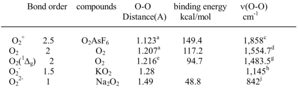
between oxygen atoms is 1.207 A. (see Table 1) However, when O 2 is ionized
by loss of an electron from one of the p* antibonding orbitals, the resulting O 2 + is
1.123 A, a considerable decrease, indicative of stronger bonding in the ion. The
bond lengths of O 2 - (a radical anion) and O 2 2- are 1.28 and 1.49 A, respectively,
confirming the fact that electrons have been added to antibonding orbitals.
Table 1. Electronic and structural properties of oxygen and its derivatives Bond order compounds O-O binding energy n(O-O)
Distance(A) kcal/mol cm -1 O 2 + 2.5 O 2 AsF 6 1.123 a 149.4 1,858 c O 2 2 O 2 1.207 a 117.2 1,554.7 d O 2 ( 1 D g ) 2 O 2 1.216 e 94.7 1,483.5 g O 2 - 1.5 KO 2 1.28 1,145 h O 2 2- 1 Na 2 O 2 1.49 48.8 842 j
a