Significance of Coronary Artery Calcium Score in the Target Lesion Evaluated by Multi-detector Computed Tomography for Selecting
Treatment of Rotational Atherectomy in Patients with Coronary Artery Disease
Teruo SEKIMOTO*1), Yuji HAMAZAKI1), Yasushi AKUTSU1, 2), Koshiro SAKAI1), Ryota KOSAKI1) Hiroyuki YOKOTA1), Tenjin NISHIKURA1), Hiroaki TSUJITA1), Seita KONDO3), Satoshi HOSOKAWA1), Shigeto TSUKAMOTO1), Kyoichi KANEKO1), Mitsunori MUTO1), Masayuki SAKURAI3), Yusuke KODAMA1), Takehiko SAMBE2, 4),
Naoki UCHIDA2, 4), Shinichi KOBAYASHI2) and Youichi KOBAYASHI1)
Abstract : We investigated whether coronary artery calcium score (CAC) in the tar- get lesion on the multidetector computed tomography angiography (CTA) predicts the addition of the Rotational atherectomy (Rota) during percutaneous coronary intervention (PCI). Lesion CAC on CTA were evaluated with quantitative coro- nary analysis (QCA) on coronary angiography for predicting the Rota treatment in 114 consecutive patients (165 target lesions) with first PCI (68 9 years old, females: 17.6%). Rota was added in 8 patients (11 lesions). The lesion length and diameter stenosis on QCA, and lesion length and lesion CAC on CTA were the primary factors associated with the addition of Rota. Using the cut-off value based on receiver operating characteristic analysis, the sensitivity and specificity for predicting the Rota based on QCA was 72.7% in 8 of 11 lesions (vessels) with Rota and the specificity was 74% in 114 of 154 without Rota in the lesion length of ≥ 23 mm (χ2=10.9, p=0.001), and 54.5% in 6 of 11 lesions with Rota and the specificity was 79.2% in 122 of 154 without Rota in the diameter stenosis of ≥ 83% (χ2=6.6, p=0.01). Those based on CTA were 90.9% in 10 of 11 lesions with Rota and 77.3% in 119 of 154 without Rota in the lesion length of ≥ 34 mm
(χ2=24.1, p<0.001), and 90.9% in 10 of 11 with Rota and 88.3% in 136 of 154 without Rota in the lesions with CAC ≥ 453 (χ2=45.7, p<0.001). Lesion CAC on CTA is most predictive of addition of Rota during PCI.
Key words : coronary artery disease, rotational atherectomy, multidetector computed tomography angiography, calcification, quantitative coronary angiographic analysis.
Original
1)Division of Cardiology, Department of Medicine, Showa University School of Medicine, 1-5-8 Hatanodai, Shinagawaku, Tokyo 142-8666, Japan.
2)Department of Internal Medicine (Cardiology), Clinical Research Institute for Clinical Pharmacology & Therapeutics, Showa University Karasuyama Hospital.
3)Cardiovascular Center, Showa University Koto-Toyosu Hospital.
4)Department of Clinical Pharmacology, Showa University School of Medicine.
* To whom corresponding should be addressed.
Introduction
Calcified coronary lesions represent an advanced stage in the atherosclerotic process whereby a soft plaque is converted to a fibrocalcific plaque. The presence and severity of coronary artery lesion calcification impact the risk of subsequent coronary events and the success rate of percuta- neous coronary intervention (PCI) in the obstructive coronary artery disease (CAD). Treatment of calcific lesions is challenging with higher rates of procedural failure, stent under-deployment, lower postprocedual minimal luminal diameter and acute gain, and an elevated risk of reste- nosis1, 2). Rotational atherectomy (Rota) has emerged to be a representative device for such calcified lesions. Rota can facilitate smooth stent delivery and stent expansion through lesion modification3-5). However, practice guidelines recommend the use of Rota for the treatment of heavily calcified or severely fibrotic lesions that cannot be bypassed by a balloon or adequately dilated before planned stenting during the PCI6).
On the other hand, electron beam tomography is a noninvasive method to quantify the extent of calcium formation in the coronary arteries. Coronary artery calcium score (CAC) such as Agatston score7) measured using multidetector computed tomography coronary angiography
(CTA) can be used as an indicator of atherosclerotic burden8).
In the present study, we retrospectively investigated whether quantitative calculating the CAC score in the target lesion by CTA could predict necessary of the use of Rota, before PCI in the CAD patients.
Subjects and methods Populations
For this retrospective cohort study we used the information in consecutive 150 patients (208 target lesions) with CAD who was suspected by the CTA and underwent the percutaneous coronary intervention (PCI) within 6 months after the CTA from December 2010 to April 2014 at Showa university Hospital. The CAD was defined as the presence of at least one coronary lesion with ≥ 75% luminal diameter stenosis of coronary artery on CAG, and PCI was planned in our hospital. The patients with acute coronary syndrome or myocardial infarction who under- went the intervention or coronary artery bypass grafting in the past were excluded from the present study. Of those, 114 consecutive patients (165 target lesions) with first PCI (the mean ages standard deviations : 68 9 years old, females : 17.6%) were finally selected in the pres- ent study. All patients gave written informed consent before the interventions, and the protocol was approved by our Institutional Review Board. Data collected retrospectively included : demo- graphics, existing medical diagnoses, risk factors for cardiovascular disease, medications, previous cardiac procedures, and prior history of cardiac events.
QCA measurement on CAG and PCI with and without Rota
The coronary artery stenosis was assessed by quantitative coronary analysis (QCA) with CAAS software (Version 5.10, Pie Medical Imaging, Maastricht, Netherlands)9). The target
vessel, target lesion and lesion length (mm) on CAG were defined as the stenotic site of more than 75% using CAAS system. The minimum vascular diameter (mm), control vascular diam- eter (mm), and percent diameter stenosis (%) were also measured into target lesion.
All patients were fully informed about the alternative treatment options and PCI-related risks before they provided written informed consent to undergo the procedure. Furthermore, when the intravascular ultrasound (IVUS) catheter could not be passed through the severe lesions, when IVUS detected near-circumferential calcification, or when the lesion did not expand fol- lowing balloon angioplasty, the final decision for Rota use was left to the discretion of the attending physician who was unaware of the calcified plaque conditions with CTA images. Rota was performed with the Rota system (Boston Scientific Corporation, Natick, MA, U.S.A.), with burr sizes from 1.25 to 2.25 mm. The recommended burr speed was 180,000-200,000 rpm with each sequence being less than 30 seconds, and care was taken to prevent any drop in rotational speed ≥ 5000 rpm. A continuous intracoronary infusion containing nicorandil, nitroglycerin and heparin was used to prevent and control slow-flow. It was advised to use incremental burr sizes to achieve a burr-to-artery ratio of at least 0.7. PCI was performed through the femoral artery approach by inserting a 7 Fr or 8 Fr guiding catheter under 10000 IU heparin intravenous administration by use of the approved systems (balloon length 10-40 mm). The technique to achieve an optimal angiographic result was determined by the operator. The procedural success was defined as angiographically proven residual stenosis less than 50% and stenosis reduction of at least 20%10). The use of stents for bail out (flow-limiting dissections, severe recoil, vessel closure) or unsatisfactory results (residual diameter stenosis > 50%) was excluded from the present study.
CTA before CAG
CTA study was performed using a 128-slice scanner (Somatom Definition AS +; Siemens Medical Solutions, Forchheim, Germany) before CAG. The routine protocol for patients with suspected CAD included CAC measurement followed by noninvasive coronary angiography. Cor- onary calcium score was measured in a non–contrast-enhanced scan, quantified using the Calcium Score module of Syngo software (Siemens Medical Solutions) and expressed in Agatston units.
Then, CTA studies were performed with a contrast-enhanced ECG gating, administering a β- blocker (a bolus injection of landiolol hydrochloride at a dose of 0.125 mg/kg) if the heart rate went above 70 beat/min. Nitroglycerine was administered sublingually immediately before the bolus injection of contrast media (Omnipaque, 350 mg iodine/ml, Daiichi Sankyo Co, Ltd, Tokyo, Japan) into the antecubital vein, followed by a 20 ml saline solution chaser bolus. The amount of the contrast media used for the scan was determined according to the patientʼs body weight and scan time [the total dose = (scan time+4 seconds) 0.07 body weight]. The scan was gated to the cardiac cycle through ECG synchronization with the following parameters : col- limation width, 64 0.6 mm ; rotation time, 330 ms ; tube voltage, 120 kV ; effective tube current, 800 mA ; table feed, 11.5 mm/rotation ; and pitch 0.3. ECG-gated data sets were finally recon- structed with a slice thickness of 2.0 mm at the point of the cardiac cycle for every 5% of R-R
interval.
Analysis of CTA and CAC measurements
Scans were analyzed off-line on a dedicated workstation (Leonardo, Siemens Medical Solution)
to measure the CAC and the stenotic degrees of coronary artery. Each identified coronary artery lesion was assessed for stenosis severity along multiple longitudinal transverse, and oblique axes with the use of multiplanar reconstructions, thin-slab maximum intensity projections, and the curved reconstruction technique. The coronary lesionʼs severity was assessed by the maximum percent luminal diameter stenosis observed in any plane. The Agatston score, both per patient, per vessel, and target lesion7, 11) were measured for each patient. The lesion length including calcified plaque was measured on the longitudinal images. Two experienced observers, who were unaware of the patientʼs clinical information, evaluated the CTA images independently.
Calcified plaque patterns on CTA
Sharp reconstruction images were used for all plaque calcification analyses. Window width and level settings were adjusted to the investigatorʼs discretion to optimally visualize the coronary artery lumen and to identify plaque calcification. Calcium plaque within CAD lesions were defined as occupying at least three contiguous pixels with a density of > 130 Hounsfield units (HU).
Statistical analysis
For descriptive purposes, the target lesions were divided into 2 groups on the basis of inter- ventional treatment such as PCI with and without Rota. All continuous variables are presented as mean SD. Differences between groups were tested using an unpaired Studentʼs test or χ2 test. The associations between the predictors, such as calcium plaque pattern of the target lesion on CTA, were formally tested by multivariable logistic regression analysis for CT one- cross section of target lesion and one target lesion of coronary artery. All multivariable analyses employed the forward stepwise method, with entry and removal probability values set at 0.5.
Pearsonʼs correlation analysis was performed to assessment dependence. All statistical analysis was completed using IBM SPSS for Windows version 20 (SPSS Inc., an IBM Company, Chicago, IL). A probability value of < 0.05 was considered significant.
Results
A case with Rota
The CAG showed the coronary artery stenosis of > 75% in middle lesion of right coronary artery visually in 73-years-old woman (Fig. 1). QCA showed the following parameters of the 23.6 mm of length of target lesion, 2.98 mm of reference diameter, 0.72 mm of minimal lumen diameter, and 76 % of diameter stenosis. The catheter of IVUS did not pass through this target lesion, which indicated an extensively solid plaque with calcification. The treatment of Rota was achieved at bar size of 1.75 mm, and the coronary stenting with ballooning was achieved. The retrospective measurement of CAC on CTA was 2743.1 in the patient, 1698.1 in the target ves-
sel, and 1034 in the target lesion. The length of calcified plaque on CTA was 39.9 mm on the longitudinal images.
Patients and lesion characteristics
In the present study, 106 patients underwent PCI without Rota and 8 patients with Rota, while in 4 patients the treatment as changed from PCI to Rota. Furthermore, 4 patients had PCI in one vessel and Rota in other vessel (These patients were evaluated as having the Rota). Among patient cohort, there was no significant difference in age, gender, risk factors, and medi- cations between the patients with and without Rota (Table 1).
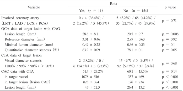
The PCI was involved in 5 vessels with LMT, 72 vessels with LAD, 37 vessels with LCX, and 51 vessels with RCA. There is no significant difference in the involved coronary artery area between the vessels with and without Rota (p = 0.71)(Table 2). Concerning every vessels, the target lesions with Rota were significantly longer lesion length (28.6 mm vs 20.5 mm, p<0.01)
and severe diameter stenosis (83.9% vs 78.1%, p=0.05) on QCA, and higher CAC of target vessel and lesion (1078 vs 357 and 826 vs 176, p<0.001 in each) and longer lesion length (45 mm vs 26.4 mm, p<0.001) on CTA than those without Rota.
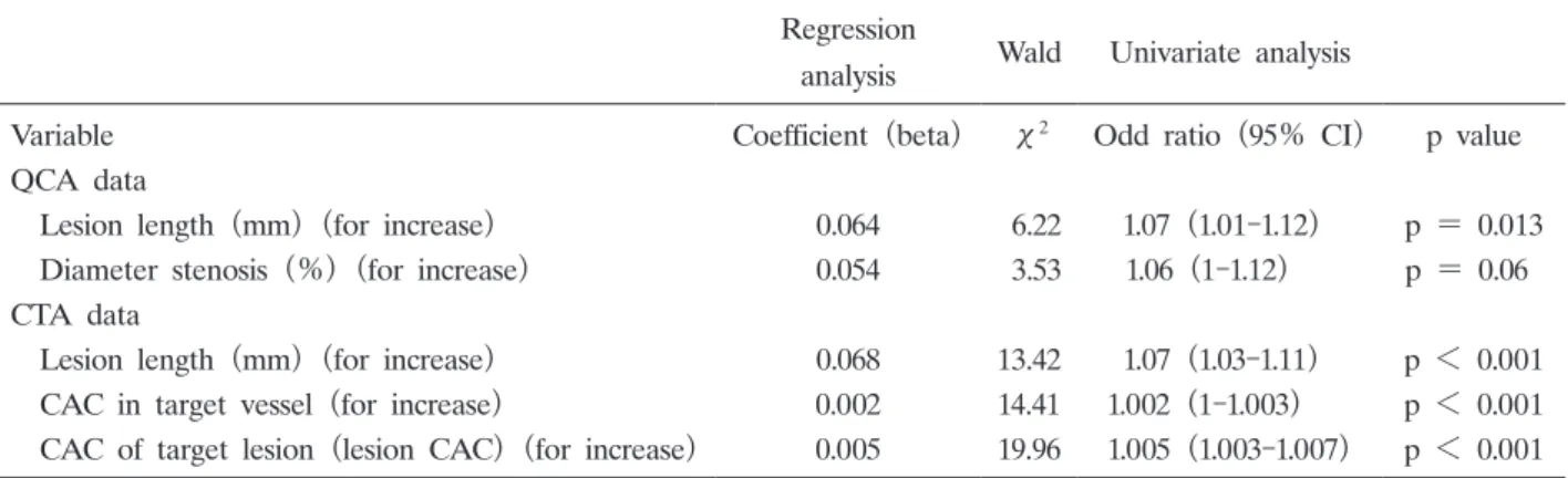
The factors for predicting the Rota
For continuous variables, the lesion length on QCA, and lesion length, and CAC in vessel and lesion on CTA were the primary factors associated with the Rota (Table 3). The lesion length was dependent on the diameter stenosis based on QCA with CAG (γ= 0.591, p<0.001), and the lesion length on CTA was dependent on CAC in vessel and lesion based on CTA (γ = 0.514 and 0.537, p<0.001 in each).
Fig. 1. The CAG showed the coronary artery stenosis of more than 75 % in the middle lesion of right coronary artery visually in 73 years old woman (Fig. 1). The catheter of IVUS did not pass through this target lesion, and the coronary artery ballooning failed despite of high pressure, which indicated extensively solid plaque with calcification. The treatment of Rota was achieved successfully, and the coronary stenting with ballooning was achieved. The lesion CAC on CTA showed high score.
Table 1. Patient characteristics
Variable Rota
p value Yes (n = 8) No (n = 106)
Age (years) 73 5 67 10 p = 0.07
Female 1 (12.5%) 21 (19.8%) p = 0.61
Diabetes 6 (75%) 41 (38.7%) p = 0.21
Hypertension 4 (50%) 76 (71.7%) p = 0.2
Hyperlipidemia 7 (87.5%) 53 (50%) p = 0.16
Current smoking 7 (87.5%) 59 (55.7%) p = 0.08
Familial history 2 (25%) 16 (15.1%) p = 0.41
History of hemodialysis 1 (12.5%) 4 (3.8%) p = 0.25
LVEF (%) 51.4 23.2% 60.1 15.5% p = 0.14
Medications before MDCT
Use of beta blocker 1 (12.5%) 7 (6.6%) p = 0.53
Use of angiotensin-II receptor blocker 5 (62.5%) 52 (49.4%) p = 0.46
Use of statin 5 (62.5%) 38 (35.6%) p = 0.13
Use of aspirin 1 (12.5%) 22 (20.8%) p = 0.57
Abbreviations ; PCI=Percutaneous coronary intervention, LVEF = Left ventricular ejection fraction.
Table 2. Vessel characteristics of CTA and CAG (QCA)
Variable Rota
p value Yes (n = 11) No (n = 154)
Involved coronary artery
(LMT / LAD / LCX / RCA)
0 / 4 (36.4%) / 2 (18.2%) / 5 (45.5%)
5 (3.2%) / 68 (44.2%) /
35 (22.7%) / 46 (29.9%) p = 0.71 QCA data of target lesion with CAG
Lesion length (mm) 28.6 8.1 20.5 9.7 p = 0.008 Reference diameter (mm) 3.01 0.46 2.99 0.63 p = 0.92 Minimal lumen diameter (mm) 0.49 0.25 0.66 0.33 p = 0.1 Quantitative diameter stenosis (%) 83.9 0.09 78.1 0.1 p = 0.05 CTA data of target lesion
Visual diameter stenosis
(100% / 99% / 90%/ > 90%)
2 (18.2%) / 0 / 6 (54.5%) / 3 (27.3%)
15 (9.7) /10 (6.5%) /
92 (59.7%) / 37 (24%) p = 0.68
CAC data with CTA 51.4 23.2% 60.1 15.5% p = 0.14
in target vessel 1078 516 357 469 p < 0.001 in target lesion (lesion CAC) 826 324 176 234 p < 0.001 Lesion length (mm) 45 12.3 26.4 13.2 p < 0.001 Abbreviations ; LMT = Left Main Trunk, LAD = Left Anterior Descending Coronary Artery, LCX = Left Circumflex Coronary Artery, RCA = Right Coronary Artery, QCA = Quantitative Coronary Analysis on Coronary Angiography, CAG = Coronary Angiography, CAC = Coronary Artery Calcium score, CTA = Multidetector Com- puted Tomography Angiography.
Table 3. Factors for predicting Rota selection in continuous valuables Regression
analysis Wald Univariate analysis
Variable Coefficient (beta) χ2 Odd ratio (95% CI) p value
QCA data
Lesion length (mm)(for increase) 0.064 6.22 1.07 (1.01-1.12) p = 0.013 Diameter stenosis (%)(for increase) 0.054 3.53 1.06 (1-1.12) p = 0.06 CTA data
Lesion length (mm)(for increase) 0.068 13.42 1.07 (1.03-1.11) p < 0.001 CAC in target vessel (for increase) 0.002 14.41 1.002 (1-1.003) p < 0.001 CAC of target lesion (lesion CAC)(for increase) 0.005 19.96 1.005 (1.003-1.007) p < 0.001 Abbreviations ; Rota = Rotational atherectomy, QCA = Quantitative coronary analysis on coronary angiography, CTA = Multidetector computed tomography angiography, CAC = Coronary artery calcium score.
Fig. 2. Based on ROC analysis, the optimal thresholds for the Rota selection in vessel were at the lesion length of 23 mm (A) and stenosis diameter of 83% (B) on QCA, and at the lesion length of 34 mm (C) and lesion CAC of lesion of 453 (D). The sensitivity and specificity based on CTA were 90.9% in 10 of 11 lesions with Rota and 77.3% in 119 of 154 without Rota in the lesion length of ≥ 34 mm, and 90.9% in 10 of 11with Rota and 88.3% in 136 of 154 without Rota in the lesion CAC ≥ 453 (χ2 = 45.7, p<0.001). The AUC for the prediction of Rota treatment was 0.953 in the lesion CAC on CTA, and it was the largest of the predictive factors.
Using the cut-off value based on ROC analysis (Fig. 2), the sensitivity and specificity for predicting the Rota based on QCA was 72.7% in 8 of 11 lesions (vessels) with Rota and the specificity was 74% in 114 of 154 without Rota in the lesion length of ≥ 23 mm, and 54.5%
in 6 of 11 lesions with Rota and the specificity was 79.2% in 122 of 154 without Rota in the diameter stenosis of ≥ 83%. Similarly, the sensitivity and specificity based on CTA were 90.9%
in 10 of 11 lesions with Rota and 77.3% in 119 of 154 without Rota in the lesion length of ≥ 34 mm, and 90.9% in 10 of 11with Rota and 88.3% in 136 of 154 without Rota in the lesion CAC ≥ 453 (χ2 = 45.7, p<0.001). The area under ROC curve (AUC) in the lesion CAC for predict- ing the Rota was the largest in the predictive factors.
Discussion
In the present study, we showed that the target lesion CAC based on CTA was the most predictive of addition of the Rota during PCI.
Wang et al12) demonstrated that a high CAC score (> 300) was a significant predictor for major adverse cardiac events (MACE) in patients with stable angina pectoris who underwent PCI, which indicated the complexity of PCI because of severely calcified plaque ; however, that study did not clarify the best tool for the decision of Rota use.
Practice guidelines recommend the use of Rota for the treatment of heavily calcified or severely fibrotic lesions that cannot be bypassed by a balloon or adequately dilated before planned stenting13). Despite the significant reduction of restenosis by Drug-eluting stent (DES), several factors including diabetes, hemodialysis, and severe calcification have been reported to be high-risk factors for restenosis after DES implantation14-20). Several studies have shown that DES implantation after the use of Rota reduced in the incident of target lesion revasculariza- tion (TLR)21) and major adverse cardiac events22-24). It is that lesion modification by the Rota could yield smooth stent delivery, prevent damage to the polymer coating25), and provide optimal stent expansion, all of which could help DES be effective, even for calcified lesions. In addition, procedural success rates of over 90% and complications less than 5% are routinely achieved after Rota of calcified lesions26-28). In the present study, no patient experienced emergency adverse events during PCI with the use of Rota such as a perforation of coronary artery and acute myocardial infarction, dissection of a coronary artery.
Development of CTA has elevated the non-invasive diagnosis of coronary heart disease to a new level. Particularly, in diagnosing calcification of coronary arteries, CTA has incomparable accuracy and allows quantification ; and the CAC score calculated using the automatic analysis software is favorably correlated, quantitatively, to the area of atherosclerotic plaques7). However, arterial wall calcification may adulterate the results of CTA, reducing the specificity and positive predictive value. Diederichsen et al29) reported that the diagnostic accuracy of CTA declined in patients with high CAC (Agatston score > 400) and suggested that patients with CS > 400 should not undergo CTA. On the other hand, Lau et al30) found that a high CAC improved the sensitivity of CTA in detecting a stenotic coronary artery, without compromising specificity.
Furthermore, in cross-sectional images, Cerci R et al31) reported that 79% of the plaques with ≥
180 calcium arc had <50% diameter of stenosis as defined by QCA and that 77% of lesions without visible residual lumen had 50% or greater lumen narrowing by QCA. It is important for the operator to predict the target lesions which are heavily calcified and undilatable with balloon technology before PCI17) because the attempts to treat these heavily calcified lesions with ballooning and stenting often lead to vessel dissection or incomplete stent deployment with adverse outcomes. In the present study, we found that the target lesion CAC based on CTA was the most predictive of Rota addition during PCI with a sensitivity of 90.9% and the speci- ficity of 88.3% in the lesion CAC ≥ 453 (χ2=45.7, p<0.001).
In conclusion, lesion CAC on CTA is most predictive of addition of Rota during PCI. The evaluation of CAC score on CTA is useful for indicating the addition of Rota.
The addition of Rota is recommended to be performed only when the heavily calcified lesion was not crossed by a balloon catheter or adequately dilated before stent implantation6). All the published prospective random control study that evaluated the various catheter-based coronary interventional devices excluded patients with severely calcified lesions6). Therefore, the evidence base for best PCI practices in patients with severely calcified lesions comes from nonrandomized single-arm studies although several retrospective studies have shown that the use of Rota before the attempt of stenting with ballooning is safe and reasonable in patients with severely calcified lesions. In the present study, the indication of Rota treatment was evaluated from CTA data retrospectively because of these reasons. Our results are based upon a single center experience with patients referred for CAD which may limit the more general application applicability of the findings. We also investigated the lesion calcified plaque on CTA as a predictor of Rota treatment in a limited number of patients. Thus, the evaluation of severely calcified plaque on CTA to decide the addition of Rota should be prospectively studied in the near future by a multicenter study.
Hemodialysis is generally known to cause the severe coronary artery calcifications, but only 5 patients with the history of hemodialysis was performed first PCI in the present study because the other patients with hemodialysis had been repeated PCI. Thus, the PCI procedure for those patients with hemodialysis should be also evaluated in future studies, independent of patients with only atherosclerosis.
Conflict of interest disclosure
The authors have declared no conflict of interest.
References
1) Vavuranakis M, Toutouzas K, Stefanadis C, et al. Stent deployment in calcified lesions: can we overcome calcific restraint with high-pressure balloon inflations? Catheter Cardiovasc Interv. 2001;52:164-172.
2) Virmani R, Farb A, Burke AP. Coronary angioplasty from the perspective of atherosclerotic plaque: morphologic predictors of immediate success and restenosis. Am Heart J. 1994;127:163-179.
3) Mints GS, Potkin BN, Keren G, et al. Intravascular ultrasound evaluation of the effect of rotational atherectomy in obstructive atherosclerotic coronary artery disease. Circulation. 1992;86:1383-1393.
4) Kovach JA, Mintz GS, Pichard AD, et al. Sequential intravascular ultrasound characterization of the mechanisms of rotational atherectomy and adjunct balloon angioplasty. J Am Coll Cardiol. 1993;22:1024-1032.
5) Ellis SG, Popma JJ, Buchbinder M, et al. Relation of clinical presentation, stenosis morphology, and operator technique to the procedural results of rotational atherectomy and rotational atherectomy-faciliated angioplasty.
Circulation. 1994;89:882-892.
6) Levine GN, Bates ER, Blankenship JC, et al. 2011 ACCF/AHA/SCAI Guideline for Percutaneous Coronary Intervention: executive summary: a report of the American College of Cardiology Foundation/American Heart Association Task Force on Practice Guidelines and the Society for Cardiovascular Angiography and Interventions.
Circulation. 2011;124:2574-2609.
7) Agatston AS, Janowitz WR, Hildner FJ, et al. Quantification of coronary artery calcium using ultrafast computed tomography. J Am Coll Cardiol. 1990;15:827-832.
8) Janowitz WR, Agatston AS, Kaplan G, et al. Differences in prevalence and extent of coronary artery calcium detected by ultrafast computed tomography in asymptomatic men and women. Am J Cardiol. 1993;72:247-254.
9) Ishibashi Y, Grundeken MJ, Nakatani S, et al. In vitro validation and comparison of different software packages or algorithms for coronary bifurcation analysis using calibrated phantoms: Implications for clinical practice and research of bifurcation stenting. Catheter Cardiovasc Interv. 2015;85:554-563.
10) Dill T, Dietz U, Hamm CW, et al. A randomized comparison of balloon angioplasty versus rotational atherectomy in complex coronary lesions (COBRA study). Eur Heart J. 2000;21:1759-1766.
11) Moselewski F, OʼDonnell CJ, Achenbach S, et al. Calcium concentration of individual coronary calcified plaques as measured by multidetector row computed tomography. Circulation. 2005;111:3236-3241.
12) Wang FF, Han JL, He R, et al. Prognostic value of coronary artery calcium score in patients with stable angina pectoris after percutaneous coronary intervention. J Geriatr Cardiol. 2014;11:113-119.
13) Task Force on Myocardial Revascularization of the European Society of Cardiology (ECS) and the European Association for Cardio-Thoracic Surgery (EACTS), European Association for Percutaneous Cardiovascular Inter- ventions (EAPCI). Wijns W, et al. Guideline on myocardial revascularization. Eur Heart J. 2010;31:2501-2555.
14) Yanagi D, Shirai K, Mori K, et al. Possible predictors of target lesion revascularization after drug-eluting stent implantation. J Cardiol. 2007;49:63-67.
15) Ishio N, Kobayashi Y, Takebayashi H, et al. Impact of drug-eluting stents on clinical and angiographic outcomes in dialysis patients. Circ J. 2007;71:1525-1529.
16) Gaku N, Kengo T, Aoki J, et al. Clinical and angiographic outcomes of sirolimus-eluting stents implantation in Japanese patients in daily practice. Circ J. 2006;70:1367-1371.
17) Aoyama T, Ishii H, Toriyama T, et al. Sirolimus-eluting stents vs bare metal stents for coronary intervention in Japanese patients with renal failure on hemodialysis. Circ J. 2008;72:56-60.
18) Kawaguchi R, Tsurugaya H, Hoshizaki H, et al. Impact of lesion calcification on clinical and angiographic outcome after sirolimus-eluting stent implantation in real-world patients. Cardiovasc Revasc Med. 2008;9:2-8.
19) Ortolani P, Balducelli M, Marzaroli P, et al. Two-year clinical outcomes with drug-eluting stents for diabetic patients with de novo coronary lesions: results from a real-world multicenter registry. Circulation. 2008:117:923-930.
20) Lemos PA, Hoye A, Goedhart D, et al. Clinical, angiographic, and procedural predictors of angiographic restenosis after sirolimus-eluting stent implantation in complex patients: an evaluation from the Rapamycin-Eluting Stent Evaluated At Rotterdam Cardiology Hospital (RESEACH) study. Circulation. 2004;109:1366-1370.
21) Kubota T, Ishikawa T, Nakano Y, et al. Retrospective comparison of clinical and angiographic outcomes after sirolimus-eluting and bare-metal stent implantation in 312 consecutive, nonrandomized severely calcified lesions using a rotablator. Int Heart J. 2011;52:65-71.
22) Mezilis N, Dardas P, Ninios V, et al. Rotablation in the drug eluting era: immediate and long-term results from a single center experience. J Interv Cardiol. 2010;23:249-253.
23) Furuichi S, Sangiorgi GM, Godino C, et al. Rotational atherectomy followed by drug-eluting stent implantation in calcified coronary lesions. EuroIntervention. 2009;5:370-374.
24) Abdel-Wahab M, Baev R, Dieker P, et al. Long-term clinical outcome of Rotational atherectomy followed by drug-elutingstent implantation in complex calcified coronary lesions. Catheter Cardiovasc Interv. 2013;81:285-291.
25) Kuriyama N, Kobayashi Y, Yamaguchi M, et al. Usefulness of rotational atherectomy in preventing polymer dam- age of evorolimus-eluting stent in calcified coronary artery. JACC Cardiovasc Interv. 2011;4:588-589.
26) Serruys PW, de Jaegere P, Kiemeneij F, et al. A comparison of balloon-expandable-stent implantation with balloon angioplasty in patients with coronary artery disease. Benestent Study Group. N Engl J Med. 1994;331:489-495.
27) Maclsaac AI, Whitlow PI, Cowley MJ, et al. Angiographic predictors of outcome of coronary rotational atherec- tomy from the completed multicenter registry. J Am Coll Cardiol. 1994;Special Issue:353A.
28) Leon MB, Kent KM, Pichard AD, et al. Percutaneous transluminal coronary rotational angioplasty of calcified lesions. Circulation. 1991;84:II521.
29) Diederichsen AC, Petersen H, Jensen LO, et al. Diagnostic value of cardiac 64-slice computed tomography: impor- tance of coronary calcium. Scand Cardiovasc J. 2009;43:337-344.
30) Lau GT, Ridley LJ, Schieb MC, et al. Coronary artery stenoses: detection with calcium scoring, CT angiography, and both methods combined. Radiology. 2005;235:415-422.
31) Cerci R, Vavere AL, Miller JM, et al. Patterns of coronary arterial lesion calcification by a novel, cross-sectional CT angiographic assessment. Int J Cardiovasc Imaging. 2013;29:1619-1627
[Received January 5, 2015 : Accepted January 19, 2015]