dur i ng c anc er c hem
ot her apy 2016
著者
H
or i e Shi geo, O
ya M
ot ot s ugu, N
angaku M
as aom
i ,
Yas uda Yos hi nar i , Kom
at s u Yas uhi r o, Yanagi t a
M
ot oko, Ki t agaw
a Yuko, Kuw
ano H
i r oyuki ,
N
i s hi yam
a H
i r oyuki , I s hi oka Chi kas hi , Takai s hi
H
i r om
as a, Shi m
odai r a H
i deki , M
ogi Aki r a, Ando
Yui c hi , M
at s um
ot o Koj i , Kadow
aki D
ai s uke, M
ut o
Sat or u
j our nal or
publ i c at i on t i t l e
Cl i ni c al and exper i m
ent al nephr ol ogy
vol um
e
22
num
ber
1
page r ange
210- 244
year
2018- 02
権利
( C) The Aut hor ( s ) 2017. Thi s ar t i c l e i s an
open ac c es s publ i c at i on
Thi s ar t i c l e i s di s t r i but ed under t he t er m
s of
t he Cr eat i ve Com
m
ons At t r i but i on 4. 0
I nt er nat i onal Li c ens e ( ht t p: / / c r ea
t i vec om
m
ons . or g/ l i c ens es / by/ 4. 0/ ) , w
hi c h
per m
i t s unr es t r i c t ed us e, di s t r i but i on, and
r epr oduc t i on i n any m
edi um
, pr ovi ded you gi ve
appr opr i at e c r edi t t o t he or i gi nal aut hor ( s )
and t he s our c e, pr ovi de a l i nk t o t he Cr eat i ve
Com
m
ons l i c ens e, and i ndi c at e i f c hanges w
er e
m
ade.
U
RL
ht t p: / / hdl . handl e. net / 2241/ 00151224
G U I D E L I N E
Guidelines for treatment of renal injury during cancer
chemotherapy 2016
Shigeo Horie1,2•Mototsugu Oya3•Masaomi Nangaku4•Yoshinari Yasuda5• Yasuhiro Komatsu6•Motoko Yanagita7•Yuko Kitagawa8• Hiroyuki Kuwano9• Hiroyuki Nishiyama10•Chikashi Ishioka11•Hiromasa Takaishi12•
Hideki Shimodaira13•Akira Mogi9•Yuichi Ando14•Koji Matsumoto15• Daisuke Kadowaki16•Satoru Muto1,2
Published online: 30 August 2017
ÓThe Author(s) 2017. This article is an open access publication
1. Introduction
Advances in cancer drug therapy have led to improvements in the outcomes of cancer patients, as well as increasing numbers of patients undergoing anticancer chemotherapy and molecularly targeted drug therapy. One adverse event associated with cancer drug therapy is nephrotoxicity, which impedes effective cancer therapy and diminishes the quality of life of cancer patients. Consequently, onco-nephrology has emerged as a new clinical field concerned with the management of nephrotoxicity in cancer drug therapy, creating expectations for advanced expertise and the accumulation of accurate evidence. However, while patients with renal impairment have heretofore undergone planning regarding administration of cancer drug therapy,
procedures for nephropathy prevention, and measures for treatment of drug-induced nephrotoxicity in clinical set-tings based on tradition, experimental rules, and informa-tion from clinical trials, the soundness of the evidence for these practices has been uncertain.
Over the past 10 years, estimated glomerular filtration rate (eGFR) has replaced creatinine clearance in the assessment of renal function; in addition, research has revealed the pathologies of and risk factors for chronic kidney disease (CKD) and acute kidney injury (AKI). The objectives of the guidelines presented here are to support improvements in the results of cancer drug therapy and the quality of life of cancer patients through application of these advances in clinical nephrology and the practice of evidence-based treatment.
& Shigeo Horie [email protected]
1 Department of Urology, Juntendo University Graduate
School of Medicine, 2-1-1, Hongo, Bunkyo-ku, Tokyo 113-8421, Japan
2 Department of Advanced Informatics for Genetic Disease,
Juntendo University Graduate School of Medicine, Tokyo, Japan
3 Department of Urology, Keio University School
of Medicine, Tokyo, Japan
4 Division of Nephrology and Endocrinology,
The University of Tokyo Graduate School of Medicine, Tokyo, Japan
5 Department of CKD Initiatives/Nephrology, Nagoya
University Graduate School of Medicine, Nagoya, Japan
6 Division of Nephrology, Department of Medicine,
St. Luke’s International Hospital, Tokyo, Japan
7 Department of Nephrology, Kyoto University Graduate
School of Medicine, Kyoto, Japan
8 Department of Surgery, Keio University School of Medicine,
Tokyo, Japan
9 Department of General Surgical Science, Gunma University
Graduate School of Medicine, Gunma, Japan
10 Department of Urology, Faculty of Medicine, University of
Tsukuba, Ibaraki, Japan
11 Department of Clinical Oncology, Institute of Development,
Aging and Cancer, Tohoku University, Miyagi, Japan
12 Keio Cancer Center, Keio University School of Medicine,
Tokyo, Japan
13 Department of Clinical Oncology, Institute of Development,
Aging and Cancer, Tohoku University, Miyagi, Japan
14 Department of Clinical Oncology and Chemotherapy,
Nagoya University Hospital, Aichi, Japan
15 Division of Medical Oncology, Hyogo Cancer Center,
Hyogo, Japan
16 Department of Clinical Pharmacology, Faculty of
Pharmaceutical Sciences, Kumamoto University, Kumamoto, Japan
For these guidelines, we have assembled a group of Japanese experts on cancer drug therapy and nephrology to select highly important clinical questions that are fre-quently encountered in everyday practice. These guidelines ultimately comprise 16 clinical questions in two chap-ters regarding assessment of renal function and prevention of nephropathy during cancer drug therapy, thereby determining the level of evidence to support clinical assessments and elucidating the nature of current standard treatments. However, in drafting these guidelines, we dis-covered a number of clinical issues (evidence gaps) regarding cancer drug therapy and renal impairment. For example, 1) there is very little clinical research on cancer drug therapy and nephropathy to begin with; 2) many clinical trials continue to use creatinine clearance to assess renal function; 3) in assessments of renal function in large populations, there is a vast discrepancy between eGFR and measured values of GFR; and 4) it remains unknown whether body surface area corrections of drug doses are appropriate for elderly patients (who have reduced muscle mass) or obese patients. These and other evidence gaps must be resolved for the sake of future research.
These guidelines were drafted with reference to the ‘‘Minds Treatment Guideline Creation Companion 2014’’ using the Minds Guideline Creation support tool ‘‘GUIDE’’. We would like to express our profound grati-tude to Doctors Tsuguya Fukui and Takeo Nakayama of Minds for their roles as advisors in the creation of our guidelines.
We would also like to take this opportunity to express our appreciation to the many young physicians of the systematic review team for their contributions in drafting structured abstracts.
The primary significance of treatment guidelines is their application in daily clinical practice. We would appreciate any criticisms or ideas that would be useful in future revisions of these guidelines.
Shigeo Horie, M.D. Professor and Chairman, Department of Urology
Juntendo University, Graduate School of Medicine
2. On the Occasion of Publication
Cancer has been the leading cause of death among Japanese people for many years; currently, cancer is responsible for approximately 30% of all deaths in Japan. As the Japanese population ages, this figure will continue to increase year after year. Therefore, further development of treatment measures against cancer is undoubtedly one of the most crucial issues for the Japanese population. One such measure
is drug therapy, which is widely performed. Many anticancer drugs are strongly associated with effects on various organs; a sufficient understanding of these associations is a prereq-uisite for effective and successful cancer drug therapy. Unfortunately, there have been no guidelines regarding cancer drug therapy in relation to associations with indi-vidual organs. Medical staffs and indiindi-viduals involved in the treatment of cancer have a great interest for the relevance of the anti-cancer agent and a kidney. However, no previous guidelines exist that systematically described the association between cancer drug therapy and the kidneys.
In addition to chronic kidney disease, the concept of acute kidney injury has rapidly become widespread in recent years. As renal function assessment methods and biomarkers continue to develop, evolutions in nephropathy concepts are being observed.
Against this backdrop, the Japanese Society of Nephrology, the Japan Society of Clinical Oncology, the Japanese Society of Medical Oncology, and the Japanese Society of Nephrology and Pharmacotherapy have jointly published the ‘‘2016 Guidelines for the Treatment of Nephropathy in Cancer Pharmacotherapy’’; the timely and fascinating publication of these guidelines marks a major step in the development of cancer pharmacotherapy. This is truly a document that individuals involved in cancer treatment have long awaited. I sincerely hope that this document will be used appropriately and effectively by all individuals who work on cancer treatment.
Lastly, I would like to express my deep gratitude to everyone involved in the drafting of these guidelines. Seiichi Matsuo, MD. PhD.
President, Japanese Society of Nephrology (President, Nagoya University)
As the Japanese population continues to age, physicians engaged in cancer pharmacotherapy increasingly encounter patients with organ dysfunction due to comorbid diseases; however, there is a lack of information regarding appro-priate cancer pharmacotherapy for cancer patients with comorbid nephropathy. Currently, package inserts for the majority of anticancer drugs contain no clear information regarding administration in patients with chronic kidney disease. Although nephropathy is a major adverse event elicited by cancer pharmacotherapy, regimens for the pre-vention of nephropathy are currently modified based on the experience of individual physicians and the customs of individual facilities.
guidelines also discuss supportive therapy for maintenance dialysis patients and patients with specific comorbidities. I believe that these guidelines will prove useful in daily clinical practice.
As part of its duties as a multidisciplinary academic society, the Japan Society of Clinical Oncology has been engaged in the formulation of guidelines for common supportive therapies for the treatment of cancer of various organs. Our society considers it greatly significant to have had the opportunity to participate in the formulation of these guidelines, which will contribute to improvements in the quality of treatment for patients with renal impairment. Lastly, I would like to express my profound gratitude to Doctor Shigeo Horie, President of the Guideline Prepara-tion Committee, for his tireless leadership in the drafting of these guidelines, as well as the many others who devoted their efforts to drafting the guidelines.
Yuko Kitagawa, M.D., Ph.D., F.A.C.S.
Chairman of Board of Directors, Japan Society of Clinical Oncology
Professor and Chairman, Department of Surgery, Keio University School of Medicine
Cancer is reported to afflict one in every two Japanese people and kill one in every three. As the Japanese popu-lation continues to age, the number of elderly cancer patients is likely to continue to increase. Consequently, an increase is also expected in the number of cancer patients with comorbidities such as nephropathy.
In the use of anticancer drugs for cancer patients with nephropathy, consideration must be given to the possibility of the enhancement of adverse events owing to diminished excretion, as well as the possibility that the toxicity of anti-cancer drugs will exacerbate nephropathy. However, as effec-tive anticancer drugs are not used based solely on comorbid nephropathy, the therapy cannot be considered appropriate.
The performance of cancer pharmacotherapy in patients with nephropathy requires knowledge of not only oncol-ogy, but also nephrology. I believe that the joint creation of these guidelines by the Japanese Society of Nephrology, kidney specialists with the Japanese Society of Nephrology and Pharmacotherapy, and cancer therapy specialists with the Japan Society of Clinical Oncology is incredibly important and significant for the performance of appro-priate pharmacotherapy in cancer patients with nephropa-thy. These guidelines establish crucial clinical questions and provide clear descriptions about these questions.
I anticipate that these guidelines will be utilized effec-tively by physicians, pharmacists, and nurses throughout Japan, and that they will be useful in the performance of appropriate anticancer drug therapy in cancer patients with nephropathy.
Yuichiro Ohe
National Cancer Center Hospital Department of Thoracic Oncology
The Japanese Society of Nephrology and Pharmacotherapy strives to foster ‘‘medical professionals who responsibly offer effective, safe, and the most appropriate drug therapy optimized to the individual patient’’. Since the society was founded, it has worked toward fulfilling the following four major objectives: 1) to ensure the proper use of drugs and prevention of toxic side effects in patients with decreased renal function, 2) to prevent renal function deterioration and cardiovascular complications through proper medication guidance, 3) to provide appropriate drug therapy to dialysis patients with complications, and 4) to prevent drug-induced renal damage caused by nephrotoxic agents and drugs inducing renal ischemia. The Japanese Society of Nephrology and Pharmacotherapy was granted the oppor-tunity to create the ‘‘2016 Guidelines for the Treatment of Nephropathy in Cancer Pharmacotherapy’’ alongside the Japanese Society of Nephrology, the Japan Society of Clinical Oncology, and the Japanese Society of Medical Oncology. The joint creation of these guidelines aligns with our own society’s goals, filling me with profound pride.
Similar to antibacterial agents and nonsteroidal anti-in-flammatory drugs (NSAIDs), anticancer drugs can easily cause drug-induced nephropathy. The renal function of a patient receiving anticancer drugs fluctuates easily due to the effects of various factors such as the patient’s condition, activity level, and age. Anticancer drug pharmacokinetics, anticancer drug interactions, and conceptions of patients’ renal function are the fortes of our society, which specializes in nephrology and pharmacotherapy. In order to exert our specialized capacity, we recently established a Committee for the Formulation and Drafting of Guidelines. Going forward, with this committee at the center of our efforts, we hope to use our specialized perspective in relation to nephrology and pharmacotherapy to contribute to the drafting and revision of various types of practice and therapeutic guidelines.
In conclusion, I earnestly hope that the use of these guidelines will lead to the implementation of safer, more effective cancer drug therapy in all medical care settings through the prevention of anticancer drug-induced irre-versible nephropathy, as well as the reduction and pre-vention of side effects, achieved by the establishment of appropriate dosages for patients with decreased renal function, including elderly patients.
Sumio Hirata,
President of the Japansese Society of Nephrology and Pharmacotherapy.
3. Background
Nephropathy is a major potential adverse event in cancer drug therapy. Anticancer chemotherapy, particularly in patients with comorbid chronic kidney disease, requires sufficient examination of the balance of the potential therapeutic benefit with the risk of decreased renal func-tion. However, cancer drug therapy in clinical settings has been performed based solely on physicians’ experience and instincts, a situation that calls for evidence-based guidelines.
The objective of the present guidelines was to draft clinical questions (CQs) and recommendations for those CQs to be specifically applied in real-world clinical prac-tice. The overwhelming diversity of drugs used to treat cancer involves equally diverse nephropathy pathologies and dose adjustments. In establishing CQs, we have attempted be as comprehensive as possible. These guide-lines take into account consistency with not only existing guidelines, but also guidelines on acute kidney injury treatment currently under production (Japanese Society of Nephrology, Japanese Society for Dialysis Therapy, Japan Society for Blood Purification in Critical Care, Japanese Society of Intensive Care Medicine, Japanese Society for Pediatric Nephrology, etc.).
In 2016, Japanese Society of Nephrology, Japan Society of Clinical Oncology, Japanese Society of Medical Oncology, and The Japanese Society of Nephrology and Pharmacotherapy established the Committee Of this guideline drafting group, which published Guidelines for treatment of renal injury during cancer chemotherapy 2016 in Jpn J Nephrol. 2016; 58:985-1050. This is the English version of that report. Chairman: Shigeo Horie.
4. Guideline objectives, assumed users, and social
significance
This document includes guidelines regarding nephropathy in patients undergoing cancer drug therapy. These guide-lines are intended to serve as a basis for assessing CQs that are likely to be frequently encountered in daily practice; they have been written for physicians, pharmacists, nurses, and all other medical personnel engaged in the treatment of cancer. The objective of the development of these guide-lines was to support clinical assessments by obtaining answers as specific as possible regarding questions encountered in real-world practice by cancer specialists in order to convey current standard views and specifics of practice. However, we ultimately treat not cancer, but rather cancer patients; rather than performing individual
medical acts uniformly, treatment should sufficiently respect each patient as an individual.
It is hereby specified that these guidelines do not contain assessment criteria for medical disputes or medical lawsuits.
5. Patients targeted by the guidelines
These guidelines are intended for the treatment of all adult cancer patients and not for pediatric cancer patients. The target of these guidelines is nephropathy directly caused by cancer drug therapy; the guidelines do not apply to, for example, nephropathy resulting from other causes in long-term cancer survivors.
6. Administrative framework
The drafting of these guidelines is characterized pri-marily by the participation of members from four dif-ferent academic societies: the Japanese Society of Nephrology, the Japan Society of Clinical Oncology, the Japanese Society of Medical Oncology, and the Japanese Society of Nephrology and Pharmacotherapy. The drafting of these guidelines brought together nearly all of the principal groups currently engaged in cancer treat-ment and kidney disease in Japan, thereby allowing us to integrate all views currently standard in Japan. Further-more, these guidelines were drafted in reference to the ‘‘Minds Treatment Guideline Creation Companion 2014’’ using the Minds Guideline Creation support tool ‘‘GUIDE’’. Therefore, Doctors Tsuguya Fukui and Takeo Nakayama of Minds participated as advisors. We would like to take this opportunity to express out pro-found gratitude for their unerring advice to the drafting committee and their efforts in keeping our discussions focused.
7. Drafting method
8. Systematic review
We requested literature searches from the Japan Medical Library Association, on behalf of the systematic review team, with our searches open to all types of literature abstracted from the keywords. We searched for literature published from 1970 to 2014; the databases searched were PubMed, Ichushi-Web, and the Cochrane Library. Evi-dence was assessed in accordance with the Minds Treat-ment Guideline Creation Companion 2014 (Table1). The systematic review team performed primary screening and secondary screening, and drafted an assessment sheet. All CQ database search results and literature assessment sheets were posted on each academic society’s website. Please feel free to refer to these posts as necessary.
9. Drafting of recommendations
Recommendation grades were determined based on the overall evidence assessments of the systematic review team with consideration for the trade-offs and balances between benefits and harm/side effects/risks. These recommenda-tion grades were determined communally by the guideline drafting committee via informal consensus; the reasons underlying the committee’s assessments were recorded. Recommendation strength was rated on a scale of 1-4 as described below.
1) Strongly recommended
2) Weakly recommended (suggestion) 3) Weakly advised against (suggestion) 4) Strongly advised against
10. Outside assessment
These guidelines are posted on the websites of the four academic societies that collaborated to author them (the Japanese Society of Nephrology, the Japan Society of Clinical Oncology, the Japanese Society of Medical Oncology, and the Japanese Society of Nephrology and Pharmacotherapy); the guidelines were opened to public comments. All comments and our responses are posted on each society’s website. Following publication, these
guidelines are scheduled to be assessed by the Appraisal of Guidelines for Research & Evaluation (AGREE) II instrument.
11. Issues in drafting of the guidelines
11.1 Assessment of renal function during cancer drug therapy
There is no established method for assessing renal function during cancer drug therapy. Although serum creatinine levels and eGFR, which are used to assess renal function in real-world clinical settings, are generally recognized to be somewhat problematic, there is currently no established method for assessing renal function before and after cancer drug therapy. The same is naturally true for proxy markers.
11.2 Diversity of anticancer drugs
The term ‘‘anticancer drug’’ covers an extremely large number of drugs. Each drug exerts different effects on renal function; discussing these individual effects is not the purpose of these guidelines. In order to introduce CQs frequently encountered in real-world cancer treatment, we have centered our discussion on widely used drugs. Wider varieties of cancer and drugs will be set aside as topics for future consideration.
11.3 Relationship to medical economics
For these guidelines, we did not examine issues in medical economics; therefore, the creation of the guidelines and the determination of recommendation levels were unaffected by concerns related to medical economics.
11.4 Reflection of patients’ opinions
It has been recommended that patients’ opinions be reflected in the creation of these guidelines. However, at the drafting stage, we were unable to construct a frame-work for incorporating patients’ opinions.
12. Sources of funding and conflicts of interest
All committee members involved in drafting these guide-lines have submitted conflict of interest declarations in accordance with the regulations of their respective aca-demic societies; these declarations are managed by each society’s secretariat. These guidelines have been drafted based purely on scientific grounds and assessment, as well as public interest. Individual committee members’ conflicts
Table 1 Assessment and definitions of overall evidence strength in systematic review
of interests associated with business-academia collabora-tions are managed properly in compliance with the Policy of Conflict of Interest in Clinical Research adopted by academic societies related to internal medicine.
The burden of funding the creation of the present guidelines was borne by the Japanese Society of Nephrology and the three related collaborating societies (the Japan Society of Clinical Oncology, the Japanese Society of Medical Oncology, and the Japanese Society of Nephrology and Pharmacotherapy). Funds were used for the drafting committee members’ transportation expenses, meeting site expenses, and meal expenses. These funds were not used for remunerations to the guideline drafting committee or the systematic review team.
13. Summary of guidelines
13.1 Assessment of renal function before and after cancer drug therapy
CQ1: Is eGFR recommended for assessment of renal function for the adjustment of anticancer drug dosages?
Recommendation grade: Weakly recommended (suggestion) Recommendations
1. When assessing renal function for adjusting anticancer drug doses, eGFR is recommended if the patient’s condition is normal for their age and gender, i.e., if the patient is not malnourished, severely emaciated, or severely obese.
2. For patients whose muscle mass differs markedly from standard values due to malnourishment or severe emaciation, eGFR may not accurately reflect GFR. In such cases, rather than estimating GFR from serum Cr levels, combination with another method is recom-mended, such as measurement of GFR based on urine collection.
3. For drugs for which doses are fixed regardless of the patient’s condition, the dose should be adjusted in accordance with creatinine clearance (Ccr) or eGFR (mL/min) without correcting for body surface area. 4. For drugs for which the dose is determined by body
surface area in accordance with the patient’s condition, it is reasonable to use Ccr corrected for body surface area (per 1.73 m2) or eGFR corrected for body surface area (mL/min/1.73 m2).
5. In the Cockcroft-Gault equation, Ccr (mL/min) is calculated using serum Cr values determined with the Jaffe´ method. When using Cr values determined with an enzymatic method, as is the standard in Japan, 0.2 is added to the actual Cr value.
Summary
When assessing renal function for adjusting anticancer drug doses, eGFR is recommended if the patient’s condi-tion is normal for their age and gender, i.e., if the patient is not malnourished, severely emaciated, or severely obese. For patients whose muscle mass differs markedly from standard values due to malnourishment or severe emacia-tion, eGFR may not accurately reflect GFR. In such cases, rather than estimating GFR from serum Cr levels, combi-nation with another method is recommended, such as measurement of GFR based on urine collection. For agents for which doses are fixed regardless of patient condition, the dose should be adjusted in accordance with creatinine clearance (Ccr) or eGFR (mL/min) without correcting for body surface area. For agents for which the dose is deter-mined by body surface area in accordance with the patient’s condition, it is reasonable to use Ccr corrected for body surface area (per 1.73 m2) or eGFR corrected for body surface area (mL/min/1.73 m2). In the Cockcroft-Gault equation, Ccr (mL/min) is calculated using serum Cr values determined with the Jaffe´ method. When using Cr values determined with an enzymatic method, as is the standard in Japan, 0.2 is added to the actual Cr value. Background and Objectives
In order to conduct anticancer chemotherapy safely and effectively, it is important to establish appropriate doses to elicit maximum anticancer effects and minimize side effects. When renal function is impaired, renally excreted drugs accumulate in the kidneys, potentially resulting in serious side effects; therefore, anticancer drug doses must be adjusted in accordance with renal function.
Estimated GFR is used to assess renal function. Outside of Japan, GFR is measured based on clearance of chro-mium (Cr) 51-labeled ethylenediaminetetraacetic acid and iodine (I)-125 sodium iothalamate, which are the respective GFR substances EDTA and iothalamate marked with radioisotopes of chromium and iodine, respectively [1]; in Japan, the gold standard is inulin clearance [1]. However, measurement of GFR requires urine collection following intravenous injection of exogenous clearance substances marked with inulin or radioactive material, thus making testing cumbersome. Therefore, Ccr and GFR are typically estimated based on serum Cr. Although various formulas have been devised for estimating GFR (Note 1) [2–8], most of these are intended for patients with chronic kidney disease and healthy individuals; their efficacy for cancer patients has not been sufficiently verified.
Cockcroft-Gault equation. In 2010, the United States Food and Drug Administration (FDA) published guidance for pharma-cokinetics research in patients with impaired renal function [9]. In addition to the conventional use of Ccr based on the Cockcroft-Gault equation, the guidance document also proposed the use of eGFR based on the Modification of Diet in Renal Disease (MDRD) equation; consequently, for drugs developed in the future, dose adjustments based on eGFR may become the norm. Proposed revised guidelines from the European Medicines Agency (EMA) also describe assessment of renal function using eGFR based on the MDRD equation and the Chronic Kidney Disease Epi-demiology Collaboration (CKD-EPI) equation [10]. In Japan, the Guideline for Clinical Evaluation of Oral Hypoglycemic Agents published by the Ministry of Health, Labour and Welfare stipulates that renal function indicators (eGFR, Ccr, etc.) are recommended for assessment of clinical trials [11]. Although this guideline is not related to drug dose adjustments, it shows that eGFR may be used frequently to assess renal function in clinical trials in Japan going forward.
The objectives of this draft are to examine existing findings on renal function assessment in the administration of anticancer drugs, and to determine the usefulness and limitations of this assessment in real-world settings. Commentary
Renal excretion of drugs occurs by glomerular filtration and tubular excretion; however, because there is no simple method for quantitatively assessing the drug excretion function of renal tubules, drug dose adjustments are typi-cally based on GFR. Likewise, in the development of novel agents, doses are often established based on GFR or on Ccr, which reflects GFR. Therefore, GFR has been estab-lished as the reference for adjusting doses of anticancer drugs.
Measurement of GFR requires measurement of the clearance of a substance that is completely filtered by glomeruli, does not bind to proteins, is not metabolized in the body, and is not secreted or reabsorbed by renal tubules. In Japan, the gold standard is inulin clearance; other countries, however, measure clearance of substances such as 51Cr-EDTA, I-125 sodium iothalamate, or iohexol. Although Ccr is sometimes measured in place of GFR, measurement of Ccr (enzymatic method) yields values 20–30% higher than measurements of GFR based on inulin clearance. This discrepancy arises from the fact that Cr is not only filtered by glomeruli, but also secreted by renal tubules; consequently, GFR&Ccr90.715 [12]. The use
of these methods in clinical settings is constrained by the need for administration of reagents and urine collection, as well as a certain length of time before results are reported. These constraints have resulted in the development of
equations for estimating GFR and Cr based on serum Cr levels.
Conventionally, drug doses have generally been adjus-ted using Ccr as estimaadjus-ted with the Cockcroft-Gault equation. However, because Ccr estimates are higher than GFR values, several different equations have been devel-oped for the accurate estimation of GFR; these equations are now also used to adjust drug doses [13]. Most equations for calculating eGFR and Ccr were developed for use in healthy individuals and CKD patients; few such equations are intended for use in cancer patients. Although the Wright formula [5], the Martin formula [6], and the Jelliffe equation [7] are intended for the estimation of GFR in cancer patients, no method has been developed for esti-mating GFR specifically in Japanese cancer patients. Therefore, in regard to the CQ of whether eGFR is rec-ommended for assessment of renal function for the adjustment of anticancer drug doses, we conducted litera-ture searches upon establishing the following two ques-tions: ‘‘Is eGFR based on serum Cr values an appropriate substitute for the gold standard of GFR based on clearance of inulin, 51Cr-EDTA, or I-125 sodium iothalamate?’’ and ‘‘Is eGFR an appropriate substitute for conventional Ccr calculated with the Cockcroft-Gault equation?’’ We found 12 studies that compared actual GFR to eGFR [14–25], three studies that compared actual Ccr to eGFR [26–28], and three studies that compared Ccr as calculated with the Cockcroft-Gault equation to formulas for eGFR and other such predictive formulas [29–31].
Results are inconsistent among studies that have examined the validities of various predictive formulas for cancer patients; this lack of consistency is assumed to potentially lead to the overestimation and underestimation of true GFR within a certain range. Overestimation of GFR can result in excessive doses of anticancer drugs and increased risk of side effects, while underestimation of GFR can lead to insufficient doses of anticancer drugs and a consequent attenuation of anticancer action. Few studies have compared actual GFR to eGFR as calculated with the Japanese Society of Nephrology’s equation in Japanese cancer patients; thus, further research is desirable. Research is also necessary to assess the usefulness of equations for estimating GFR based on serum cystatin C rather than serum Cr. Most studies compare eGFR to the gold standard of actual GFR; no studies have examined therapeutic effects and side effects resulting from admin-istration of anticancer drugs based on eGFR. Research is also needed on clinical outcomes comparing the use of eGFR to the use of actual GFR or the use of Ccr as esti-mated with the Cockcroft-Gault equation.
anticancer drug dose adjustment can be considered unnecessary. However, in the adjustment of doses based on data from clinical trials, it is safe to use the same renal function assessment methods and predictive equations. No matter which predictive equation is used, for patients with a markedly abnormal condition whose renal function necessitates anticancer drug dose adjustment or who are borderline for such adjustment, rather than using eGFR based on serum Cr value, it is safer to use a combination of other methods such as actual GFR based on urine collec-tion (Note 2) and GFR as estimated based on cystatin C. Although actual GFR based on urine collection and inulin clearance is preferable, when these are difficult to imple-ment, GFR can be approximated by multiplying Ccr (en-zymatic method) by 0.715 [12].
When performing dose adjustments in accordance with Ccr or GFR, the following point must be noted: when assessing Ccr and GFR, the decision of whether to correct for body surface area is related to the method of measuring serum Cr value using the Cockcroft-Gault equation.
Drug doses are either fixed (mg/day) regardless of the patient’s condition (body weight and body surface area) or tailored to the patient’s condition (body weight and body surface area). In the use of agents for which the dose is fixed regardless of condition, the dose is adjusted in accordance with Ccr or eGFR (mL/min) without correcting for body surface area (Note 3). In regard to this point, the Japanese Society of Nephrology-edited 2012 CKD Practice Guide recommends the following: ‘‘When using renally excreted agents for patients with diminished renal function, renal function should be assessed with eGFR (mL/min) without correcting for body surface area, doses should be reduced, and administration intervals should be prolonged’’ [13]. The EMA Guideline on the evaluation of the pharmacokinetics of medicinal products in patients with decreased renal function also recommends that GFR be measured and recorded without correcting for body surface area [10]. On the other hand, in the use of agents for which doses are established based on body surface area (mg/m2) and body weight (mg/kg), it is reasonable to use Ccr corrected for body surface area (per 1.73 m2) or eGFR corrected for body surface area (mL/min/1.73 m2). Doing so is reasonable because when using Ccr or GFR per mL/min for correction in the use of agents for which the dose is adjusted in accordance with body surface area, the double-counting of patient condition leads to excessive doses for large-bodied patients and insufficient doses for small-bodied patients. Ccr values as calculated with the Cockcroft-Gault equation are in mL/min without correcting for body surface area, whereas in the MDRD equation and the Japanese Society of Nephrology eGFR equation, Ccr values are corrected per
1.73 m2 body surface area (mL/min/1.73 m2). Therefore, caution is necessary when applying these equations.
In Japan, Cr values are often measured with an enzy-matic method; however, it must be noted that the Cock-croft-Gault equation uses Cr values determined with the Jaffe´ method. In the Jaffe´ method, Cr values are 0.2 mg/dL higher than Cr values determined with an enzymatic method; therefore, when calculating Cockcroft-Gault Ccr using Cr values determined with an enzymatic method, 0.2 is added to the enzymatic test Cr value.
Although some patients who undergo cancer drug ther-apy for urinary tract tumors possess only one kidney, eGFR reflects the aggregate function of both kidneys; therefore, eGFR can also be used for patients with only a single kidney.
* Note 1: Renal function estimation equations 1) Cockcroft-Gault equation [2]
Estimated Ccr mLð =minÞ ¼ ð140 ageÞ
body weight kgð Þ ð72 serum CrÞ
For women, the above value is multiplied by 0.85. The serum Cr value is determined with the Jaffe´ method. For serum Cr values determined with an enzymatic method, 0.2 is added to the value.
2) Japanese Society of Nephrology eGFR equation [3] eGFR mL=min=1:73 m2 ¼194 serum Cr 1:094
age 0:287
For women, the above value is multiplied by 0.739. 3) MDRD equation [4]
eGFR mL=min=1:73 m2
¼175 serum Cr 1:154
ðageÞ 0:203
ð0:742 for women½ Þ ð1:212 for black patients½ Þ
4) Wright formula [5]
eGFR mLð =minÞ ¼ f½6580 ð38:8 ageÞ body surface area ½1 0:168
ðmen 0; women 1Þg= serum Cr The serum Cr value is determined with the Jaffe´ method. See Note 2 for the formula for estimating body surface area.
5) Martin formula [6]
eGFR mLð =minÞ ¼ f163 body weight kg½
½1 ð0:00496 ageÞ ½1 0:252
6) Jelliffe equation [7]
Estimated Ccr mL=min=1:73 m2
¼ ½98 16 ageð 20Þ=20=serum Cr
Used for patients aged 20-80 years. For women, the above value is multiplied by 0.9.
7) CKD-EPI equation [8] eGFR mL=min=1:73 m2
¼141 ðserum Cr=jÞa 0:993 age
jis 0.9 for men and 0.7 for women.
ais-1.209 when serum Cr is larger thanj; otherwise,a
is-0.411 for men and-0.329 for women.
For women, the above value is further multiplied by 1.018.
For black patients, the above value is further multiplied by 1.159.
Note: The unit of serum Cr values is lmol/L in the Wright formula and Martin formula, and mg/dL in all other equations
* Note 2: Measurement of actual GFR based on urine collection
When renal function must be assessed accurately, measurement of inulin clearance is recommended. There is a standard method and a simple method for doing so. In the standard method, saline solution containing 1% inulin is continuously infused; urine and midpoint blood are col-lected three times at 30-minute intervals, and the mean of the three clearances is calculated. In the simple method,
urine is collected for approximately 1 hour under contin-uous infusion of inulin, and clearance is determined from blood collected before and after urine collection. The simple inulin clearance method is shown in a Fig.1 [13]. Measurement of inulin clearance requires approximately 700 mL of additional fluid intake; thus, care must be taken to avoid excessive body fluid volume.
* Note 3: GFR not corrected for body surface area Estimated GFR (mL/min/1.73 m2) predicts GFR for a standard body surface area; it does not represent actual GFR in individual patients. In patients whose condition differs greatly from the standard condition for their age and sex, eGFR over-estimates or underover-estimates actual GFR. Therefore, when establishing drug doses, renal function must be assessed using GFR without correcting for body surface area (mL/min).
‘‘Not correcting for body surface area’’ means deter-mining the actual GFR of individual patients rather than correcting GFR units per 1.73 m2. Values yielded by estimation equations are already corrected per 1.73 m2 body surface area; therefore, GFR without correcting for body surface area is calculated as follows after determining the individual patient’s body surface area:
GFR not corrected for body surface area mLð =minÞ ¼ eGFR mL=min=1:73 m2
1:73
patients body surface area m2
The DuBois formula [32], shown below, is a typical formula for estimating body surface area.
Start of saline including 1 % inulin administration
h / L m 0 0 1 h
/ L m 0 0 3 n o i t a r t s i n i m d a e r o f e b
-15 min 30 min 45 min 60 min 75 min 90 min 105 min 120 min
Drinking 500 mL Drinking 180 mL
Blood collection Blood collection
Complete urination Urine collection
Fig. 1 Simple inulin clearance method. 1) Complete urine collection 45 minutes after initiating inulin administration. Blood collection during urination. 2) Urine sampling upon urge to urinate at approximately 60 minutes of urine collection. Blood collection
Body surface area m2 ¼ 0:007184
body weight kgð Þ0:425 height cmð Þ0:725
CQ2: Is biomarker-based assessment recommended for early diagnosis of anticancer drug-induced AKI?
Recommendation grade: Weakly recommended (suggestion) Recommendation
At present, we cannot strongly recommend biomarker-based assessment for early diagnosis of anticancer drug-induced AKI. Although urine protein, urinary albumin, serum cystatin C, b2 microglobulin, urinary NAG, and urinary L-FABP can be measured as biomarkers of AKI in Japan, we cannot strongly recommend these or any other measurements as biomarkers of AKI.
Summary
In the last several years, several novel biomarkers of AKI have been reported. However, none of these biomarkers have yet demonstrated sufficient reliability, sensitivity, or rapidity in testing and assessment to be used in daily clinical practice.
Background and Objectives 1) Diagnostic criteria for AKI
In 2004, the Acute Dialysis Quality Initiative proposed the first uniform diagnostic criteria for AKI. These criteria focus on serum Cr and urine collection, which can be easily
measured at any facility; the criteria are divided into 5 levels of renal dysfunction described by the acronym RIFLE (Risk, Injury, Failure, Loss, End-stage kidney dis-ease) (Table2) [33]. Furthermore, in 2007, the Acute Kidney Injury Network (AKIN) proposed its own classifi-cation (Table3) [34]. The AKIN classification defines the diagnostic criteria for AKI as a 1.5-fold increase or an increase of C0.3 mg/dL in serum Cr within 48 hours, or reduced urinary output (\0.5 mL/kg/h) over the span of 6
hours; severity is classified into 3 levels based on the degree of serum Cr increase and urinary output reduction. Severity is also assessed based on serum Cr increase and urinary output reduction within 1 week.
2) Background and objectives
Anticancer drugs frequently result in kidney injury; they are considered to account for 15% of all cases of drug-induced kidney injury, the third-leading cause of these injuries, following antibacterial agents and non-steroidal anti-inflammatory drugs [35]. Anticancer drugs are also known to result in AKI; AKI occurred in 36% of a group of 537 patients with acute myeloid leukemia or high-risk myelodysplastic syndrome who underwent induction chemotherapy, while 61.7% of patients who developed ESRD died [36]. A separate study reported an extremely high mortality rate of 73% among cancer patients with comorbid AKI [37]. Anticancer drug-induced AKI not only increases the risk of CKD and ESRD, but also requires adjustment of anticancer drug doses due to decreased renal
Table 2 Acute Dialysis Quality Initiative (ADQI) AKI diagnostic criteria (RIFLE classification). Crit Care. 2004;8:R204-12. (DOI 10.1186/ cc2872)ÓBellomo R, et al.; licensee BioMed Central Ltd. 2004. http://ccforum.com/content/8/4/R204
Diagnostic criteria based on serum Cr or GFR Diagnostic criteria based on urine output (UO)
Risk Increased serum CrC1.5-fold the normal value, or GFR decrease[25% normal value UO\0.5 mL/kg/h for 6 hours
Injury Increased serum CrC2-fold the normal value, or GFR decrease[50% normal value UO\0.5 mL/kg/h for 12 hours
Failure Increased serum CrC3-fold the normal value, GFR decrease[75% normal value, or serum Cr C4 mg/dL with acute riseC0.5 mg/dL
UO\0.3 mL/kg/h for 24 h, or
anuria for 12 hours Loss Need for renal replacement therapy for 4 weeks
ESRD Need for dialysis for longer than 3 months
Table 3 AKIN AKI diagnostic criteria (AKIN classification) Crit Care. 2007;11:R31. (DOI 10.1186/cc5713) ÓMehta RL, et al.; licensee BioMed Central Ltd. 2007
Stage Diagnostic criteria based on serum Cr Diagnostic criteria based on urine output (UO)
1 Increase in serum CrC1.5-2-fold from baseline orC0.3 mg/dL UO\0.5 mL/kg/h for more than 6 hours
2 Increase in serum CrC2-3-fold from baseline UO\0.5 mL/kg/h for more than 12 hours
3 Increase in serum Cr[3-fold from baseline, or serum CrC4 mg/dL with acute increaseC0.5 mg/dL
UO\0.3 mL/kg/h for 24 hours, or anuria for
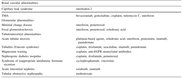
function, thus interfering with the impending administra-tion of the next anticancer drug. Due to the wide variety of chemotherapy regimens, AKI presents with a wide variety of clinical symptoms. Examples of anticancer drug-induced AKI are shown in Table4[38,39]. A classic example of a tubular disorder-inducing drug is platinum-based agents, which primarily result in disorders in the tubulointersti-tium. For example, cisplatin is known to cause AKI in approximately one-third of patients [40]. The anti-VEGF antibody bevacizumab is well known to cause vascular disorder and induce TMA.
Although the emergence of the RIFLE and AKIN classifications, which are based on serum Cr and urinary output, have resulted in significant advances in the diagnosis of AKI, many issues remain. Serum Cr is affected by several factors such as age, body weight, sex differences, other agents, muscle metabolism, protein intake, and hypervolemia; thus, it is deeply flawed as a biomarker of AKI [41,42]. In addition, elevated serum Cr does not manifest until 48-72 hours after the initial occurrence of nephrotoxicity, thus hindering prompt AKI diagnosis and therapeutic intervention [42]. To compensate for the flaws of serum Cr, the usefulness of many novel biomarkers of AKI has been examined. However, the clinical use of novel biomarkers of AKI still faces high hurdles due to the need to establish threshold values in accordance with sex differences, age differences, and primary diseases [43].
The objectives of this guideline are to examine the latest findings regarding biomarkers for AKI induced by anticancer drugs, and to determine the usefulness and
limitations of these biomarkers in real-world clinical settings.
Commentary
The biomarkers discussed in this draft can be assessed objectively and serve as indicators of pharmacological responses to biological changes, histological changes, and therapeutic interventions [44]. Biomarkers of anticancer drug-induced AKI must be immune to interference from all types of treatment. Potential roles for biomarkers include: 1) risk assessment, 2) early diagnosis, 3) classification of disease stage, 4) differential diagnosis, 5) indication of therapeutic effects, and 6) determination of prognosis. Anticipation is particularly high for the practical applica-tion of biomarkers that enable earlier diagnosis than do serum Cr and eGFR.
The present draft divides biomarkers into two cate-gories: those that can be used in clinical practice and are covered by health insurance in Japan, and those that can-not. Also, in 2010, the Predictive Safety Testing Consor-tium’s Nephrotoxicity Working Group submitted results for drug toxicity studies and analyses of biomarker per-formance to the FDA and the European Medicines Evalu-ation Agency; these results presented Kidney Injury Molecule-1 (Kim-1), urinary albumin, urine protein, b2 microglobulin, serum cystatin C, clusterin, and trefoil factor 3 (TFF-3) as biomarkers related to renal function safety [45]. Although the objective of this report is limited to safety assessments, we felt it necessary to discuss the usefulness of the above 7 biomarkers as biomarkers of anticancer drug-induced AKI.
Table 4 Examples of anticancer drug-induced AKI (includes only anticancer drugs covered by insurance in Japan) Kidney Int. 2015;87:909-17, Clin J A Soc Nephrol. 2012;7:1713-21.Ó[2012] Modified from the American Society of Nephrology
Renal vascular abnormalities
Capillary leak syndrome interleukin-2
TMA bevacizumab, gemcitabine, cisplatin, mitomycin C, interferon Glomerular abnormalities
Minimal change disease interferon, pemetrexed
Focal glomerulosclerosis interferon, pemetrexed, zoledronic acid Tubulointerstitial abnormalities
Acute tubular necrosis platinum-based agents, zoledronic acid, interferon, pentostatin, imatinib, pamidronate
Tubulitis (Fanconi syndrome) cisplatin, ifosfamide, azacitidine, imatinib, pamidronate Magnesium wasting cisplatin, anti-EGFR monoclonal antibodies
Nephrogenic diabetes insipidus cisplatin, ifosfamide, pemetrexed Syndrome of inappropriate antidiuretic hormone
secretion
cyclophosphamide, vincristine
1. Biomarkers for which measurement is covered
by health insurance
a) Urinary albumin
Urinary albumin levels increase as a result of enhanced glomerular permeability and impaired proximal tubular reabsorption. In fact, short-term and long-term adminis-trations of nephrotoxic anticancer drugs have been reported to increase levels of urinary microalbumin [46]. However, urinary albumin levels are known to increase not only as a result of AKI, but also due to factors such as fever, exer-cise, dehydration, diabetes, and hypertension; thus, the specificity of urinary albumin as a biomarker of AKI is considered limited [47].
b) Urine protein
In detection of glomerular disease, urine protein is said to be superior to BUN and serum Cr in terms of diagnostic performance [48]; however, urine protein is reported to have low specificity as a biomarker of AKI [49], and its usefulness has not been established.
c) Serum cystatin C
Cystatin is the most important cysteine protease inhibitor in the human body. Cystatin C, a 13-kDa protein secreted by all nucleated cells, is characterized by the fact that it does not bind to plasma proteins. Therefore, cystatin C is freely filtered by the renal glomeruli; after being reabsorbed by the proximal tubules, more than 99% of it is degraded by the endocytic receptor megalin [50]. Unlike Cr, cystatin C is not secreted by the renal tubules into urine, and its levels are not dependent on sex or muscle mass. In patients with mild to moderate renal impairment, serum cystatin C is well correlated with GFR [51]; thus, cystatin C can be used to detect nephrotoxicity at an early stage with greater sensitivity than serum Cr, thus making serum cystatin C a potentially useful biomarker of AKI [52]. However, serum cystatin C is limited in the following two ways: 1) it is affected by diabetes, high levels of corticosteroids, thyroidism, inflammation, hyperbilirubinemia, and hyper-triglyceridemia [53]; 2) when GFR reaches\15 mL/min/
1.73 m2, the increase in serum cystatin C slows and levels off at 5-6 mg/L [54]. Beno¨hr et al. [55] have demonstrated that serum cystatin C levels are significantly elevated on day 5 following cisplatin administration compared to 3 days prior to administration. At present, serum cystatin C has not been established as a useful biomarker of anti-cancer drug-induced AKI. Although measurement of serum cystatin C is covered by health insurance, measurement of urinary cystatin C is not.
d)b2 microglobulin
b2 microglobulin is a polypeptide comprising 99 amino acids with a molecular weight of 11,800; it is distributed on the surface of nucleated cells throughout the body as the L chain of the major histocompatibility complex HLA class I antigen. b2 microglobulin passes freely through the glomerular basement membrane and is almost completely reabsorbed by the proximal tubule; in tubular disorders, however, decreased reabsorption leads to increased excre-tion of b2 microglobulin in urine, thus making b2 microglobulin a potentially useful marker of AKI. In fact, urinaryb2 microglobulin has been reported to increase 4-5 days earlier than does serum Cr in tubular disorders [56]. However, in aciduria and at room temperature, b2 microglobulin is extremely unstable, thus limiting its use-fulness as a biomarker [57].
e) NAG
In the kidneys, NAG is a glycolytic enzyme present in lysosomes and produced in the endoplasmic reticula of proximal tubule cells. Tubular disorders result in increased excretion of NAG in urine, thus making urinary NAG a potentially useful marker of AKI; urinary NAG is reported to demonstrate abnormal values 12 hours to 4 days earlier than does serum Cr [58]. Goren et al. [59] compared concentrations of NAG before and after cisplatin admin-istration in 12 patients. In their investigation, concentra-tions of NAG increased following cisplatin administration, reached their peak on day 3, and subsequently decreased. In an examination of NAG and b2 microglobulin in 8 patients before and after cisplatin administration, Ikeda et al. [60] reported that b2 microglobulin reached peak levels on day 3 and decreased to pretreatment levels in 1 week, although only 1 patient demonstrated increased NAG for 2 weeks. However, urinary NAG activity is inhibited by many nephrotoxic substances, magnesium, and endogenous urea [61]. Furthermore, urinary NAG levels are increased not only in AKI, but also in rheumatoid arthritis [62], impaired glucose tolerance [63], and hyper-thyroidism [64]; thus, the specificity of urinary NAG for AKI is considered low.
f) Urinary L-FABP
following cardiovascular surgery are reported to demon-strated an increase in urinary L-FABP immediately after surgery [67], while a high urinary L-FABP value is reported to be an independent predictor of AKI [68]. As a biomarker of AKI, L-FABP compares favorably with Kim-1, NGAL, and NAG [69]. In Japan, assessment of L-FABP for the diagnosis of AKI is covered by health insurance. However, there has been very little investigation of the usefulness of L-FABP as a biomarker of anticancer drug-induced AKI in humans; further study is necessary going forward.
2. Biomarkers for which measurement is
not covered by health insurance
a) Urinary Kim-1
Kidney Injury Molecule-1 is a transmembrane glycoprotein produced in the proximal tubule during kidney injury; for 12 hours following renal ischemia, excretion of the extra-cellular domain of Kim-1 into urine is increased [70]. In animal models of cisplatin-induced nephrotoxicity, levels of Kim-1 increased earlier than did levels of serum Cr, indicating that Kim-1 is a useful biomarker of tubular disorders [71]. In addition, a systematic review reported fluctuation of Kim-1 within 24 hours of kidney injury [72]. The United States FDA has approved Kim-1 as a marker of AKI. Tekce et al. [73] compared levels of serum Kim-1 and urinary Kim-1 prior to cisplatin administration and at days 1, 3, and 5 after cisplatin administration in 8 patients with AKI and 14 patients without AKI with an eGFRC90 mL/ min. On day 1, there were no significant differences between the groups in serum Cr, eGFR, or serum Kim-1; however, urinary Kim-1 levels were significantly higher in the AKI group. On day 3, the two groups demonstrated significant differences in serum Cr, eGFR, and urinary Kim-1; however, there were no significant differences in serum Kim-1. Thus, urinary Kim-1 demonstrates potential as an early marker of cisplatin-induced AKI. However, the stability of Kim-1 is markedly reduced in urine; thus, further study of urinary Kim-1 is considered necessary [74].
b) NGAL
Neutrophil gelatinase-associated lipocalin (NGAL) is a 25-kDa glycoprotein secreted primarily by activated neu-trophils; under normal circumstances, 100% of NGAL is reabsorbed by the proximal tubule. In tubular disorders, NGAL is expressed in the ascending limb of the loop of Henle and in part of the collecting ducts; due to increased excretion into blood and urine, NGAL demonstrates
abnormal values 2-4 hours following AKI. A meta-analysis of more than 2,500 cases found that NGAL is a useful marker not only for the diagnosis of AKI, but also for renal prognosis [75]. Peres et al. [76] reported that following the administration of cisplatin, the group of patients with AKI demonstrated higher NGAL levels than the non-AKI group; however, this difference was not significant. Gaspari et al. [77] also compared NGAL levels between a group of 12 AKI patients and a group of 12 non-AKI patients at 1 and 4 hours and at 1, 2, 3, 7, and 15 days after cisplatin administration. Although a significant difference between the AKI group and the non-AKI group in serum Cr was first observed on day 3 following cisplatin administration, a significant difference in NGAL was first observed on day 1. Therefore, NGAL may enable detection of cisplatin-in-duced AKI earlier than does serum Cr.
c) Clusterin
Clusterin, a 76-80-kDa glycoprotein, is assumed to exert an anti-apoptotic renoprotective effect in kidney injury. Uri-nary clusterin is reported to be superior to BUN and serum Cr in the detection of proximal tubular injury [48]. How-ever, insufficient research has been done on clusterin in regard to human AKI, and the usefulness of clusterin as a biomarker of anticancer drug-induced AKI is unknown.
d) Urinary TFF-3
Urinary excretion of TFF-3 is reduced in AKI. Although urinary TFF-3 has been demonstrated to be a useful marker of AKI in animal models, its usefulness in humans has not been sufficiently examined [78].
e) Endothelin-1
Endothelin-1 is a 21-amino acid protein that possesses a vasoconstrictor effect; in the kidneys, it is expressed in mesangial cells and collecting ducts. Takeda et al. [79] measured urinary endothelin-1-like immunoreactivity/Cr before and 1 and 2 weeks after cisplatin treatment; these authors reported that urinary endothelin-1-like immunore-activity/Cr was significantly increased at 1 and 2 weeks after cisplatin treatment compared to pretreatment levels. Following cisplatin treatment, b2 microglobulin/Cr and endothelin-1-like immunoreactivity/Cr peaked on day 2 and subsequently declined, whereas NAG/Cr peaked on day 6.
The usefulness of various biomarkers has been exam-ined in animal models [80]; however, there is a significant dearth of evidence in humans. The following circumstances account for the near-absence of evidence: 1) The lack of uniformity in diagnostic criteria for AKI leads to differ-ences among reports. 2) Although many reports have examined AKI overall, very few reports have focused on anticancer drug-induced AKI. 3) For many biomarkers, evident threshold values have not been established, making assessment in individual studies difficult. 4) Cancer drug therapy often combines multiple drugs. Different drugs induce nephrotoxicity via different mechanisms, while some drugs (cisplatin, etc.) are assumed to act via multiple mechanisms; therefore, assessment with a single biomarker may not be valid (in fact, a study has demonstrated the usefulness of a combination of multiple biomarkers [49]). 5) When using serum, the possibility cannot be ruled out that what is being assessed is not AKI, but rather the effects of anticancer drugs throughout the body. Furthermore, in such cases, the effects of other factors, such as age and past history of CKD and other complications, are unknown.
Although biomarkers enhance our understanding of drug-induced AKI, much remains unknown regarding their contribution to the diagnosis of AKI. When conducting anticancer chemotherapy, nephrologists must determine when biomarkers are necessary, which biomarkers are useful, how to interpret biomarker data, and how to utilize biomarker data on an individual basis in treatment for each patient.
13.2 Prevention of decreased renal function during cancer drug therapy
(1) Overview
CQ3: Is reduction of anticancer drug doses recom-mended for mitigating toxicity in patients with decreased renal function?
Recommendation grade: Weakly recommended (suggestion) Recommendation
When using drugs that lead to an increased risk of adverse drug events in patients with decreased renal function, dose reduction is recommended. However, when the goal is to cure cancer, doses must ultimately be determined with consideration of the balance between risks and benefits. Summary
When using agents that lead to an increased risk of adverse drug events in patients with decreased renal function, dose reduction is recommended. However, when the objective is to cure cancer, doses must ultimately be determined with consideration of the balance between risks and benefits.
Background and Objectives
The kidneys are an elimination pathway for many anti-cancer drugs and their metabolites; therefore, renal impairment can delay the excretion and metabolism of anticancer drugs, potentially resulting in increased toxicity and thus necessitating consideration of dose reduction [81]. For patients with decreased renal function, dose reduction is also sometimes considered for anticancer drugs that are metabolized in the liver. For example, dose reduction is considered necessary when irinotecan is administered to dialysis patients [82–85]. For sorafenib, as well, a drug that is primarily metabolized in the liver, some believe that dose reduction should be considered [86]. The present draft summarizes evidence related to dose reduction and pre-sents principles for dose reduction for major anticancer drugs.
Commentary
Answering CQ3 requires studies comparing frequencies of adverse drug events between normal doses and reduced doses in patients with decreased renal function; however, the search formula used in the present guidelines yielded no relevant literature. Such studies present ethical issues and are considered difficult to conduct. Much of the available evidence [87–90] comes from studies that com-pared the frequencies of adverse drug events in patients with normal renal function and patients with decreased renal function (reduced doses) [87–90]. However, there are very few such studies; thus, the quality of the evidence is judged to be extremely low (D: Almost no confidence in effect estimates).
Consideration of the balance between benefits and risks is particularly important in determining recommendation levels, but due to the paucity of evidence regarding the efficacy of treatment with reduced doses, our recommen-dation is weak.
However, in real-world clinical settings, attempts have been made to reduce doses in accordance with renal function and to control plasma drug concentrations; these attempts have yielded a small number of studies and guidelines that serve as references. One such attempt with carboplatin dosing is the Calvert formula, which calculates doses using target AUC and Ccr as estimated with the Cockcroft-Gault equation based on the results of a phase I clinical trial (see CQ10 for details) [91]. Another study has reported a revised Calvert formula based on data from Japanese patients [92].
books with information on dose reduction for anticancer drugs [94]. Outside of Japan, the United States FDA [95] and the European Medicines Agency [96] have published guidelines calling for the inclusion of methods of admin-istration for patients with decreased renal function in package inserts for all types of drugs; these publicly available package inserts may also serve as a reference for dose reduction.
For anticancer drugs, the therapeutic range and the toxic range are extremely close to each other. Therapeutic drug monitoring is considered useful for preventing toxicity in such cases; in fact, therapeutic drug monitoring has been proven effective in randomized clinical trials for some anticancer drugs [97,98]. However, at present, attempting to measure blood concentrations of anticancer drugs is not standard practice.
A realistic desirable approach for patients with decreased renal function is to begin anticancer drug administration by referring to the above-mentioned dose adjustment guidelines, monitor adverse events more clo-sely than normal, and consider adjusting doses in future
treatment. In patients for whom the objective is to cure cancer, doses must ultimately be determined in consider-ation of the balance between risks and benefits.
(2) Platinum-based drugs
CQ4: Is risk factor assessment recommended for pre-dicting cisplatin-induced AKI?
Recommendation grade: Weakly recommended (suggestion) Recommendation
Reported predictors of cisplatin-induced AKI include hypoalbuminemia; smoking; female sex; age (1.03-fold increase in risk per year of age); concomitant use of other anticancer drugs; comorbid cardiovascular disease or dia-betes; advanced cancer; and total cisplatin dose. In order to prevent cisplatin-induced AKI, risk factors should be assessed prior to drug administration.
Summary
Reported predictors of cisplatin-induced AKI include hypoalbuminemia; smoking; female sex; age (1.03-fold
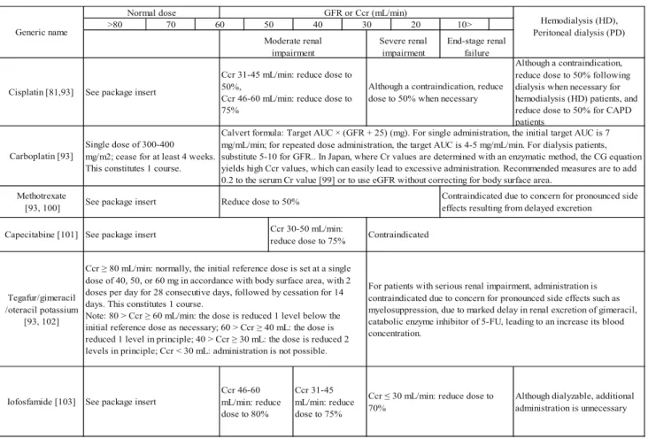
Table 5 Dose reduction methods for major anticancer drugs in patients with decreased renal function
CAPD: continuous ambulatory peritoneal dialysis, CG: Cockcroft-Gault
Although dialyzable, additional administration is unnecessary
Hemodialysis (HD), Peritoneal dialysis (PD) Generic name
Cisplatin [81,93]
20 10>
Normal dose GFR or Ccr (mL/min)
Moderate renal impairment
Severe renal impairment
End-stage renal failure 70
>80 60 50
Although a contraindication, reduce dose to 50% following dialysis when necessary for hemodialysis (HD) patients, and reduce dose to 50% for CAPD patients
Ccr ≥ 80 mL/min: normally, the initial reference dose is set at a single dose of 40, 50, or 60 mg in accordance with body surface area, with 2 doses per day for 28 consecutive days, followed by cessation for 14 days. This constitutes 1 course.
Note: 80 > Ccr ≥ 60 mL/min: the dose is reduced 1 level below the initial reference dose as necessary; 60 > Ccr ≥ 40 mL: the dose is reduced 1 level in principle; 40 > Ccr ≥ 30 mL: the dose is reduced 2 levels in principle; Ccr < 30 mL: administration is not possible. Carboplatin [93]
Methotrexate [93, 100]
Capecitabine [101]
Tegafur/gimeracil /oteracil potassium
[93, 102]
Contraindicated
For patients with serious renal impairment, administration is contraindicated due to concern for pronounced side effects such as myelosuppression, due to marked delay in renal excretion of gimeracil, catabolic enzyme inhibitor of 5-FU, leading to an increase its blood concentration.
Single dose of 300-400 mg/m2; cease for at least 4 weeks. This constitutes 1 course.
Calvert formula: Target AUC × (GFR + 25) (mg). For single administration, the initial target AUC is 7 mg/mL/min; for repeated dose administration, the target AUC is 4-5 mg/mL/min. For dialysis patients, substitute 5-10 for GFR.. In Japan, where Cr values are determined with an enzymatic method, the CG equation yields high Ccr values, which can easily lead to excessive administration. Recommended measures are to add 0.2 to the serum Cr value [99] or to use eGFR without correcting for body surface area.
See package insert Reduce dose to 50% Contraindicated due to concern for pronounced side
effects resulting from delayed excretion
Ccr 30-50 mL/min: reduce dose to 75% See package insert
30
Iofosfamide [103]
See package insert
Ccr 31-45 mL/min: reduce dose to 50%,
Ccr 46-60 mL/min: reduce dose to 75%
Although a contraindication, reduce dose to 50% when necessary
See package insert
Ccr 31-45 mL/min: reduce dose to 75% Ccr 46-60
mL/min: reduce dose to 80%
Ccr ≤ 30 mL/min: reduce dose to 70%
increase in risk per year of age); concomitant use of other anticancer drugs; comorbid cardiovascular disease or dia-betes; advanced cancer; and total cisplatin dose. However, among existing studies, there is no consistent definition of AKI, there are no clear threshold values for risk factors, and there are no established measures for cases with risk factors. Thus, many issues remain for further investigation. Background and Objectives
Cisplatin, a key drug in treatment for many types of cancer, is one of the most commonly used anticancer drugs. However, cisplatin is known to produce side effects such as myelosuppression, intestinal toxicity, and neurotoxicity; another crucial side effect, nephrotoxicity, is a potential subsequent cisplatin dose-limiting factor. One-third of patients receiving cisplatin are presumed to have comorbid AKI [104], which often results in the limitation of subse-quent doses of cisplatin. Furthermore, AKI sometimes develops into chronic tubulointerstitial fibrosis and irre-versible chronic tubulopathy, which may further progress to CKD [105,106]. The present draft examines risk factors that may serve as predictors of cisplatin-induced AKI. Commentary
Cisplatin-induced renal injury is considered to manifest primarily as proximal tubular injury, particularly in the S3 segment [107]. Cisplatin is absorbed from the basolateral surface into cells and injures mitochondrial DNA, thereby activating apoptosis. Intracellular accumulation of cisplatin results in inflammation, oxidative stress, and ischemic injury [105]. Hypomagnesemia is also considered to cause renal injury. Magnesium is thought to be involved in active transport mechanisms in the renal tubules. Sobrero et al. have supposed that hypomagnesemia leads to an increased concentration of cisplatin in renal tubular cells, thereby causing proximal tubular injury [108].
In an investigation by de Jongh et al. [109] of weekly-dose cisplatin for 400 patients with locally advanced or metastatic cancer, 36% of patients received cisplatin alone, 49% received cisplatin ? etoposide, and 15% received cisplatin?paclitaxel. A total of 116 patients (29%) demonstrated a reduction in Ccr of C
25%, while 29 patients (7%) were unable to continue cisplatin due to nephrotoxicity. Independent predictors of post-cisplatin nephrotoxicity as determined by mul-tivariate analysis were paclitaxel coadministration (odds ratio [OR] 4.0, p = 0.001), hypoalbuminemia (OR 3.5, p = 0.006), smoking (OR 2.5, p = 0.002), female sex, and old age. According to age group, the risk of
nephrotoxicity was 26% among patients aged \ 48
years, and increased with age to 35% for patients aged 48-62 years and 41% for patients aged[62 years; the
risk of nephrotoxicity increased 1.03-fold per year (OR 1.03, p = 0.007). Regarding gender, the risk of nephrotoxicity was twice as high for women as for men (OR 2.0, p = 0.025). Another study reported that cis-platin excretion capacity is lower in women than in men [110]; however, the cause of this difference is unknown. The involvement of smoking in nephrotoxi-city has been surmised to be the effect of oxidant stress [111]; however, one possibility that cannot be ruled out is that smoking causes cardiovascular disease, which secondarily leads to post-cisplatin nephrotoxicity. Also, in hypoalbuminemia, an increased concentration of unbound cisplatin is considered to enhance nephrotox-icity [109]. The cited study, which defines nephrotoxi-city as a reduction in Ccr of C 25%, is not strictly an assessment of predictors of AKI.
In an investigation of 425 patients treated with cisplatin (total dose 220 mg/m2 [median]), Stewart et al. [112] reported that in multivariate analysis, the factors that pre-dicted maximum increases in serum Cr up to 4 weeks after cisplatin treatment were serum albumin, serum potassium, body surface area, and number of administrations. How-ever, this study contains flaws: renal function was assessed with serum Cr alone, and the authors’ method of assessing maximum increases in serum Cr up to 4 weeks after cis-platin treatment is neither a well-established method nor period for assessment. Furthermore, anticancer drugs were used in combination with many other drugs; thus, the degree to which cisplatin contributes to changes in renal function is unknown.
In an examination of 1,721 patients treated with cis-platin, Mizuno et al. [113] found, in multivariate analysis, that cancer stage 4 diagnosis (OR 1.8, p = 0.011) and total cisplatin dose were risk factors for moderate AKI (1.5-1.9-fold increase in serum Cr within 7 days of cisplatin treat-ment), while comorbid cardiovascular disease, comorbid diabetes mellitus, and cancer stage 4 diagnosis were risk factors for severe AKI (C 2.0-fold increase in serum Cr within 7 days of cisplatin treatment).