Abstract: We examined the role of Tie2 in regulating wound healing after tooth extraction. Wistar rats underwent maxillary incisor tooth extraction, and immunodetection techniques were used to determine Tie2 expression in the healing wound. The wound was initially filled with blood coagulum containing densely aggregated erythrocytes, leukocytes, fibrin, and endothelial progenitor cells, indicating that blood vessel formation started in the socket. Tie2 was detected on monocytic cell membranes. On day 3, fibroblastic cells proliferated in the coagulum, small vessels appeared by day 5, and new bone formed in the vessel-rich area. Robust woven bone trabeculae were present around vessels by day 7, and woven bone and osteoclast-like giant cells were present on day 10. Woven bone surrounded sinusoidal capillary- like vessels. Full-length (140-160 kDa) Tie2 was not detected at any time, although Tie2 fragments were present in the healing wound. N-terminus- and C-terminus-specific Tie2 antibodies detected 40-kDa and 60-kDa fragments or 70-kDa and 50-kDa frag- ments, respectively. The levels of these fragments decreased during the first 3 days and started to increase by day 5-10. The Tie2 extracellular domain initially inhibited angiogenesis, and its degradation relieved inhibition of new vessel formation. The onset of vessel formation in the wound may be induced by
scattered endothelial progenitor cells.
(J Oral Sci 57, 313-318, 2015)
Keywords: Tie2; angiopoietin receptor; receptor tyrosine kinase; tooth-extraction socket;
wound healing; neovascularization.
Introduction
During the healing of a tooth extraction socket, blood vessel formation establishes the major pathways for transport and delivery of regulatory factors, oxygen, and nutrients and elimination of waste. Neovascularization is therefore essential for proper wound healing, which requires the activity of angiogenic and growth factors that engage cell-surface receptor tyrosine kinases. For example, the receptor tyrosine kinase Tie2 is a type I transmembrane protein that serves as the receptor for angiopoietins (1-6). It comprises an extracellular domain (ECD) with three epidermal growth factor-like domains situated between the first and second immunoglobulin- like loops. This second loop is followed by three fibronectin type III repeats upstream of the transmem- brane region (2,4) (Fig. 1, left). In normal embryonic and adult tissue, signaling by the Tie2 ligands angiopoi- etin-1-4 is crucial in the attachment (stabilization) and detachment (destabilization) of endothelial cells and their surrounding support cells (pericytes) during angio- genesis and vascular remodeling (7-14). After paracrine production of angiopoietin-1 by pericytes, this protein binds to Tie2 expressed by endothelial cells and sustains the mutual attachment of these two cell types (9,15).
In contrast, autocrine production of angiopoietin-2 by endothelial cells in response to local hypoxia antagonizes Original
Temporal expression in rats of receptor tyrosine kinase Tie2 during early wound healing after tooth extraction
Hirotaka Sato
1), Shuya Kasai
2), and Chihaya Maesawa
2)1)
Department of Pathology, Division of Anatomical and Cellular Pathology, Iwate Medical University, Iwate, Japan
2)
Institute of Biomedical Science, Department of Tumor Biology, Iwate Medical University, Iwate, Japan
(Received June 4, 2015; Accepted August 6, 2015)
Correspondence to Dr. Hirotaka Sato, Department of Pathology, Division of Anatomical and Cellular Pathology, Iwate Medical University, 2-1-1 Nishitokuta, Yahaba-cho, Shiwa-gun, Iwate 028-3694, Japan
Fax: +81-19-908-8018 E-mail: [email protected] doi.org/10.2334/josnusd.57.313
DN/JST.JSTAGE/josnusd/57.313
Tie2 signaling (16,17), with Tie1-Tie2 complex forma- tion (18), causing detachment of endothelial cells from pericytes. This detachment in turn enables endothelial cells to proliferate in the presence of vascular endothelial growth factor (14,19,20).
Surprisingly little is known about the role of angiopoi- etin/Tie2 signaling during neovascularization in healing tooth-extraction sockets. Here, we utilized a rat model of this process to analyze wound healing, using histological and immunodetection techniques.
Materials and Methods
Animals and wound models
Forty-eight 10-week-old male Wistar rats underwent right maxillary incisor tooth extraction under general anesthesia by intraperitoneal (i.p.) administration of pentobarbital sodium (40 mg/kg). Groups of eight rats each were killed by an i.p. overdose of pentobarbital sodium at 12 h and 1, 3, 5, 7, and 10 days after surgery.
Right craniomaxillary tissues were dissected. This study was approved by the Ethical Committee on Animal Experiments of Iwate Medical University (15-002).
Tissue preparation
One-half of the dissected right craniomaxillary tissues were fixed in 4% paraformaldehyde (PFA), decalcified with 10% Na
2EDTA (pH 7.5) for 2 weeks, dehydrated
using an ascending series of alcohol concentrations, and embedded in paraffin. Serial sections (thickness, 6 µm) were prepared on the sagittal plane for histological examination, stained routinely with hematoxylin and eosin (H-E), and analyzed using the immunohisto- chemical techniques described below. The lateral socket walls were removed from the remaining tissues, and their entire contents were harvested for Western blot analysis.
Immunohistochemistry
The sections were deparaffinized using Hemo-Clear (FALMA, Tokyo, Japan), hydrated, and subjected to antigen retrieval by incubation at 95°C for 5 min in 10 mM sodium citrate buffer, pH 6.0. After blocking endogenous peroxidase activity with 0.3% hydrogen peroxidase in methanol, immunohistochemistry was performed using a polymeric enzyme-labeled antibody method (N-Histofine Simple Stain MAX PO kit; Nichirei Biosciences Inc., Tokyo, Japan) at room temperature in humidified chambers. The substrate was 3’3-diaminoben- zidine tetrahydrochloride (DAB; Wako, Osaka, Japan).
The sections were counterstained with hematoxylin. The specificities of the Tie-2 antibodies were confirmed previ- ously (21,22). Normal rabbit immunoglobulin G (IgG;
Santa Cruz Biotechnology, Inc., Santa Cruz, CA, USA), diluted to an equivalent protein concentration, served as a negative control in place of the primary antibody.
70kDa
50kDa
Epitope Tie2(C20) C-terminus
FN III TM
TK TK
Fig. 1 The structure of Tie2 (left), epitopes recognized by anti-Tie2 antibodies (right), and proteolytic fragments of Tie2 detected by the antibodies (right). Ig, immunoglobulin domain; EGF, EGF-like repeats; FN III, fibronectin type III domains;
TM, transmembrane; TK, tyrosine kinase domain.
Western blotting
Wound tissues and HUVEC cells as the control were homogenized using a sonicator (Branson Sonifier Cell Disruptor, Model S-150D; Danbury, CT, USA) for 30 s at an output frequency of 22.5 kHz with the probe intensity gradation set at 2-3 in cold RIPA buffer (50 mM Tris- HCl pH 8.0, 150 mM NaCl, 0.5% sodium deoxycholate, 0.1% SDS) containing a mixture of protease inhibitors (Complete Protease Inhibitor Cocktail, Roche Diag- nostics GmbH, Penzberg, Germany). The homogenate was centrifuged at 15,000 rpm for 10 min at 4°C, and the supernatant was used for Western blotting analysis.
Protein concentrations were determined using the Bradford assay (23), with bovine serum albumin as the standard. Electrophoresis of the extracted samples (20 µg/10 µL) was performed using a 10
%SDS-polyacryl- amide gel according to the method of Laemmli (24).
After electrophoresis, the protein was electroblotted onto a polyvinylidene fluoride (PVDF) membrane (Hybond- P, GE Healthcare UK Ltd, Buckinghamshire, UK). The PVDF membranes were incubated overnight at 4°C in 5% nonfat dry milk in 0.2% Tween-PBS (to reduce nonspecific binding of the antibodies), incubated for 2 h using the primary antibodies described below, and incu- bated for 1 h using the N-Histofine kit described above.
The ECL Plus detection kit (Amersham Bioscience UK Ltd., Buckinghamshire, UK) and X-ray film (Fuji Photo Film, Tokyo, Japan) were used to visualize the antibody- antigen complexes. The amount of protein on the blots was estimated using ImageJ 1.48 (Rasband, W.S., ImageJ, U. S. National Institutes of Health, Bethesda, Maryland, USA, http://imagej.nih.gov/ij/, 1997-2012) and is expressed as the mean value of four different wound sets for each time point. The mean value of each fragment was converted to a relative ratio and plotted.
The data were normalized using the mean value of the 50-kDa band 12 h after extraction as standard.
Primary antibodies
The rabbit anti-Tie2 (C-20) polyclonal antibody (anti-Tie2-C; Santa Cruz Biotechnology, Inc.) was raised against a peptide at the intracellular carboxy terminus of Tie2 (amino acid residues 1103-1122), and the rabbit anti-Tie2 (H-176) polyclonal antibody (anti-Tie2-N;
Santa Cruz Biotechnology, Inc.) was raised against a recombinant protein corresponding to the extracellular domain of Tie2 (amino acid residues 25-200). They were used as primary antibodies for immunohistochemistry and Western blotting (Fig. 1, right) and were diluted to 1:200 for each technique.
NB
A B C
D E F
NB NB
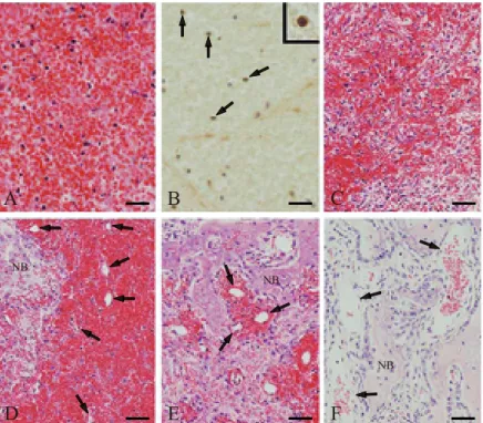
Fig. 2 Histological analysis of extraction sockets during healing. (A) Erythrocytes, leukocytes, and fibrin in the blood coagulum immediately, 12 h, and 1 day after tooth extraction. (B) By day 1, detection of Tie2 expression by monocytic cells (arrows and inset) in the blood coagulum using anti-Tie2-C. (C) Proliferation and random distribution of spindle-shaped fibroblastic cells in the coagulum on day 3. (D) Microvessels (arrows) and new bone formation in the coagulum on day 5. (E) Robust woven bone formed around the vessels (arrows) on day 7. (F) Sinusoidal capillary-like vessels (arrows) surrounded by woven bone on day 10. NB, newly formed bone. Scale bars: (A) 25 µm, (B) 100 µm (inset, 7.8 µm), (C-F) 50 µm.
Results
Histological findings
The tooth-extraction socket was analyzed at 12 h and 1 day post-extraction. The socket was filled with blood coagulum composed of densely aggregated erythrocytes, leukocytes, and fibrin (Fig. 2A). Using the anti-Tie2-C, we detected Tie2 expression on the cell membrane in some monocytic cells (Fig. 2B). Three days after extrac- tion, proliferating spindle-shaped fibroblastic cells were distributed randomly in the blood coagulum (Fig. 2C).
Numerous small vessels appeared in the coagulum by day 5, and new bone formation was observed in the vessel- rich area across the socket (Fig. 2D). Robust formation of trabeculae of woven bone was seen around the vessels by day 7 (Fig. 2E), and the socket was filled with extensively deposited woven bone accompanied by osteoclast-like giant cells by day 10. The areas surrounded by this woven bone included sinusoidal capillary-like vessels, which were fewer and larger than those observed on day 7 (Fig. 2F). Using an anti-Tie2-N that recognizes its extracellular domain, Tie2 was not detected in the socket at any time after extraction.
Western blot and quantitative analysis of Tie2 expression in tooth extraction wounds
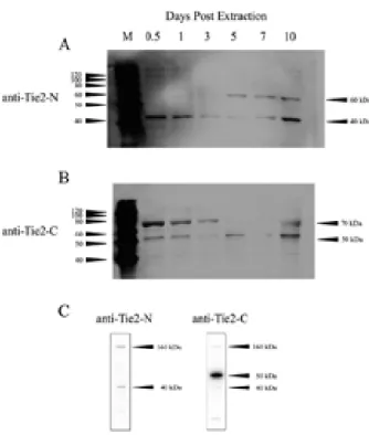
Western blot analysis detected Tie2 fragments in the healing extraction wound. The anti-Tie2-N, which recog- nizes the N-terminal region of the ECD, detected 40- and 60-kDa fragments (Fig. 3A), and the anti-Tie2-C, which recognizes the intracellular C-terminal domain (ICD), detected 70- and 50-kDa fragments (Fig. 3B). Analysis of lysates prepared from HUVEC cells showed that the anti-Tie2-N detected 160- and 40-kDa fragments and that the anti-Tie2-C detected 160-, 50-, and 40-kDa fragments (Fig. 3C).
Quantitative analysis of the four bands detected by the antibodies (Fig. 4) revealed that the amount of the 40-kDa fragment derived from the ECD detected by anti-Tie2-N decreased rapidly from 12 h to 3 days, started to increase by day 7, and then rose sharply until day 10. In addition, the 60-kDa fragment derived from the ECD, which was detected by anti-Tie2-N, first appeared on day 5, and the amount increased from days 7 through 10. Finally, the amounts of the 70- and 50-kDa fragments detected by anti-Tie2-C rapidly declined from 12 h to 3 days and then increased sharply from days 7 to 10.
Fig. 3 Western blot analysis of Tie2 expression in the tooth- extraction wound. (A) Anti-Tie2-N detected 60- and 40-kDa ECD fragments. (B) Anti-Tie2-C detected 70-kDa EDC/ICD and 50-kDa ICD fragments. (C) In HUVEC cell lysates, anti-Tie2-N detected 160- and 40-kDa fragments, and anti-Tie2-C detected 160-, 50- and 40-kDa fragments. M, molecular weight standards.
Fig. 4 Quantitative analysis of Western blots of Tie2 in tooth extraction wounds (n = 4). Relative ratio of each band at the indicated times, using the mean value of the ICD 50-kDa (Tie2-C) fragment at 12 h after extraction as reference. The level of the ECD 40-kDa (Tie2-N) fragment rapidly decreased from 12 h to 3 days and increased after 5 days. The ECD 60-kDa (Tie2-N) frag- ment was first detected on day 5 and its levels increased thereafter.
Levels of the 70-kDa ECD/ICD and 50-kDa ICD (Tie2-C) frag- ments decreased from 12 h to day 7 but increased sharply on day 10.
Days Post Extraction
0 1 2 3 4 5 6 7 8 9 10
0
tion wound sockets as proteolytic cleavage fragments derived from its EC and IC domains. The EC domain regulates angiogenesis by sequestering its ligands (angiopoietins) (20,21). According to its predicted amino acid sequence, the present Western blotting analyses—
using antibodies that recognize epitopes at its N- and C-termini, respectively (Fig. 1)—indicate that full-length Tie2 (140-160 kDa) is proteolytically cleaved into frag- ments representing the 40- or 60-kDa N-terminal regions (anti-Tie2-N) from the ECD and other fragments of 70- or 50-kDa fragments encompassing the C-terminal region (anti-Tie2-C). Further, the data indicate that the 40-kDa ECD fragment detected by the anti-Tie2-N represents the N-terminal region of the 60-kDa ECD fragment. These fragments correspond to those detected in our analysis of peripheral blood (data not shown).
The 60-kDa fragment derived from the ECD, which was detected 5-10 days after tooth extraction, may be present in newly formed tissue and is cleaved in periph- eral blood to generate the N-terminal 40-kDa fragment of the ECD. The functions of the 60-kDa and 40-kDa ECD fragments are unknown. Some evidence indicates that a soluble form of the Tie2 ECD, which includes the ligand binding site, acts as a Tie2 inhibitor or as a reservoir of angiopoietins (25,26). In either case, the soluble form would inhibit the blood vessel formation required for healing of the tooth-extraction wound.
Our kinetic analysis of the appearance of each frag- ment (anti-Tie2-N, 60 kDa and 40 kDa; anti-Tie2-C, 70 kDa and 50 kDa) after tooth extraction (Fig. 4) suggests that each proteolytic fragment was degraded early during wound healing to allow the angiogenesis required for wound healing. Thus, proteolysis of each of these fragments caused levels to decrease immediately after extraction to a minimum level, or a level slightly above the detection limit, until day 3. The resulting lack of Tie2 expression in the wound would therefore allow angiogenesis to proceed until at least day 5, after which the fragments reappeared at readily detectable levels.
The blood coagulum in the extraction socket contains endothelial progenitor cells (EPCs); thus, blood vessel formation started sporadically in the socket. Immunohis- tochemical analysis revealed expression of Tie2 ICD on the membranes of monocytic cells scattered around the coagulum in the early socket. These may be EPCs (27).
Because microvessels started to appear sporadically in the 5-day socket, vessel formation may originate from the EPCs in the coagulum.
We conclude that temporal expression of Tie2 in the
inhibition of new vessel formation. Moreover, onset of vessel formation in the extraction socket may be induced by EPCs scattered across the extraction socket. The roles of the soluble form of the Tie2 ECD and of other growth factor receptors expressed by cells in the healing socket remain to be determined.
Acknowledgments
This study was supported in part by a Grant-in-Aid for Strategic Medical Science Research Center from the Ministry of Education, Culture, Sports, Science and Technology of Japan, 2010-2014.
Conflicts of interest
No competing financial interests to declare.
References
1. Dumont DJ, Yamaguchi TP, Conlon RA, Rossant J, Breitman ML (1992) tek, a novel tyrosine kinase gene located on mouse chromosome 4, is expressed in endothelial cells and their presumptive precursors. Oncogene 7, 1471-1480.
2. Dumont DJ, Gradwohl GJ, Fong GH, Auerbach R, Breitman ML (1993) The endothelial-specific receptor tyrosine kinase, tek, is a member of a new subfamily of receptors. Oncogene 8, 1293-1301.
3. Runting AS, Stacker SA, Wilks AF (1993) tie2, a putative protein tyrosine kinase from a new class of cell surface receptor. Growth Factors 9, 99-105.
4. Sato TN, Qin Y, Kozak CA, Audus KL (1993) Tie-1 and tie-2 define another class of putative receptor tyrosine kinase genes expressed in early embryonic vascular system. Proc Natl Acad Sci U S A 90, 9355-9358.
5. Eklund L, Olsen BR (2006) Tie receptors and their angio- poietin ligands are context-dependent regulators of vascular remodeling. Exp Cell Res 312, 630-641.
6. Jeltsch M, Leppänen VM, Saharinen P, Alitalo K (2013) Receptor tyrosine kinase-mediated angiogenesis. Cold Spring Harb Perspect Biol 5, doi: 10.1101/cshperspect.a009183.
7. Dumont DJ, Gradwohl G, Fong GH, Puri MC, Gertsenstein M, Auerbach A et al. (1994) Dominant-negative and targeted null mutations in the endothelial receptor tyrosine kinase, tek, reveal a critical role in vasculogenesis of the embryo. Genes Dev 8, 1897-1909.
8. Sato TN, Tozawa Y, Deutsch U, Wolburg-Buchholz K, Fujiwara Y, Gendron-Maguire M et al. (1995) Distinct roles of the receptor tyrosine kinases Tie-1 and Tie-2 in blood vessel formation. Nature 376, 70-74.
9. Suri C, Jones PF, Patan S, Bartunkova S, Maisonpierre PC, Davis S et al. (1996) Requisite role of angiopoietin-1, a ligand for the TIE2 receptor, during embryonic angiogenesis.
Cell 87, 1171-1180.
10. Maisonpierre PC, Suri C, Jones PF, Bartunkova S, Wiegand SJ, Radziejewski C et al. (1997) Angiopoietin-2, a natural
13. Stratmann A, Risau W, Plate KH (1998) Cell type-specific expression of angiopoietin-1 and angiopoietin-2 suggests a role in glioblastoma angiogenesis. Am J Pathol 153, 1459- 1466.
14. Yancopoulos GD, Davis S, Gale NW, Rudge JS, Wiegand SJ, Holash J (2000) Vascular-specific growth factors and blood vessel formation. Nature 407, 242-248.
15. Hanahan D (1997) Signaling vascular morphogenesis and maintenance. Science 277, 48-50.
16. Oh H, Takagi H, Suzuma K, Otani A, Matsumura M, Honda Y (1999) Hypoxia and vascular endothelial growth factor selectively up-regulate angiopoietin-2 in bovine microvas- cular endothelial cells. J Biol Chem 274, 15732-15739.
17. Mandriota SJ, Pyke C, Di Sanza C, Quinodoz P, Pittet B, Pepper MS (2000) Hypoxia-inducible angiopoietin-2 expres- sion is mimicked by iodonium compounds and occurs in the rat brain and skin in response to systemic hypoxia and tissue ischemia. Am J Pathol 156, 2077-2089.
18. Marron MB, Singh H, Tahir TA, Kavumkal J, Kim HZ, Koh GY et al. (2007) Regulated proteolytic processing of Tie1 modulates ligand responsiveness of the receptor-tyrosine kinase Tie2. J Biol Chem 282, 30509-30517.
19. Holash J, Wiegand SJ, Yancopoulos GD (1999) New model
of gastric ulcers in rats. World J Gastroenterol 9, 788-790.
22. Abramovich D, Rodriguez Celin A, Hernandez F, Tesone M, Parborell F (2009) Spatiotemporal analysis of the protein expression of angiogenic factors and their related receptors during folliculogenesis in rats with and without hormonal treatment. Reproduction 137, 309-320.
23. Bradford MM (1976) A rapid and sensitive method for the quantitation of microgram quantities of protein utilizing the principle of protein-dye binding. Anal Biochem 72, 248-254.
24. Laemmli UK (1970) Cleavage of structural proteins during the assembly of the head of bacteriophage T4. Nature 227, 680-685.
25. Reusch P, Barleon B, Weindel K, Martiny-Baron G, Gödde A, Siemeister G et al. (2001) Identification of a soluble form of the angiopoietin receptor TIE-2 released from endothelial cells and present in human blood. Angiogenesis 4, 123-131.
26. Harris AL, Reusch P, Barleon B, Hang C, Dobbs N, Marme D (2001) Soluble Tie2 and Flt1 extracellular domains in serum of patients with renal cancer and response to antiangiogenic therapy. Clin Cancer Res 7, 1992-1997.
27. Asahara T, Murohara T, Sullivan A, Silver M, van der Zee R, Li T et al. (1997) Isolation of putative progenitor endothelial cells for angiogenesis. Science 275, 964-967.