THE THESIS OF DOCTOR OF PHILOSOPHY
Study on a Virtual Reality-based
Training System for the Vascular
Interventional Surgery
Jin Guo
Graduate School of Engineering Kagawa University
Abstract I
Ph.D. thesis of Dr. Jin Guo
Abstract
Cardiovascular and cerebrovascular diseases are the leading causes of deaths worldwide. Based on a World Health Organization survey, it is estimated that, till 2020, coronary artery disease and cerebral hemorrhage are still the first and second causes of death of human being. Catheter-based endovascular interventional procedures have been extensively adopted for treatment and diagnosis of various vascular diseases. During the procedures, a flexible catheter having a pre-bent shape with natural curvature at the tip is directed inside the patient's vascular structure, from the femoral artery to the target position, to cure the vascular diseases by manipulating the tail part of the catheter. The gradually increasing success rate of catheter-based interventions, combined with their less blood loss and minimal damage to healthy tissue, has lead to a significant growth in the number of procedures performed annually. However, it is still a challenging work to position a catheter in a target vessel branch within the highly complicated and delicate vascular structure. The interventionalists have to carry out the operations by imagining the spatial relationship between the catheter and its surrounding vessels based on their experience and feelings of hands due to the lack of intuitive visual feedback. Additionally, complicated vessel shapes and multi contacts between the catheter and the blood vessel walls lead to significant deterioration in the feasibility and maneuverability of positioning the
II Study on a Virtual Reality-based Training System for the VIS
catheter. The catheter manipulation skills of experienced interventionalists are therefore critical to the success of the endovascular catheterization.
Compared to conventional training methods on cadavers, animals, phantoms or real patients, the VR-based simulators provide not only the flexible and repeatable scenarios but also the possibility to precisely assess training progress. The virtual reality-based simulators for training in Vascular Interventional Surgery (VIS) were presented in this paper. This application allows generating realistic physical-based models of catheters and blood vessels, and enables surgeons to touch, feel and manipulate virtual catheter inside a vascular model through the same surgical operation mode as is used in actual VIS. Experimental results show that the error rate is in an acceptable range and that simulators can be used for surgery training.
Additionally, most of the existing controllers for virtual reality-based simulators introduced commercially available interfaces (e.g. PHANTOM Omni) or a remote controller for game machines (e.g. Nintendo Wii) or robotic control consoles which are manipulated, unfortunately, not in a manner that is in accordance with conventional catheter operation practices. Furthermore, few researchers focused on the controller designs with much consideration of the natural catheter manipulation skills obtained from manual catheterization. A new master controller for endovascular catheterization that can allow the
Abstract III
Ph.D. thesis of Dr. Jin Guo
interventionalists to apply conventional pull, push and twist of the catheter as used in current practice was presented in this paper. A catheter sensing unit (used to measure the motion of the catheter) and a force feedback unit (used to provide a sense of resistance force) were both presented. A camera was used to allow a contactless measurement that avoids additional friction and the force feedback in the axial direction was provided by the magnetic force generated between the permanent magnets and the powered coil. Performance evaluation of the controller was evaluated by first conducting comparison experiments to quantify the accuracy of the catheter sensing unit, and then conducting several experiments to evaluate the force feedback unit. From the experimental results, the minimum and the maximum errors of translational displacement were 0.003 mm and 0.425 mm respectively. The average error was 0.113mm. In terms of rotational angles, the minimum and the maximum error were 0.39º and 7.2º respectively. The average error was 3.61º. The force resolution was approximately 25mN and a maximum current of 3A generated an approximately 1.5 N force. Based on analysis of requirements and state of the art in computer-assisted and robot-assisted training systems for endovascular catheterization, a new master controller with force feedback interface was proposed to maintain the natural endovascular catheterization skills of the interventionalists.
IV Study on a Virtual Reality-based Training System for the VIS
reality-based method for reducing visual feedback transmission times based on an Internet-based tele-operative robotic catheter operating system was designed for vascular interventional surgery, to reduce radiation-exposure times, and afford unskilled surgeons the opportunity to learn basic catheter/guidewire skills, while allowing experienced physicians to perform surgeries cooperatively. Remote surgical procedures, limited by variable transmission times for visual feedback, have been associated with deterioration in operability and vascular wall damage during surgery. At the patient’s location, the catheter shape/position was detected in real time and converted into three-dimensional coordinates in a world coordinate system. At the operation location, the catheter shape was reconstructed in a virtual-reality environment, based on the coordinates received. The data volume reduction significantly reduced visual feedback transmission times; coordinate values were easily synchronized with control data and haptic signals. Remote transmission experiments, conducted over inter-country distances, demonstrated the improved performance of the proposed prototype.
Contents V
Ph.D. thesis of Dr. Jin Guo
Contents
Abstract ... I Contents ... V List of Figures ... IX Acknowledgements ... XV Declaration ...XVII Chapter 1 Introduction ... 19 1.1 Preface ... 19 1.2 Background ... 20 1.3 Literature review ... 25 1.4 Contributions ... 35 1.5 Thesis structure ... 38Chapter 2 Overview of the virtual reality-based training system ... 41
2.1 The previous virtual reality-based simulators ... 42
2.2 The proposed VR-based training system ... 50
2.3 The interaction between the vascular model and the catheter model ... 52
2.4 Summary ... 54
Chapter 3 A hybrid method for 3D vascular reconstruction ... 57
3.1 A fast 3D reconstruction method ... 57
VI Study on a Virtual Reality-based Training System for the VIS
3.2.1 Enhancement and cleaning ... 65
3.2.2 Level set segmentation ... 65
3.2.3 Skeletonization and centerline extraction ... 70
3.2.4 Surface reconstruction ... 71
3.3 Summary ... 77
Chapter 4 Physics-based models for the virtual reality-based training system ... 79
4.1 Physics-based modeling of blood vessels ... 79
4.2 Physics-based modeling of the catheter ... 81
4.3 Collision detection ... 83
4.4 The performance evaluation of the deformation model of the blood vessel walls and the simulated catheter ... 85
4.5 Summary ... 88
Chapter 5 A new haptic controller for training in Vascular Interventional Surgery ... 90
5.1 Overview of the proposed master controller ... 91
5.2 Catheter sensing methods ... 93
5.3 Design of the force feedback unit ... 102
5.4 Control system mechanisms ... 106
5.5 Experimental results ... 109
5.5.1 Evaluation of feature extraction algorithm ... 109
5.5.2 Evaluation of catheter sensing unit ... 109
Contents VII
Ph.D. thesis of Dr. Jin Guo
5.7 Summary ... 119
Chapter 6 The 2D and 3D models for reducing the time delay of visual feedback based on virtual reality techniques ... 122
6.1 2D Model for reducing the time delay of visual feedback ... 122
6.1.1 The Implementation Method at the slave side ... 122
6.1.2 The Implementation Method at the master side ... 124
6.1.3 Experimental results ... 124
6.2 3D models for reducing the time delay of visual feedback based on virtual reality techniques ... 128
6.2.1 Structure of the Proposed Method ... 129
6.2.2 Two-camera System Calibration ... 131
6.2.3 Two-camera System Rectification ... 134
6.2.4 Feature Extraction and Stereo Matching ... 140
6.2.5 3-D Coordinate Reconstruction ... 146
6.2.6 Experimental setup ... 157
6.2.7 Experimental results ... 160
6.3 Discussion ... 167
6.4 Summary ... 169
Chapter 7 Conclusions and future work ... 172
7.1 Conclusions ... 172
7.2 Future work ... 175
References ... 177
VIII Study on a Virtual Reality-based Training System for the VIS
Appendix I ... 197 Appendix II ... 222 Biographic Sketch ... 242
List of Figures IX
Ph.D. thesis of Dr. Jin Guo
List of Figures
Figure1- 1 Vascular Interventional Surgery (VIS) ... 22
Figure1- 2 the procedure of catheter ablation [Padalino13] ... 22
Figure1- 3 the procedure of aneurysm coil embolization [Spiotta12] .. 23
Figure1- 4 the procedure of stent delivery ... 24
Figure1- 5 the procedure of drug delivery ... 24
Figure1- 6 the Sensei Robotic Catheter System [HansenMedical] ... 27
Figure1- 7 the Stereotaxis Niobe II [Stereotaxis] ... 29
Figure1- 8 the Angio Mentor™ System [Simbionix, Ltd.] ... 30
Figure1- 9 the Procedicus Vascular Interventional System Trainer [VIST, MenticeAB] ... 31
Figure1- 10 The structure of the thesis... 39
Figure 2- 1 The first virtual-reality-based training system ... 42
Figure 2- 2 The improved virtual-reality-based training system ... 43
Figure 2- 3 The design of the master controller for the VR-based training system. ... 44
Figure 2- 4 The whole structure of VR-based training system ... 45
Figure 2- 5 the conceptual principle of the controller ... 47
Figure 2- 6 the schematic description of the working components of an optical mouse ... 47 Figure 2- 7 The overview of the proposed VR system for
X Study on a Virtual Reality-based Training System for the VIS
catheterization training ... 50
Figure 2- 8 The proposed VR-based system for endovascular catheterization training ... 51
Figure 2- 9 the coordinates of tip of catheter in Figure 2-12 ... 52
Figure 2- 10 The recorded collision vectors ... 53
Figure 2- 11 The recorded collision vectors-2 ... 54
Figure 2- 12 The Endo Vascular Evaluator model (EVE) ... 55
Figure 3- 1 The whole structures of all the blood vessels ... 59
Figure 3- 2 The specific blood vessel ... 60
Figure 3- 3 The normal CT file ... 60
Figure 3- 4 The advanced CT file ... 61
Figure 3- 5 The results of noise removement ... 61
Figure 3- 6 The reconstruction result of the blood vessels inside the bones ... 62
Figure 3- 7 The reconstruction result of the only bones... 63
Figure 3- 8 The 3D CT dataset of the specific patient ... 64
Figure 3- 9 The selection of the start point and the end point of the desired blood vessel branch... 67
Figure 3- 10 The cloud points’ model based on the level sets algorithm ... 68
Figure 3- 11 The selction of another segment of the desire branch vessels ... 69
List of Figures XI
Ph.D. thesis of Dr. Jin Guo
Figure 3- 12 The connection between the two branch blood vessels ... 69
Figure 3- 13 The reconstruction results based on the refined algorithm ... 70
Figure 3- 14 The extraction results of the centerlines ... 71
Figure 3- 15 The illustration of reflective (A with AF ) and rotational (A with AR) symmetries. ... 74
Figure 3- 16 The 15 basic intersection topologies ... 75
Figure 3- 17 The surface reconstruction based on the marching cube algorithm ... 75
Figure 3- 18 The rendering results of the surface model after the surface refinement ... 76
Figure 3- 19 The comparison results before and after surface refinement ... 76
Figure 4- 1 The simulation images of catheter: (a) the real catheter. (b) the simulated catheter in VR system ... 82
Figure 4- 2 The collision detection between the simulated catheter and the blood vessel walls ... 85
Figure 4- 3 The vectors attached to the vertices of the blood vessel walls ... 86
Figure 4- 4 The deformation of the blood vessel walls ... 87
Figure 4- 5 The deformation of the simulated catheter ... 87
XII Study on a Virtual Reality-based Training System for the VIS
Figure 5- 2 The relationship between the pixel units and physical units
... 94
Figure 5- 3 The markers attached to a 2-mm-diameter catheter. ... 95
Figure 5- 4 The design of the force feedback unit ... 100
Figure 5- 5 The design of the bearing with a grasper ... 102
Figure 5- 6 The schematic of the control system ... 104
Figure 5- 7 The results of the feature detection algorithm ... 105
Figure 5- 8 The process of capturing feature points for the rotational calculation. ... 107
Figure 5- 9 The results of the insertion and extraction motion ... 111
Figure 5- 10 The results of the rotation motion ... 112
Figure 5- 11 The results of pull/push experiments with different velocities ... 113
Figure 5- 12 The results of pull/push experiments with different displacement. ... 114
Figure 5- 13 The results of the rotation experiments with different velocities. ... 114
Figure 5- 14 The experimental setup for the evaluation of the force feedback unit ... 115
Figure 5- 15 The calibration results between the current and the force .... ... 116
Figure 5- 16 The comparative results between the require force and the measured force ... 117
List of Figures XIII
Ph.D. thesis of Dr. Jin Guo
Figure 6- 1 The structure of the surgeon console………125 Figure 6- 2 The path of the tip of the catheter in the slave side……..128 Figure 6- 3 The coordinates of the tip of the catheter in the slave
side………128 Figure 6- 4 The reconstructed path of the tip of catheter in the master
side………129 Figure 6- 5 The general structure of Internet-based tele-surgical
systems………..130 Figure 6- 6 The procedure for two-camera calibration………...132 Figure 6- 7 The chessboard used for two-camera calibration……….135 Figure 6- 8 The original pictures captured by the two-camera
system………140 Figure 6- 9 The rectified images based on the proposed algorithm…141 Figure 6- 10 The stereo figure for the two cameras after stereo
rectification………...142 Figure 6- 11 The surgical procedure using the proposed catheter
operating system………...143 Figure 6- 12 The red markers attached to a 2-mm-diameter
catheter………..143 Figure 6- 13 The rectified images based on the proposed
algorithm………...144 Figure 6- 14he extraction results for the red markers……….…150 Figure 6- 15The insertion experiment inside a straight
XIV Study on a Virtual Reality-based Training System for the VIS
Figure 6- 16 The insertion experiment inside a straight
pipe-Frame3………..153 Figure 6- 17 The insertion experiment inside a bent pipe-Frame1 ....155 Figure 6- 18 The insertion experiment inside a bent pipe-Frame 2…157 Figure 6- 19 The actual distance and calculated distance…..……….165 Figure 6- 20 The error distance for the catheter tip……….165 Figure 6- 21 The remote transmission experiments from Takamatsu to
Beijing………...……166 Figure 6- 22 The remote transmission experiments from Takamatsu to
Harbin………166 Figure 6- 23 The remote transmission experiments from Takamatsu to
Acknowledgements XV
Ph.D. thesis of Dr. Jin Guo
Acknowledgements
The author wishes to express his great gratitude to his supervisor Professor Shuxiang Guo for his invaluable guidance, support and friendly encouragement throughout my Ph.D. course and for providing me with first class resources.
The author would like to thank Prof. Hideyuki Hirata and Prof. Keisuke Suzuki. Thanks them for their valuable advices and suggestion on my research. Without their help, I can hardly finish this thesis.
Also the author would like to acknowledge the efforts of his laboratory members, especially Dr. Nan Xiao and Ms. Maoxun Li whose time and expertise were greatly appreciated.
This research was supported by Kagawa University Specially Promoted Research fund 2012.
Finally, the author gives special appreciation to his parents for their love, patience and support.
Declaration XVII
Ph.D. thesis of Dr. Jin Guo
Declaration
I hereby declare that this submission is my own work and that to the best of my knowledge and belief. It contains no material previously published or written by another person nor material which to a substantial extent has been accepted for the award of any other degree or diploma of the university or other institute of higher learning, except where due acknowledgment has been made in the text.
Chapter1 Introduction 19
Ph.D. thesis of Dr. Jin Guo
Chapter 1
Introduction
1.1 Preface
In this thesis, the virtual reality-based simulators for surgeons’ training in vascular interventional surgery were proposed and developed. These simulators were capable of generating the realistic virtual-reality environment of blood vessels according to a patient’s special computed tomography (CT) or magnetic resonance imaging (MRI). In addition, the new master controller had the ability to allow the surgeons to directly employ a real catheter as the controller maintaining the surgeons’ conventional operating skills in an actual surgery. It can also allow the surgeons to carry out interventions with force feedback in the axial direction, which provides the surgeon with a sense of resistance.
(1) To reconstruct a realistic three-dimensional vascular model, a median filter algorithm was employed to reduce the noise of CT images. Next was to segment the blood vessel walls from the CT or MRI images based on the level set algorithm. Then the 3D surface of a vascular segment from CT or MR images was rebuilt by using marching cube algorithm. Finally, centerlines of a vascular segment
20 Study on a Virtual Reality-based Training System for the VIS
were computed and the surface of a vascular segment was smoothed. (2) To maintain the natural endovascular catheterization skills of the interventional radiologist, a camera was employed to detect the axial and radial motions of a catheter simultaneously with the ability to allow interventional radiologists to use their dexterous skills while performing catheter-based interventions. Additionally, a force feedback unit was developed to provide the interventionalists with a sense of resistance force.
(3) To realize the distance teaching and learning, the virtual reality-based method was developed to effectively reduce the transmission time of visual feedback due to the significant reduction in data volume using coordinates instead of actual images.
1.2 Background
With the quickening pace of modern life, the brain diseases of people are increasing, such as cerebral aneurysm and infarction and so on. The traditional surgery spends patients a lot of operation time and has long recovery time; the burden on patients is heavy. Vascular Interventional Surgery (VIS, Shown in Figure 1-1) is a revolutionary surgical technique, in which surgery is done using precise medical devices and viewing equipment inserted through a small incision instead of making a large incision to expose the operation site. A lot of diagnosis and medical surgery with an endoscope or a catheter are performed for
Chapter1 Introduction 21
Ph.D. thesis of Dr. Jin Guo
vascular interventional surgery recently. The procedure of catheter ablation is shown in Figure 1-2 and the Figure 1-3 presents the coil embolization surgery. The diagram of the stent delivery is shown in Figure 1-4, and the Figure 1-5 shows the drug delivery surgery. The main advantage of this technique is to reduce trauma to healthy tissue since this trauma is the leading cause of patient’ pain and scarring and a prolonged hospital stay. Fast recovery and a short hospital stay also reduce the cost of surgery and radiation exposure. A critical disadvantage of this surgery technique, however, is its complexity, requiring extensive training efforts of the interventionalists to achieve competency because the arteries through which the catheter passes are extremely intricate and delicate. It is difficult for novice surgeons to insert the catheter from femoral to the target position within the patient’s vascular structure especially the blood vessel of patient’s brain. In addition, repeated insertion of the catheter through several tests could tear a blood vessel and cause bleeding instantaneously, while excessive pressure could rupture blood vessels. For practical and ethical reasons, realistic virtual-reality simulators provide a promising alternative method compared to other available alternatives such as anesthetized animals, human cadavers and patients. VR-based simulators enable novice interventionalists to learn basic wire and catheter handling skills and provide expert practitioners with the opportunities to rehearse new operation procedures prior to performing them on a patient.
22 Study on a Virtual Reality-based Training System for the VIS
Figure1- 1 Vascular Interventional Surgery (VIS)
Chapter1 Introduction 23
Ph.D. thesis of Dr. Jin Guo
Figure1- 3 the procedure of aneurysm coil embolization [Spiotta12] Additionally, distance teaching/training has become indispensable to novice surgeons around the world. As an example, the use of this advanced minimally invasive technique in Canada has been severely hampered by the inability to provide adequate training and support to community surgeons, many of who live in remote regions of the country [Anvari07]. If a remote surgical system is in place, a competent surgeon can work cooperatively to assist in a surgical procedure that takes place in another region [Mitsuishi07]. However, due to long transmission times and the variable quality of the visual image feedback, these surgeons may be exposed to prolonged periods of radiation [Guo12]. A more efficient, safe, and reliable method is needed to disseminate new surgical knowledge worldwide.
24 Study on a Virtual Reality-based Training System for the VIS
Figure1- 4 the procedure of stent delivery
Chapter1 Introduction 25
Ph.D. thesis of Dr. Jin Guo
Based on aforementioned background, in this paper, a virtual reality-based catheterization training system for vascular interventional surgery was proposed and developed. The simulators were capable of extracting blood vessel walls from the specific patients’ CT or MRI images. Physics-based deformation model of the catheter and the blood vessel walls were designed and developed in the virtual reality-based training system. A new master controller with force feedback was designed and developed to maintain the natural endovascular catheterization skills of the interventional radiologist. For distance training and teaching, a newly developed method was described, based on virtual reality and image-processing technology.
1.3 Literature review
Compared to conventional surgical methods, vascular interventional surgery has many advantages. Therefore, Catheter-based endovascular interventional procedures have been extensively adopted for treatment and diagnosis of various vascular diseases. During the procedures, interventionalists manipulate the tail part of the catheter to target branch vessels to cure the vascular diseases [Guiatni13]. Thus complicated vessel shapes and multi contacts between the surgical instrument and the vessel walls inevitably lead to significant deterioration in the feasibility and operability of positioning the catheter [Srimathveeravalli10].
26 Study on a Virtual Reality-based Training System for the VIS
To afford novice interventionalists the opportunity to learn basic catheter manipulation skills, robot-assisted and computer-assisted catheterization training is two promising methods. Two commercially available endovascular simulators are the Procedicus Vascular Interventional System Trainer (shown in Figure 1-9) [VIST,
MenticeAB][Aggarwal06] (VIST, Mentice AB, Gothenburg, Sweden)
and the Angio MentorTM (shown in Figure 1-8) [Simbionix,
Ltd.][Benjamin14] (Simbionix, Ltd., Cleveland, OH, U.S.), which
allows novice interventionalists to conduct the comprehensive and goal-oriented training and education program before treating real patients including catheter/wire manipulating, stent deployment and angioplasty balloon inflation in the domain of renal, carotid, aortoiliac and peripheral vascular structures [Desser07]. The Sensei robotic navigation system (shown in Figure 1-6) [HansenMedical][Saliba08] (Hansen Medical, Mountain View, Calif) and the Niobe magnetic navigation system (shown in Figure 1-7) [Stereotaxis][Chun08] (Stereotaxis, St. Louis, Mo) are two typically commercial surgical robotic systems that have been used in the endovascular interventional surgery with electromechanical base and magnetically controlled mechanisms of action. They have been successfully used in different clinical applications including cardiac ablation and endovascular aneurysm repair [Payne12].
Chapter1 Introduction 27
Ph.D. thesis of Dr. Jin Guo
Figure1- 6 the Sensei Robotic Catheter System [HansenMedical] However, there are several limitations to the application of these commercial products. The most obvious one is the expensive cost of the systems, which makes them cost prohibitive to most private centers [Antoniou11, Das14, Binning12]. Additionally, the catheters must be integrated with their custom interfaces and navigation systems to do the catheterization operations, which cannot be widely available.
In addition to the commercially available products, many research groups have studied endovascular interventional training systems. Cercenelli et al. [Cercenelli07][Marcelli08] developed a novel tele-robotic system to manipulate standard electrophysiology catheters. However, there is no force feedback device in their controller design.
28 Study on a Virtual Reality-based Training System for the VIS
The loss of force feedback in robotic catheter systems can increase the risk of damage to the blood vessel walls. Fukuda et al. [Tanimoto97,
Negoro02, Tercero12] at Nagoya University have developed a Joy Stick
as the controller of the master system with a haptic unit to handle the manipulators of the slave system. There are two joints in the design of the master controller and each joint was connected to a motor with a wire mechanism. Another research group in [Guo12] designed a master controller to simulate the conventional push, pull and rotate motion used in current practice. However, unlike the conventional endovascular catheterization, in which interventionalists manipulate an actual catheter directly using their hands, the employment of these master controllers replaces the catheter from the interventionalists’ hand with the “handle part”, which are made of metal materials with different size and material attributes from a real catheter, to carry out catheterization training operations. The loss of the hand feelings on a real catheter unavoidably contaminates their intuitive and dexterous skills in the interventional radiology procedure. To achieve better training effects, Yogesh Thakui et al. [Thakui09] developed a novel remote catheter navigation system to directly manipulate percutaneous transluminal catheters from a location far away from the patient while allowing the interventionalists to apply conventional insertion, extraction and rotation motion in the procedure. A similar design was presented by the researchers in [Ma13]. However, these research achievements have a common defect: To measure the motion of the
Chapter1 Introduction 29
Ph.D. thesis of Dr. Jin Guo
input catheter, the catheter is needed to be clamped by the rollers coupled with encoders. To ensure the catheter does not slip in the apparatus, a force has to be applied to the rollers, which leads to the difficulties in designing the force feedback device to provide the interventionalists with a sense of resistance force.
Figure1- 7 the Stereotaxis Niobe II [Stereotaxis]
Because vascular interventional techniques have unavoidably reduced the sense of touch compared to open surgery, surgeons have to rely more on the haptic feeling generated by the interaction between blood vessels and the catheter. Even if the color and texture of blood vessels visually convey crucial anatomical information, touch is still critical in surgery. The benefits of using force feedback devices in vascular interventional surgery training through simulation have
30 Study on a Virtual Reality-based Training System for the VIS
already been recognized by several research groups and many of the companies working in this area [Basdogan01, Çakmak00, Brown02,
Tsang08, Wei09, Basdogan07, Chiang11, Takashima07, Meier01,
Aggarwal06, Neequaye07]. In these researches, however, the virtual surgical trainings were carried out without haptic feedback, or researched on a virtual model of body organs not a vascular physical model. Moreover, some achievements in this area have used commercial products or other haptic devices as a controller to in virtual vascular interventional surgery [Basdogan04]. Nevertheless, it is not convenient when surgeons drive the catheter for insertion and rotation because it does not accord with the custom of surgeons’ operations.
Chapter1 Introduction 31
Ph.D. thesis of Dr. Jin Guo
Figure1- 9 the Procedicus Vascular Interventional System Trainer
[VIST, MenticeAB]
Due to practical and ethical reasons, computer-assisted training systems have distinct advantages over traditional training methods on cadavers, animals, phantoms or real patients. The VR-based simulators are capable of providing not only the more realistic scenarios but also the flexible training courses. However, most of the existing controllers introduced commercially available interfaces [Srimathveeravalli10] (e.g. PHANTOM Omni) or a remote controller for game machines
[Wei09] (e.g. Nintendo Wii) or robotic control consoles [Lu11] which
32 Study on a Virtual Reality-based Training System for the VIS
with conventional catheter operation practices.
To address the need for providing a familiar and ergonomic setting for the interventionalists, a compact master controller capable of measuring the radial and axial motion of the input catheter simultaneously and providing force feedback while still preserving the manual catheter manipulation skills was designed. A camera was used to allow a contactless measurement that avoids additional friction and the force feedback in the axial direction was provided by the magnetic force generated between the permanent magnets and the powered coil.
Because the vascular structure through which the catheter passes is highly intricate and delicate, VIS requires extensive training by the surgeon to achieve competency. Thus, distance teaching/training has become indispensable to novice surgeons around the world. To meet these requirements, the Internet-based remote catheter operating system provides a promising method to realize participation of highly capable physicians in surgical procedures that take place in distant locals [Liu11, Arata07, Arata06]. In 2001, for example, researchers in
[Marescaux01] performed remote surgery using a surgical robot, ZEUS,
via a dedicated asynchronous transfer mode (ATM) between New York and Strasbourg, France. However, a dedicated fiber-optic network for remote surgery would be costly. Conventional existing network infrastructures, such as the Internet, provide a preferable conduit for future remote surgical procedures [Arata07].
Chapter1 Introduction 33
Ph.D. thesis of Dr. Jin Guo
Many research groups have studied robotic telesurgical systems based on the Internet [Zhou07, Al-Mouhamed05, Islam11, Xiao12,
Kim10, Xie07, Smith10]. For example, the Centre for Minimal Access
Surgery (CMAS) at McMaster University used broadband Internet and telecommunication systems to provide distance training and mentoring to community surgeons living in remote northern communities of Canada [Anvari07]. Generally, the concept of an Internet-based surgical system consists of three parts: a master system at the operation location, a slave system at the patient’s location, and a communication link, as shown in Figure 7-1. The master and slave systems are connected by transmission control protocol/Internet protocol (TCP/IP) networks for sending and receiving data, including image information, haptic feedback, and manipulator control signals.
The time delay for visual transmission is unavoidable with Internet-based remote surgical systems. The visual information is much larger than the control signals and haptic feedback. Thus, the incremental time delay for visual information is always associated with significant deterioration in the feasibility and maneuverability of surgical tools. This delay thus decreases the effectiveness of the training session during remote surgical education, and has the potential to contribute to surgical damage to the surrounding tissue during the procedure. To date, researchers have focused mainly on delayed control and force signals via the Internet [Natarajan08, Richert09]; however,
34 Study on a Virtual Reality-based Training System for the VIS
few studies have examined the time delay associated with visual transmission. Conventionally, delays in visual information retrieval have been handled by predictive methods, increased autonomy [Smith10], or high-speed image compression processes [Jayender08]. However, most prediction-based delay compensation methods rely on predicting the feedback from the remote environment, which may be difficult to model, particularly if the main feedback is a video signal. Additionally, the image compression process is time-consuming.
To solve these problems, a newly developed method was proposed, based on virtual reality and image-processing technology, for efficiently reducing visual feedback transmission times, making it easier to control the time difference between haptic signals, control data, and image information. This was achieved by reducing the amount of communication data in the image information. Several remote visual feedback transmission experiments were conducted to demonstrate the feasibility of the proposed prototype, via a cooperative surgical effort between research groups in China and Japan. The tele-operative catheter operating system proposed in this work facilitates teamwork between medical professionals worldwide; additionally, this technique minimizes radiation exposure times by reducing the transmission time for visual feedback.
Chapter1 Introduction 35
Ph.D. thesis of Dr. Jin Guo
1.4 Contributions
In this thesis, a virtual reality-based endovascular catheterization training system was designed and developed. Firstly, a hybrid 3D reconstruction method was proposed to extract the blood vessel walls from the specific patients’ CT or MRI files. Physics-based deformation models were built to simulate the behavior after the interaction between the blood vessel walls and the virtual catheter. A new master controller with force feedback for the virtual reality training system was designed and developed to allow the interventionalists to do the surgical training courses by using the natural endovascular catheterization skills. To solve the high cost and difficulties in distance teaching in Canada, a virtual reality and image-processing based method was described to improve the feasibility and maneuverability of the remote learning and traing course.
The mainly contributions of the virtual reality-based systems are as following:
(1) Flexible reconstruction methods for realistic three-dimensional vascular models based on the specific patients’ CT or MR images. Core components of virtual-reality surgical simulators and training systems are the realistic vascular models that are virtual representations of real blood vessels that display accurate displacement due to the fact that models with incorrect material properties and shapes could result in
36 Study on a Virtual Reality-based Training System for the VIS
adverse training effects. In this study, two kinds of reconstruction algorithms were proposed. One is to rebuild the vascular structure based on normal and advanced CT or MRI files. The normal images were employed to subtract their corresponding advanced images to remove human bones and other soft tissues, thereby remaining the data of blood vessels. This approach is adaptable to calculate the whole vascular structure. The other one is employed to reconstruct the 3D vascular models based on advanced CT or MRI files only according to marching cube and level sets algorithms. This approach is preferable to build a vascular segment due to its flexibility.
(2) Development of a new master controller for the virtual reality-based training system. Based on the requirements of the interventionalists and the state of the art in the design of the master controller for computer-aided and robot-aided training systems for endovascular surgery, a new master controller capable of contactless measuring the axial push-pull and twisting motions of a catheter with force feedback to the operators was proposed, whilst still preserving the natural ergonomic skills of conventional catheterization. A camera was employed to detect the motions of the catheter freely and interventionalists can manipulate the real catheter intuitively with a familiar, ergonomic setting. Force feedback to the interventionalists in axial direction was generated by Magnetic force based on the calculated forces between the vascular model and the virtual catheter in a virtual
Chapter1 Introduction 37
Ph.D. thesis of Dr. Jin Guo
reality environment or the forces from the sensing unit of the slave system. Several experiments were conducted to evaluate the performance of the new master controller and the experimental results showed that the errors of the catheter sensing unit and haptic unit of the new master controller were in an acceptable range and it can be used as a controller in a computer-aided or robot-aided training system.
(3) Design of a new prototype to reduce the transmission time of visual feedback based on virtual reality and image processing algoritms. In this part, a new prototype based on the Internet was proposed and developed, for efficiently reducing the transmission time of the visual feedback. It also can make it easier to control the time difference between haptic signals, control data, and image information by reducing the amount of communication data in the image information. Several remote visual feedback transmission experiments were conducted to demonstrate the feasibility of the proposed prototype, via a cooperative surgical effort between research groups in China and Japan. The purpose of the experiments was to evaluate the performance of the new catheter operating system and its associated transmission times for visual feedback, by comparing performance before and after the application of the proposed architecture. The maximum error for catheter reconstruction on the tip of the catheter was 0.93 mm less than 1 mm in these 10 frames. Remote transmission experiments showed that the transmission time of the visual feedback was reduced
38 Study on a Virtual Reality-based Training System for the VIS
significantly based on the newly developed method.
1.5 Thesis structure
Figure 1-10 shows the structure of the thesis. Chapter 1 is the introduction. It is composed of preface, background, literature review, research purposes, research approaches and thesis structure. In chapter 2, overview of the previous and proposed virtual reality-based training systems are compared and introduced. Chapter 3 introduces the 3D vascular reconstruction algorithms. Two reconstruction methods were proposed and developed. The specific patients’ CT or MRI images were the input of the reconstruction algorithms. Level sets and marching cubes algorithms were used to segment the blood vessel walls and to generate the surface model of the blood vessels. In chapter 4, physics-based models for the virtual reality-based training system has been described. The mass-spring model was employed to simulate the deformation behavior of the blood vessel walls to obtain better performance. The elastic-joint-based method to simulate the deformation behavior of the virtual catheter model has been introduced. In chapter 5, a new haptic controller for training in vascular interventional surgery has been developed. Several experiments were conducted to evaluate the performance of the new master controller. Chapter 6 presented the 2D and 3D models for reducing the time delay of visual feedback based on image processing and virtual reality
Chapter1 Introduction 39
Ph.D. thesis of Dr. Jin Guo
techniques. In chapter 7, conclusions and future work were given.
Chapter2 Overview of the virtual reality-based training system 41
Ph.D. thesis of Dr. Jin Guo
Chapter 2
Overview of the virtual reality-based
training system
There are several conventional methods for the training of catheterization skills such as animals, anatomic phantoms and actual patients. However, animals have the distinct anatomical differences with human being and it is very difficult and expensive to produce anatomic phantoms due to the lack of flexibility. Obviously, acting directly on the actual patients is very dangerous to both patients and trainees. Compared to conventional training methods, virtual reality-based catheterization training systems have distinct advantages such as high flexibility, high realism, low cost and without any risks. It allows not only training novice surgeons with basic catheter manipulation skills and enabling them to learn new catheterization courses but also designing a surgical pre-planning treatment in the VR system to better accomplish the operation before an actual surgery is performed.
42 Study on a Virtual Reality-based Training System for the VIS
2.1 The previous virtual reality-based simulators
Our laboratory has been working on the virtual reality-based simulators for endovascular catheterization training. The first VR-based training system is shown in Figure 2-1.Figure 2- 1 The first virtual-reality-based training system
On the master side, a surgeon operated a handle to drive a catheter for insertion or rotation, which, to the surgeon, seems to clamp catheter directly. At the same time, control instructions of the catheter operating system were transmitted to the virtual-reality environment. The catheter in the virtual-reality environment was inserted or rotated according to control commands from the master side. However, there are some limitations for this VR-based training system. It is lack of deformation for catheter and blood vessel walls and it is absence of the collision
Chapter2 Overview of the virtual reality-based training system 43
Ph.D. thesis of Dr. Jin Guo
detection between the catheter model and the 3D vascular model in the vritual reality environment. In terms of the master controller, it is incapable of using an actual catheter to do the training courses.
Thus, the deformation model of the blood vessel walls and the catheter and the collision detection algorithms were added to the virtual reality training system. The improved VR-based training system in vascular interventional surgery was shown in Figure 2-2. If the catheter contacts a blood vessel, the collision vectors between the simulated catheter and the blood vessel walls was detected, stored and the deformation steps were carried out to provide the better performance of visual feedback to the novice surgeons. Based on collision vectors and three-dimensional vascular image information, the virtual-reality environment can be used for medical training.
44 Study on a Virtual Reality-based Training System for the VIS
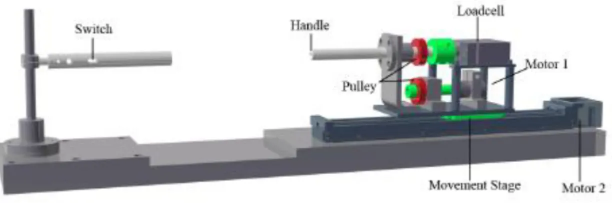
Figure 2- 3 The design of the master controller for the VR-based training system.
The surgeon’s console was the master side of the entire system, as shown in Figure 2-3. Surgeons carried out operations using the console. The switch on the left handle was used to control the two graspers at the slave side; only one switch was necessary, because the catheter was clamped by only one grasper at a time. The operator’s action was detected using the right handle. The movement part of the catheter manipulator mimicked the same motion as the right handle of the controller. The right handle measured both the axial and radial movement of the surgeon’s hand. The handle was sustained by a bearing, linked to a load cell; a pulley was attached to the handle. A dc motor (Motor 1) with an encoder generated torque feedback. A pulley which is couple to the upper one is fixed to the axle of the motor. All of the parts mentioned above were placed on a translation stage, which was driven by a stepping motor (Motor 2). Axial movement was measured as follows. The pulling/pushing force was measured by the
Chapter2 Overview of the virtual reality-based training system 45
Ph.D. thesis of Dr. Jin Guo
load cell when the operator pulled or pushed the handle. This, in turn, generated a movement output that allowed the handle to follow the surgeon’s hand. The force feedback depended on the speed of the translation stage. The displacement and speed of the translation stage were sent to the catheter manipulator side, which allowed the catheter manipulator to remain synchronized with the controller. When the operator rotated the handle, the rotation angle was measured by an encoder installed on the dc motor. The dc motor functioned in tandem with the control mode to generate a damping force to the surgeon. The damping was calculated from the torque generated by the catheter manipulator side.
46 Study on a Virtual Reality-based Training System for the VIS
However, this achievement in this area has not used a real catheter as a controller to operate the virtual vascular interventional surgery. Therefore, it is not convenient when surgeon drive the catheter for inserting and rotating because it does not accord with the custom of surgeons’ operations. Therefore, the objective of the optimization effort of the VR-based training system is to present an interventionalist’s training system based on virtual reality technology with a new mechanical structure of catheter controller for vascular interventional surgery. The training system can not only generate the realistic virtual reality environment of blood vessels according to patient’s special computed tomography (CT) or magnetic resonance imaging (MRI), but also allow unskilled doctors to drive a real catheter for training courses directly and simulate surgeon’s operating skills, insertion and rotation in real surgery (shown in Figure 2-4).
The conceptual principle of the controller which was applied to operate virtual catheter inside blood vessel model has been shown in Figure 2-5.
Chapter2 Overview of the virtual reality-based training system 47
Ph.D. thesis of Dr. Jin Guo
Figure 2- 5 the conceptual principle of the controller
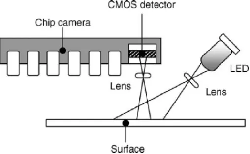
Figure 2- 6 the schematic description of the working components of an optical mouse
48 Study on a Virtual Reality-based Training System for the VIS
The catheter can be subjected to two different sets of movement during manipulation: insertion/retraction and rotation. According to the translation and rotation motion, catheter can be manipulated to reach different parts of the blood vessels. The photoelectric sensor was used to measure the information of displacement and rotation of the catheter. The basic working principle of an optical mouse was described in Figure 2-6. A single light emitting diode (LED) illuminated the surface at an angle. A lens was used to image the surface of the mouse pad onto a CMOS sensor located in the camera chip. The off-axis illumination by the LED helps to put the tiny textures on the surface in sharp contrast. The CMOS sensor typically comprises 18 pixel × 18 pixel (324 pixels in total). The optical sensor works by comparing the images of the surface that are refreshed approximately every 1500th of a second. As it is too computationally taxing to compare the images at all 324 possible overlaps, a 5 pixel × 5 pixel window, taken from the center of the second image, was normally used for the overlap matching process. This window is moved relative to the first image and the chip rates how well each of the 324 pixels matches up. These ratings was added to an overall score for the overlap. Once the chip has found the best overlap, it checks the scores of the eight pixels surrounding the center of the window. Finally, it sended the actual value of the displacement to the computer. Measurement accuracy was typically limited to the pixel spacing of the imaging sensor located in the chip. The unit of data measured by a photoelectric sensor is pixel not millimeter. Therefore,
Chapter2 Overview of the virtual reality-based training system 49
Ph.D. thesis of Dr. Jin Guo
next step was to convert pixel unit into millimeter unit. Some experiments have been carried out to find the relationship between pixel data and real displacement of catheter, as Eq. 2-1 shows:
{𝑼 = 𝒂 ∙ 𝑿
𝜽 = 𝒃 ∙ 𝒀 (Eq. 2-1) where X and Y are the sensor outputs in vertical and horizontal direction and U and 𝛉 are real displacements in Cartesian coordinate system. Moreover, a and b are two constants measured by experiments. Then it is much easier to calculate the velocity and acceleration as Eq. 2-2 shows:
𝑺𝒑𝒆𝒆𝒅 = 𝑼(𝒌)−𝑼(𝒌−𝟏)∆𝑻 (Eq. 2-2) There are two obvious advantages in mechanical design of this controller compared to other training system based on virtual reality technology. The first one is the fact that unskilled doctors can operate the real catheter directly for their training courses and the other one is that the measurement of the translational displacement and rotational angles of the catheter is contactless. Therefore, the whole structure of this controller is simple and has better maneuverability and it is extremely competent to train unskilled interventionalists due to the fact that the operation on this controller is almost the same with the custom of surgeon’s operations in an actual surgery.
50 Study on a Virtual Reality-based Training System for the VIS
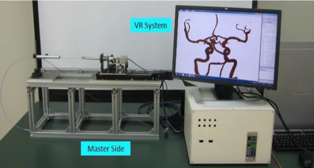
2.2 The proposed VR-based training system
The proposed VR-based system for endovascular catheterization training (shown in Figure 2-7 and Figure 2-8) comprises the foundation elements (hardware and software), the core engines (visualization, modeling, and dynamic interaction), and a master controller.
Figure 2- 7 The overview of the proposed VR system for catheterization training
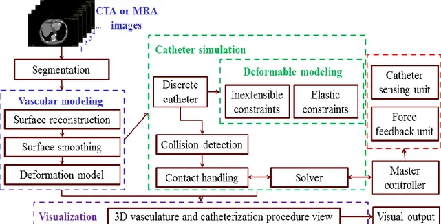
Through the controller, the operator communicates manipulation of the catheter and view to the system, which presents both visual and force feedback. The hardware includes a master controller with force feedback, which will be described in detail later (Chapter 5). Software includes segmentation algorithms for the CTA or MRA images, 3D vascular reconstruction and the physics-based model of the vascular
Chapter2 Overview of the virtual reality-based training system 51
Ph.D. thesis of Dr. Jin Guo
structure and the catheter model.
Figure 2- 8 The proposed VR-based system for endovascular catheterization training
Compared to the previous VR-based training simulators, the newly proposed endovascular catheterization training system has many advantages. The deformation models for the catheter and blood vessel walls were added to the virtual reality environment. Additionally, the collision vectors can be calculated and recorded for further analysis. Most importantly, interventionalists were allowed to directly employ an actual catheter with force feedback to carry out the surgical training courses.
52 Study on a Virtual Reality-based Training System for the VIS
2.3 The interaction between the vascular model
and the catheter model
During the training procedure, doctors operate the controller to drive catheter to insert or rotate, at the same time, the controlling instructions of the catheter operating system are transmitted to the virtual reality environment. The catheter in virtual reality environment can insert or rotate according to the controlling commands from the controller. Based on the three-dimension vascular information, doctors can decide whether to insert or rotate the catheter.
Figure 2- 9 the coordinates of tip of catheter in Figure 2-12
Figure 2-12 hows the status of the tip of catheter which is tracked in a yellow circle inside the blood vessel model when inserting and
Chapter2 Overview of the virtual reality-based training system 53
Ph.D. thesis of Dr. Jin Guo
rotating the controller to control the virtual catheter passing through a curve part and Figure 2-9 shows the three-dimensional coordinates' changes of the tip of the virtual catheter when passing through the curve part.
Figure 2- 10 The recorded collision vectors
When the collision between the simulated catheter and the blood vessel walls happened, two kinds of vectors, collision vectors in both tangential and vertical directions, were calculated and recorder, as shown in Figure 2-10. The collision vectors of the catheter tip were stored separately with the body part of the catheter, which was used to calculate the tip force feedback (shown in Figure 2-11). In addition, the
Collision vectors in vertical direction
Collision vectors in tangential direction
54 Study on a Virtual Reality-based Training System for the VIS
collision vectors of both the catheter body part and the tip part can be used to calculate the force feedback generated from the virtual reality environment.
Figure 2- 11 The recorded collision vectors-2
2.4 Summary
In this chapter, the former virtual reality-based training systems have been introduced. Compare to the previous training systems, the overview of the proposed VR-based endovascular catheterization training system was introduced according to the requirements of the software part (virtual reality environment) and the hardware part (the master controller). Finally, the experiments about the interaction between the vascular model and the catheter model were conducted. The collision vectors were calculated and recorded.
Collision vectors in body part Collision vectors in
Chapter2 Overview of the virtual reality-based training system 55
Ph.D. thesis of Dr. Jin Guo
Chapter 3 A hybrid method for 3D vascular reconstruction 57
Ph.D. thesis of Dr. Jin Guo
Chapter 3
A hybrid method for 3D vascular
reconstruction
3.1 A fast 3D reconstruction method
It is very critical to build the realistic physical-based vascular models that are virtual representations of real blood vessels that display accurate collision detection and deformation. Due to the fact that the reconstructed vascular model was generated based on the specific patients’ CT or MRI files, the shape and size were the same with the patients, which results in better training effects to the interventionalists.
In order to reconstruct a realistic three-dimensional vascular model, a median filter algorithm was applied to reduce the noise of CT images, and then use a local thresholding algorithm to realize image segmentation of CT images, finally adopting volume-rendering technology to reconstruct the vascular model.
The median filter is a nonlinear digital filtering technique, often used to remove noise that could be generated in several ways. In this case, noise is generated by the process of image collection. Median filtering is very widely used in digital image processing because, under certain
58 Study on a Virtual Reality-based Training System for the VIS
conditions, it preserves edges while removing noise. It is always used in pre-processing steps. The main idea of the median filter is to run through a signal entry by entry, replacing each entry with the median of neighboring entries. This pattern of neighbors is called a "window", which slides entry by entry over the entire signal. Set {Xij (i, j)∈I^2 }
determines the grey level of each point of an image, Sij is a filtering
window and Zij means the mid-value of a window in Sij, so
Zij=Med{Xij}=Med{X((i+r), (j+s)), (r, s)∈Sij, i, j∈I^2 } (Eq. 3-1)
The window of the median filter often chooses 3*3, 5*5 or 7*7.
Thresholding algorithms mainly based on regional technology sort image pixels using different characteristic thresholds. The segment algorithm was described as follows: First, set the original image as f (x, y), next find T1, T2, T3……TN (N >1) according to corresponding
algorithms in original images, then sort images into several parts: if f (x, y)>TN, g (x, y)=LN;
if TN-1<f (x, y)<TN, g (x, y)=LN-1;
……
if T1<f (x, y)<T2, g (x, y)=L1;
if f (x, y)<T1, g (x, y)=L0.
Volume rendering methods were employed to generate the images of a 3D volumetric data set without explicitly extracting geometric
Chapter 3 A hybrid method for 3D vascular reconstruction 59
Ph.D. thesis of Dr. Jin Guo
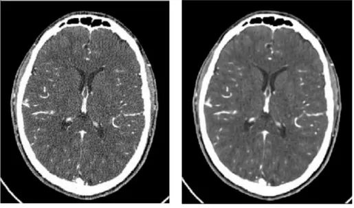
surfaces from the data. These techniques used an optical model to map data values onto optical properties, such as color and opacity. During rendering, optical properties were accumulated along each viewing ray to form an image of the data. Textures mapping to apply images, or textures, to geometric objects were applied in my case. Volume aligned texturing produces images of reasonable quality, although there is often a noticeable transition when the volume is rotated. The normal and adavanced CT files were shown in Figure 3-3 and Figure 3-4 respectively. Figure 3-5 presents the results of the noise removement.
60 Study on a Virtual Reality-based Training System for the VIS
Figure 3- 2 The specific blood vessel
Chapter 3 A hybrid method for 3D vascular reconstruction 61
Ph.D. thesis of Dr. Jin Guo
Figure 3- 4 The advanced CT file
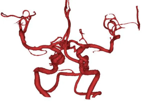
Three-dimensional reconstruction images of blood vessels are shown in Figure 3-1 for multi-branched blood vessels and interventionalists can choose part of them as research topic shown in Figure 3-2.
62 Study on a Virtual Reality-based Training System for the VIS
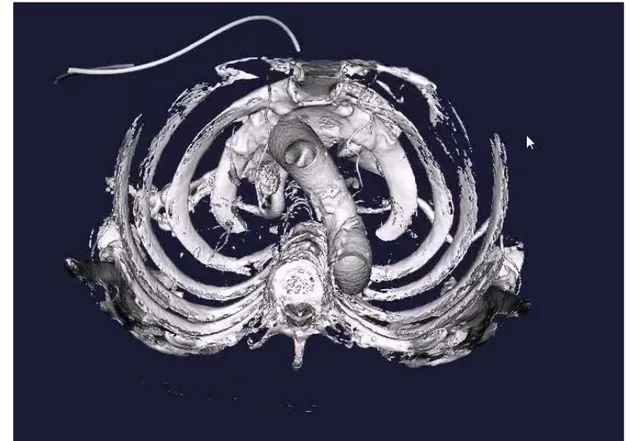
If the interventionalists are interested in the blood vessels within the bones or the onely bones, the thresholding values can be adjusted to realize these purposes. The reconstruction results of the blood vessels inside the bones were presented in Figure 3-6, which can better represent the spatial relationship between the blood vessels and the bones. The rebuilt results of the only bones were displayed in Figure 3-7.
Figure 3- 6 The reconstruction result of the blood vessels inside the bones
Chapter 3 A hybrid method for 3D vascular reconstruction 63
Ph.D. thesis of Dr. Jin Guo
3.2 A refined reconstruction method
The reconstructed surface has been integrated into a simulator and fulfills the needs of medical applications: efficient collision detection and collision response between the vascular surface and the simulated surgical tools, physics-based deformation simulation, and anatomical visualization.
64 Study on a Virtual Reality-based Training System for the VIS
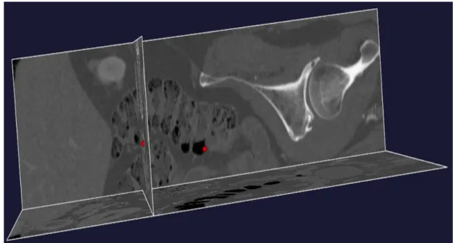
Figure 3- 8 The 3D CT dataset of the specific patient
Different techniques can be used for the segmentation of vascular structures. One of the simplest and sometimes most efficient techniques is thresholding, which usually needs some preprocessing to allow outlining tiny tubular structures. More complex methods, such as centerline enhancement and contour extraction, have more potential for computing the topological representation of a vascular network. Both techniques are very efficient for segmenting the carotids and other relatively large vessels, but have trouble outlining small vessels accurately, especially in the brain when datasets have resolutions coarser than 0.5*0.5*1mm3. In this study, scans from real patients were used from an electronic database of imaging studies. In this case, the above techniques do not perform well on the whole dataset. Regardless of the technique chosen, it is usually preceded by a noise reduction
Chapter 3 A hybrid method for 3D vascular reconstruction 65
Ph.D. thesis of Dr. Jin Guo
process, such as anisotropic diffusion filtering. The refined reconstruction technique is composed of the four following steps:
reduction of the noise of the input MRA or CTA images;
segmentation of the required blood vessels through a level set evolution, initialized from a manually selected start and end points;
reconstruction of the blood vessel surface model by using marching cubes algorithm;
surface refinement.
3.2.1 Enhancement and cleaning
To enhance the medical data set, the first step consists of filtering the images obtained from commercial medical scanners such as Computed Tomography Angiography (CTA) or Magnetic Resonance Angiography (MRA). To reduce the data noise while preserving and enhancing small vascular structures, an anisotropic filter is used.
3.2.2 Level set segmentation
The vessel contours were segmented by means of a level set evolution applied on the enhanced data set. For better efficiency, the active contour was initialized by a threshold on the image intensity. Manual seed selection and region growing methods would be time consuming and less robust since disconnected parts could be missing.
66 Study on a Virtual Reality-based Training System for the VIS
Although the level set technique is computationally more expensive than a straightforward threshold, it allows a better estimation of the contours based on both intensity values and edges. The level set evolves a surface according to three different forces: an advection force that pushes the surface towards the edges of the image; a smoothing term, proportional to the minimal curvature of the surface that keeps the surface smooth; and a balloon force, defined as [Wu11]:
m.
if I
τ
m,
if I
τ
e
(I)
F
σ -m - b1
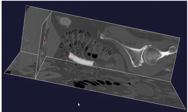
2 2 1 (Eq. 2-2)The algorithm allows the contours to expand within vascular structures; this force is defined by Eq. 2-2, and depends on the intensity of each voxel. It relies on the intensity statistics to either expand or shrink the evolving contour. I is the intensity image, m stands for the mean intensity of the vessels, r is their standard deviation, and s is a threshold that allows shrinking the contour when the position is unlikely to belong to a vessel. Figure 3-8 shows the 3D CTA dataset. The initial value of Eq. 2-2 with parameters m = 1140, r = 25 and s = 0.2 together with the image intensity histogram after the different pre-processing steps explained in the previous sections.
Chapter 3 A hybrid method for 3D vascular reconstruction 67
Ph.D. thesis of Dr. Jin Guo
Figure 3- 9 The selection of the start point and the end point of the desired blood vessel branch
The selection of the start point and the end point of the desired blood vessel branches were shown in Figure 3-9. The refined reconstruction algorithm will only deal with the vessel branch from the start point to the end point.
68 Study on a Virtual Reality-based Training System for the VIS
Figure 3- 10 The cloud points’ model based on the level sets algorithm Figure 3-10 presents the cloud points’ model after the evolution of the level sets algorithm. Additionally, other desired blood vessel branches can be calculated and integrated to the rebuilt cloud points’ model for further analysis. Figure 3-11 shows the selection of the new start point and the new end point. Finally, the newly reconstructed model was integrated with the former one, which was represented in the Figure 3-12. Finally, the whole reconstruction results based on the refined algorithm were shown in Figure 3- 13.