Title
[症例報告]Schwannomas in Ryukyu University Hospital 1983
-1991
Author(s)
Hagiwara, Keisuke; Higa, Tadashi; Maehira, Fusahiro;
Miyazato, Hajime; Nonaka, Shigeo
Citation
琉球医学会誌 = Ryukyu Medical Journal, 13(4): 369-374
Issue Date
1993
URL
http://hdl.handle.net/20.500.12001/3103
Schwannomas in Ryukyu University Hospital 1983-1991
Keisuke Hagiwara, Tadashi Higa, Fusahiro Maehira*,
Hajime Miyazato * and Shigeo Nonaka
Department of Dermatology, Research Center of Comprehensive Medicine, Faculty of Medicine, University of the Ryukyus
'Shuri Clinic of Dermatology, Naha
"Division of Dermatology, Okinawa Prefectual Naha Hospital
(Received on May 24th 1993, accepted on October 12th 1993)
key word : schwannomas
ABSTRACT
From 1983 to 1991, total 3 cases ofschwannomas have been registered in the De-partment of Dermatology of University of the Ryukyus Hospital. They comprised only 0.026% of total ouトpatients and 0.11% of new out-patients in a year. All the cases were ordinary types of schwannoma. The tumor occurred on the right thigh of a 52-year-old woman measured 20× 15 × 12cm and weighed 2250g; it is probably the largest schwannoma reported on the extremities in Japan.
INTRODUCTION
Schwannomas are known to be one of the common soft tissue tumors. According to a sur-vey on their incidence, they are the third most common following angiomas and lipomas, and
comprise roughly lO% of the benign soft tissue tumors as a whole. They occur practically in every anatomical region. Among the schwann0-mas of dermatological interest, sizes range from 0.2cm to 20cm in maximum diameter. Approx-imately 90% of the reported cases so far have
the maximum diameters less than or equal to
5cm. Only 12 cases whose maximum diameters are bigger than 5cm, have been reported in Japan since 1973. In our ouトpatient clinic, tot-al 3 cases of schwannomas have been seen since 1983. In this study, the brief outline of each case is portrayed in a chronological order of registration, and a clinical observation is dis-cussed.
CLINICAL MATERIAL
During the period from 1983 to 1991, total 3 cases of schwannomas were seen at the
De-370 Schwannomas
Table 1. Summary of three cases of schwannomas
cN:ヲAge-Dateof
(years)oclst.visit
SiteSi
(C!宗Duration
tolst.vi
(year茸10r
Histol0号ical diagnosis 43 M 1988 72 1988 52 F 1990 2×2×2 3back0.7×0.7×0.3
right
thigh20×15×1210
Antoni A+B ordinary type Antoni B ordimnary type Antoni A+B ordinary type-partment of Dermato一ogy of the University of
the Ryukyus Hospital, 2 cases in 1988 and 1 case in 1990. They comprised only 0.026% of total out-patients and 0.11% of new out-patients in a year. Table 1 shows their brief outlines.
Case 1 : A 43-year-old man was first seen, in July 1988, with a lump on the posterior aspect of his left leg. He had noticed it 3 years earlier for the first time. Since then it graduaレ Iy increased in size, though remaining asymp-tomatic throughout its growth. Physical ex-animation revealed a moderately firm, sub-cutaneous movable mass measuring 2 × 2cm. The tumor was easily enucleated. Histological findings were as follws: the tumor was encapsu-lated with fibrous membranes. Inside the mass, proliferation of the spindle-shaped cells was observed, whose nuclei were elongated. Some of those cells were arranged in so-called pallisad-ing arrangement. In some areas, aggregations of clear round cells with mucinous degeneraton were seen. The histological diagnosis was an ordinary type of schwannoma conposed of two types of tissue, Antoni types A and B. The pa-tient has been well and without recurrence for about 4.5 years following the excision.
Case 2 : A 72-year-o一d woman was first
seen, in November 1988, with a bean-sized nodulus on the back. She noticed it 3 years earlier for the first time. She told us that it was asymptomatic and the size was relatively stable. On physical examination, the tumor was
Fig. 1. Clinical appearance of Case 3 in April 1990, showing the anterior aspect of a big sub-cutaneous tumor on the medial femoral region
of the right thigh measuring 20×15×15cm.
skin-colored, soft and half dome-like with a smooth surface. It measured 7×7×3mm. His-tological findings were as follows : under the epithelium, loose, edematous, hypervascular, and relatively hypocellular tissues were seen. The constituent cells were long, spindle-shaped with nuclei of variable shapes. No mitoses were de-tected anywhere. The histologic diagnosis was Antoni B type tissue of an ordinary type of schwannoma. The patient has been well for ab-out 4 years after the operation.
Case 3 : A 52-year-old woman was first seen, in April 1990. with a man's head sized lump on her right thigh. She noticed it about 10 years earlier for the first time. It was henegg sized at that time. Since then it had
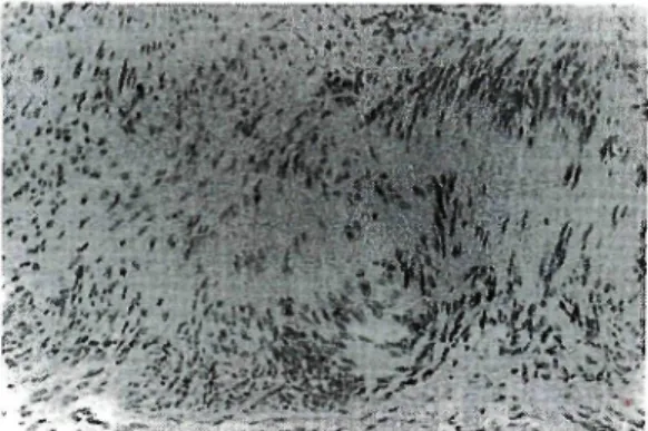
Fig. 2. Histopathology of Case 3. The section taken from the central area shows hypercellular por-tions composed of spindle-shaped cells forming so-called Verocay bodies, i.e., Antoni type A tissue. Other specimens taken from the peripheral area show hypocellular portions of edematous stroma containing few haphazardly arranged cells with nuc】ei of variable shapes, i.e., Antoni type B tissue. (Hematoxylin-eosin stain, ×200)
It remained asymptomatic throughout its
gro-wth. Physical examination revea一ed no
abnor-mahties except a big subcutaneous tumor on the medial femoral region of the right thigh, which measured 20×15× 15cm (Fig. 1). It was roughly egg-shaped, normal skin colored, elastic hard with a smooth surface, well-demarcated, slightly movable, and not adhesive to the sur-rounding tissues. Neither sensory abnormali-ties nor lymph node enlargement were detected on her body.
On admission, laboratory findings including blood analysis, chemisry tests, serologic tests and unnalysis were essentia】ly within normal limits. However, a CT examination as well as an ultrasonograph of the femoral region revealed a well-demarcated, almost homogeneous, solid ma-ss with small, round shadows of increased de-nsity that suggested some cystic degeneration inside the mass. The boundary between the
mass and the femoral muscles was clear. No in°
vasion to the muscles or to the surrounding tis-sues was detected.
Based upon the above findings, we prod visionally diagnosed the tumor as a benign soft tissue tumor, and completely excised it. The re・ moved tumor was encapsulated within semi-translucent membranes. It measured 20× 15× 12 cm and weighed 2250g. On sectioning the
tumor perpendicular to its long axis, the cross sections revealed yellow to pearl gray, multi-lobulated, intracapsular tissues with partially cystic degeneration and hemorrhages.
Histopathological findings were as follows: in general, based upon observation of all the speci-mens taken from various areas of the tumor, the specimens from the central areas were signifi-cantly hypercellular; whereas those from the peripheral areas included hypocellular portions with various degeneration. The hypercellular portions were composed of spindle-shaped cells whose nuclei were elongated and tightly packed. Thev were arranged in many areas in a
stream-ing fashion, and formed so-ca一led Verocay
bodies in some areas (Fig. 2). Essentia一ly no
mitotic figures were detected in the specimens from these areas. From these findings, it was concluded that the histological diagnosis was Antoni type A schwannoma. On the other hand, the hypocellular portions were composed of ede-matous stromas that contained relatively few, haphazardly arranged cells with nuclei of vari-able shapes. In places, fatty degeneration, cys-tic degeneration, hemorrhages and necrosis were observed. Again, there was no mitosis. These findings suggested that the histological diagnosis for these areas was Antoni B type schwannoma. In order to confirm the diagnosis, other stains such as immunoperoxidase stains against S-100 protein and neuron-specific eno-lase were utilized--. No contradictory results
were obtained. We therefore concluded that the
final diagnosis for this tumor was a mixture of two types of tissue Atltoni types A and B of an ordinary type of schwannoma.
372 Schwannomas
uneventful; her general status as well as the local wound healing were good. She has been well for approximately 3 years after the opera-tion without a recurrence.
DISCUSSION
It is widely accepted today that schwanno-mas are tumors of nerve sheath origin. They
arise most common一y from either cranial or
peripheral nerves, and develop practically in every anatomica】 region. According to the sta-tistic in 1974 about benign soft tissue tumors in Japan by Enjoji et al., schwannomas are the third most common, following hemangiomas and lipomas and comprise about lO% of benign soft tissue tumors as a whole日. However, as for the
incidence, there is enough room for controversy: some authors disagree with the figure and sug-gest that the actual incidence should be higher, indicating a statistical inadequacy of the study. In our office, there is not enough accumulation of schwannoma cases to make any comment on this issue.
In our small group of the 3 cases, the anato-irucal distribution of the tumors was as follows: one was seen on the trunk and two were seen on the lower extremities. In 1969, Das Gupta et al. studied 303 patients with benign solitary neurogemc tumors in the U.S. and reported the anatomical distribution of schwannomas as fol-lows : 44.8% of them were located in the head and neck region, 19.1% on the upper exremity. 8.5% on the trunk, 13.5% on the lower extrem・ ity and 13.8% in various unusual sites- On the other hand, in Japan, Enjoji et al. reported, in the above mentioned paper, the following dis-tribution of 825 casea of schwannoma: 29.1: of them were located in the head and neck re, gion, 20.5% on the upper extremity, 22.5% on the trunk, 21.8% on the lower extremitj, and 5.3% in miscellaneous regions.
These statistics differ. However, it is not
clear what kind of factors (e.g. races, statistical biases, etc.) are involved. There has been no study which compares the incidence of such tumors among the races.
In 1989, Iwashita reported an almost iden-tical distribution of schwannomas as that of En-joji et a】.on the basis ofareview of 1271 cases of schwannoma registered to the Histopathology Office of Kyushu University from 1951 to 1987 : 26.6% were distributed in the head and neck region, 24.0% on the trunk, 24.5% on the upper extremity and 24.2% on the lower extremity. In summarizing these statistical fi-gures, it seems safe to say that if the surface of the whole body is divided into these 4 major re-gions, the frequencies in the respective regions
are almost the same, namely 20-25%.
Male-female ratio of schwannomas in our
small group was one to two favoring females. However, Iwashita reported that sex of the pa-tients was not an important factor in schwann0-mas: it occurs equally on both sexes with the
ratio of approximately one to one'. Many
au-thors have given unanimous approvals to this statement
Ages at onset oi schwannomas were reported to range from 2 to 89 years with a mean of 46'. Das Gupta's report as well as others almost coincide with it '. Our small group showed a mean age of 55.7, which is slightly higher.
As briefly mentioned in the introduction, maximum diameters of reported cases of schwa-nnoma range from 0.2cm to 20cm with a mean of 3.1cm'. Soul stated in 1954 that those that arose on the extremities never exceeded 6cm in diameter". Harkin et al. stated in 1968 that they would rarely become bigger than 8cm'. From these assertions, it seemed reasonable to us to agree with defining giant schwannomas as those whose diameters were bigger than 5cm I. Using this working definition, we studied the
extremi-ties in Japan from 1973 to 1992. Approx-imately 90% of the reported cases so far had the maximum diameters less tham or equal to
5cm蝣蝣''蝣蝣'. Of those whose maximum diameters
are bigger than 5cm. only 12 cases in total have been reported since 1973 in Japan'. The big-gest one, 20× 12× 10cm, that occurred on the lower extremity was reported by Ichise et al. in 1973"'- One of the cases in this paper de・ scribed as case 3 was slightly bigger, i.e., 20×
15×12cm; this is probably the biggest schwan-noma on the extremities in Japan. Our small group showed an average size of 7.6cm. It is significantly bigger than the average size men-tioned above (3.1cm). This is clearly due to the large contribution of case 3.
Most of schwannomas arising on the extrem上. ties are sma】1. One reason for this may be that they are easily recognized from outside either by the patients or by their family members. On the other hand, for those that arise somewhere in the deep anatomical regions of the body such as the pelvic cavity or the retroperitoneum, it is usually not until they become big enough to cause some symptoms that the patients initially become aware of them. Incidentally, the biggest schwannoma reported in 1978 in Japan arose from the retroperitoneum, which measured 30× 30cm and weighed about 5kg to our know-ledge"- The true reason why case 3 showed such big dimensions is still unknown. However, we speculated that some peculiar character of the patient must have played a role in her pro-longed first visit to a hospital. She had never shown the tumor even to her husband before the first visit and refused to let him know her hospitahzation even after her discharge, prob-ably with some unknown anxieties.
Iwashita mentioned in the above paper that the relationships between the tumors and the nerves involved in them or responsible for the
formation of the tumors were clearly discussed
only in 22% of the registered cases. Manabe et al. reported in 1987 that the nerves involved in the tumors were identified during operation
in 70% of 105 schwannoma cases'. They speculated, however, that in the remaining 30%
of the cases with unidentified nerves, minute muscular or cutaneous branches must be
re-sponsible. We were not ab一e to detect the
nerves involved in case 3 during the operation and reached the same speculation.
There have been several proposa一s
regard-ing subtypregard-ing of schwannomas. Accordregard-ing to Iwashitas attempt, they are classified into 7 subtypes: ordinary, cellular, plexiform,
pig-′
mented, myxomatous and organoid types. The respective frequencies of these subtypes are 73% for ordinary type, 19% for degenerated type, 3% for plexiform type, 2% for organoid type, 1% for cellular type, and 1% for
myxoma-tous type. In our small group, al一 were
ordin-ary types.
To compare the frequencies of schwannomas in ouLpatient clinics of dermatology of universi-ty hospitals in japan, we looked for such statis-tics in the literature. Very few reports have been seen regarding this issue. One such report was presented indirectly by the Department of Dermatology, Kurume University, School of Medicine: 5 cases of schwannomas were seen from 1975 to 1985 . The figure is slightly bigger than ours. Other institutes including sur-gical departments reported much bigger figures. One reason for this may be that patients with schwannomas of small sizes tend to visit a sur-gical clinic rather than a dermatology clinic of a university hospital, eventually resulting in a higher incidence in the former. However, it is not clear whether schwannomas are rare in Oki-nawa. Further studies are needed such as a nation-wide survey on the incidence.
374 Schwannomas
REFERENCES
1) Enjoji, M., Iwasaki, H., and Komatsu, K.: Benign soft tissue tumors in Japan-A statis-tic analysis of 8086 tumors, Gan no Rin-sho, 20 : 594-609, 1974 (inJapanese).
2) Weiss, S. W., Langloss, J. M., and Enzinger,
F. M.: Value of S-100 protein in the
di-agnosis of soft tissue tumors with particu-lar reference to benign and malignant Schwann cell tumors, Laboratory Investiga-tion, 49 : 299-308, 1983.
3) Manabe, M., Kawaguchi, T., Amino,
KりMat-sumoto, S., Kitagawa, T., Machinami, M., Totake, M., Huruya, K., Wada, NH Isobe, Y.,
and Tako, M∴ A clinico-pathological study
on 105 cases of schwannoma deve一oped in
peripheral nerves, Rinsho Seikeigeka, 22 二
1035-46, 1987 (in Japanese).
4) Das, Gupta, T. K., Basfield, R. H., Strong, E. W., and Haidu, S. I∴ Benign solitary
schwannomas (neurilemomas), Caner, 24 355-66, 1969.
5) Iwashita, T∴ Neurilemomas of the soft
tissues-An analysis of 1271 tumors in an attempt at subtyping, Fukuoka Ishi, 80 : 355-67, 1989 (in Japanese).
6) Hu, F∴ Neurilemoma, in Demis DJ, Thiers
BH, Smith EB, Burgdorf WHC (eds) :
Clinic--al Dermatology, Lippincott, Philadelphia, 1991, unit 25-2.
7) Soul, E. H.: Primary soft tissue tumors of the extremities; Classification, histogenesis and incidence, Acad. Ortho. Surg., ll : 3-11,1954.
) Harkin,J. C, and Reed, R.J.: Tumors of the peripheral nervous system. in Atlas of Tumor Pathology, 2nd Series, Fascicle 3, p29-51, Armed Forces Institute ol Patholo-gy, Washinton, D. C, 1968.
9) Nakajima, K., Koshino, T., Morii, T., Ejima,
M., and Kushida, K∴ Giant schwannoma
arising from the tibial nerve-Two case re-ports, Kantou Seisaishi, 18 : 365-69, 1987 (in Japanese).
10) Ichise, T.: Two cases of neurilemomas arising from the peripheral nerves, Kantou Seisaishi, 4 : 56-60, 1968 (in Japanese). ll) Hiramatsu, Y., Shidahara, M., Yonezawa, Y.,
Shimizu, K., and Sekiba, K∴ A case of
re-troperitoneal neurilemoma and a statistical observation of 48 cases in Japan, Sanka to Fujinka, 49 : 110-6, 1982 (in Japanese). 12) Tanaka, H., Hachisuka, H., Mori. 0., and
Sasai, Y.: Re-investigation about tumors of neural tissues with light microscopy, Japanese Journal of Clinical Dermatology, 39(1) : 6ト4, 1985 (in Japanese).