JAIST Repository https://dspace.jaist.ac.jp/ Title X線反射率測定によるポリヘキシルチオフェン超薄膜の 構造評価 Author(s) 黒澤, 剛志 Citation Issue Date 2006-03
Type Thesis or Dissertation Text version none
URL http://hdl.handle.net/10119/3228 Rights
Description Supervisor:佐々木 伸太郎教授, 材料科学研究科, 修 士
Structural evaluation of polyhexylthiophene ultra-thin films
by X-ray reflectivity measurements
Tsuyoshi Kurosawa
School of Materials Science,
Japan Advanced Institute of Science and Technology
March 2006 1. Introduction
π-Stacking is one of the central themes in recent organic, biochemical, and polymer chemistry1). Various planar aromatic molecules form the stacked structure, in which the plane-to-plane distance is in the range of 3.4-3.8 Å1). Thin films of π-conjugated polymers are widely used for the fabrication of optoelectronic and electronic devices. More detailed studies are needed for structure evaluation of thin films. It was recently reported that nondoped HT-P3HexTh molecules formed an ordered, stacked structure with the hexyl side chains oriented perpendicularly to the surface of a SiO2/Si substrate when the polymer was spin-coated on the substrate.8)9) The nanostructure of thin films of regioregular head-to-tail poly(3-hexylthiophene-2,5-diyl) (HT-P3HexTh) was studied by X-ray reflectivity measurements.
2. Experimental
HT-P3HexTh films were offered from Prof. Yamamoto (Chemical Resources Laboratory, Tokyo Institute of Technology). Five specimens (No. 1-5) of thickness less than 10 nm were prepared by spin-coating
method on Al2O3 (001) plates. Reflectivity profiles were measured with Cu Kα radiation (wavelength λ = 1.5418 Å) by using an ordinary XRD diffractmeter with the specular reflection condition at room temperature. The range of reflection angle (2
θ
) was 1-10 °. The interference pattern from surface of thin film and the film-substrate interface is expressed byI(q) = q-4 {K1 + K2 cos(qL)}・exp(-q2A2)
where q = 4π sinθ / λ, L is film thickness and A is surface roughness (square root of square average of deflection from height of average). K1 and K2 are constants. Thickness (L) and surface roughness (A) of the films were analyzed by Fourier transformation of X-ray reflectivity profiles. Surface images were obtained by atomic force microscopy (AFM), from which surface roughness was evaluated.
3. Results and Discussion
X-ray reflectivity profile and the Fourier transformation of sample No.1 is show in Figure 1, which gives L = 1.7 nm and A ≅ 0.3 nm. This thickness corresponds to the size of the molecule (1.65 nm). The data are listed in Table 1. The roughness A = 0.3-0.4 nm increased with L. The roughness estimated by AFM was comparable to X-ray reflectivity data. The evaluated values of thickness were not discrete with a specific interval. The molecular chains were stacked without preferred orientation on the substrate.
Figure 1. (a) X-ray reflectivity profiles of sample N o . 1 a n d ( b ) t h e F o u r i e r t r a n s f o r m . L = 1.7 nm A = 0.34 nm 40 0 2.00 4.00 6.00 8.00 10.00 0 10.0 20.0 F (z ) I( q) ・ q 4 z / nm 2θ / deg (Cu Kα)
0.34
0.40
0.40
0.49
0.37
0.51
0.17
X-ray reflectivity AFM
No. thickness roughness roughness
( L / nm) (A / nm) (A / nm)
substrate
-
0.08
1
1.7
0.30
2
2.8
3
4.2
0.42
4
5.1
5
Table 1. Data of HT-P3HexTh thin films
6.5
0.43
0.44
(a)
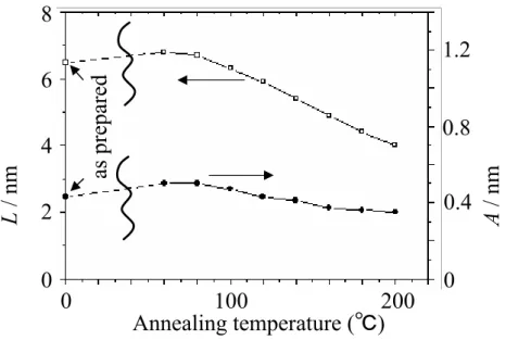
Annealing temperature (℃)
Figure 2. Effect of heat treatments on thickness (L) and surface roughness (A) of sample No. 5. The sample was annealed at each temperature for 1h in vacuum.
8 6 4 2 0 0 100 200 1.2 0.8 0.4 0 A / nm L / nm as prepared S H C O H S H C6H13 S H C O C5H11
Scheme 1. Chemical change in thiophene unit.
heat
Effect of heat treatments on L and A was investigated. The dependence of L and A on annealing temperature of No. 5 is show in Figure 2. Thickness has increased by 10-15 % while maintained during one year. This increase is due to the oxidation reaction which happens at Cα atoms of the side chains (Scheme 1.). The thickness of samples has decreased gradually by the stepwise heat treatment (Figure 2.). It was attributed to the thermal decomposition of the side chains.