Effects of peripheral administration of rat urotensinII
on circulation, and on distribution of c-Fos
immunoreactive neurons in the brain
Kiyoshige TAKAYAMA
1)*, Shunsuke OBINATA
1), Midori INOUE
1),
Ayuko NAKATA
1), Eriko WAKAMATSU
1)(Revised September 30, 2009, Accepted December 21, 2009)
Abstract : The purpose of this study was to examine the effects of intravenous injection of urotensinII on the systemic blood pressure and heart rate, and to examine whether the intraperitoneal administration of urotensinII evoked the stimulation of central neurons. Urotensin, originally isolated fish neuro-endocrine systems, is a peptide which consists of 12 amino acids. Lately human urotensinII was identified to be consisted from 11 amino acids. Receptors of urotensin have been found to highly distribute in the mammalian cadrdiovascular systems. It has been reported that urotensin evoked contraction of thoracic artery, while the dilatation of blood vessels was alternatively induced. In this study, we examined the effects of peripheral injection of rat urotensinII consisted of 14 amino acids on the circulation and on the activity of neurons in the brain. It was found that intravenous injection of rat urotensinII elicited dose-dependently decrease in blood pressure, and increase or decrease in heart rate. To examine whether an intraperitoneal injection of rat urotensinII induced an excitation of central neurons, we used immunohistochemical method to study the expression of c-Fos protein in neurons of the rat brain after intraperitoneal injection of rat urotensinII (test experiment). In the control experiment rat was intraperitoneally injected saline solution without rat urotensinII. It was found that intraperitoneal injection of rat urotensinII induced expression of c-Fos protein in several nuclei in the brain. These results suggested that rat urotensinII might exhibit physiological functions via central neuronal pathway as well as peripheral direct pathways.
Key words : Rat urotensinII, Cardiovascular responses, Brain, Neuron, c-Fos
1)Department of Laboratory Sciences, Gunma University School of Health Sciences *Author for correspondence.
Reprint address : Department of Laboratory Sciences, Gunma University School of Health Sciences, 3-39-15 showa-machi, Maebashi, Gunma 371-8514, Japan
Tel/Fax: +81 27 220 8943
Email: takayama@health.gunma-u.ac.jp INTRODUCTION
A number of physiologically active peptides derived from internal organs have been discovered in these recent 20 years1). In our laboratory we have performed
experiments on the hypotheses that these physiologically active peptides may play their role via central neurons to the target organs, not only direct actions to the organs. And we have reported several
studies of effects of some of these active peptides on physiological functions. Gastrin, well known to induce gastric acid secretion, was found to induce stimulation of neurons in the lateral habenular nucleus, central nucleus of amygdala, lateral parabrachial nucleus, solitary tract nucleus and dorsal motor nucleus of the vagus nerve2). Galanin that is known to have inhibitory action to gastric acid secretion was found to stimulate neurons in the area postrema3). Intravenous (i.v.) administration of leptin, that is noted to have pathophysiological function in relationship to obesity, was found to induce stimulation in neurons in some nuclei in the brain4). Furthermore, effects of intravenous administration of ghrelin and apelin on the central neurons were studied in our laboratory5, 6). In a series of our experiments, we have suggested that physiologically active peptides cause stimulatory influence on the central neurons to exhibit their physiological functions.
UrotensinII, originally isolated from nervous systems of fishes, is a cyclic peptide consisted of 12 amino acids7). Recently, in human was identified human urotensinII consisted of 11 amino acids, and the receptors of the peptide were found to distribute densely in the cardiovascular systems of mammals. It was reported in the experiments using rat arterial preparation that the urotensinII showed much stronger vasoconstriction than endothelin8). On the other hand, human urotensinII was reported to have vasodilatory actions9). Considering these contradictory reports, we were urged to examine what type of cardiovascular responses may be caused by the peripheral administration of rat urotensinII.
It is well known that proto-oncogene c-fos induces c-Fos protein transiently within neurons in response to various kinds of stimulations, and the induction of c-Fos protein in neurons starts about 20 min after the stimulation10, 11). So far, no studies have been reported on the expression of c-Fos protein in central neurons after peripheral or central injection of urotensinII. Second, we undertook to survey the neuronal expression of c-Fos protein throughout the CNS after intraperitoneal (i.p.) injection of urotensinII. UrotensinII was administered intraperitoneally, but not intravenously, to inhibit unexpected expression of
c-Fos protein to a minimum due to stimulation of central neurons during the operations for the cannulation of trachea, artery and vein. In the previous reports from our laboratory, the operational treatment of animals such as cannulation of artery and vein induced was found to induce much more c-Fos protein in the central neurons 5, 6).
In this study, first, cardiopulmonary responses were investigated, and secondly, distribution of c-Fos-immunoreactive (c-Fos-ir) neurons after i.p. administration of rat urotensinII was surveyed through the rat brain.
MATERIALS AND METHODS
All of the steps of the experimental procedures were conducted in agreement with the guiding principles for the care and use of experimental animals approved by the Society for Neuroscience. Experiments were done on male Wister rats weighing 250-270 g (9-10-weeks old, SLC, Japan, Shizuoka, Japan). To avoid any restlessness of a single rat, some rats were housed in a cage in quiet room under conditions of regulated illumination (LD12:12, light on at am 08:00) and constant temperature (22 ± 2℃) with ad lib water and rat chow (MF, Oriental Yeast Co., Ltd., Tokyo, Japan).
In the first experiments, rats (n=4) were anesthetized with i.p. injection of pentobarbital sodium (Abbott, USA; 50 mg/kg). The trachea was cannulated with vinyl tubing (2 mm in internal diameter). Femoral artery and vein were cannulated with polyethylene catheters (0.58 mm, internal diameter). Systemic arterial blood pressure (ABP) was measured with a pressure transducer (TP-400T, Nihon Kohden, Japan) connected to carrier amplifier (AP601G, Nihon Kohden, Japan), and heart rate (HR) was computed from pulse pressure by a cardiotachometer (AT601G, Nihon Kohden, Japan). ABP and HR were recorded on a recticorder (RJG4226, Nihon Kohden, Japan), urotensinII in 0.9% NaCl solution was administered into the femoral vein to induce cardiopulmonary responses. Rectal temperature was maintained at 37℃ with an infrared heat lamp.
In the second experiments, we undertook to examine which neurons in the CNS may express
c-Fos protein after intraperitoneal injection of urotensinII (300 μg/kg) in 1 ml of 0.9% NaCl solution using Wistar rat (n=1). One and a half hour after the urotensinII injection, rat was anesthetized by i.p. injection of pentobarbital sodium (80 mg/kg, Abbott, USA), and perfused via the left ventricle with about 20 ml of saline to flush out the blood. This was immediately followed by 100 ml of 0.5% glutaraldehyde and 4% paraformaldehyde (PFA, Merck, German) in 0.1 M phosphate buffer (PB, pH 7.4) under pressure of 100-120 mmHg, and 400 ml of 4% PFA in PB under hydrostatic pressure. The brain was removed and cut into two blocks at the level between superior and inferior colliculi. They were fixed in 4% PFA in PB for further 1.5 h at 4℃, then soaked stepwise in 10, 20, 25% sucrose in PB at 4℃. Each brain block was frozen and cut into serial transverse sections at 40 μm in thickness. The sections were collected in plates containing PB chilled by ice water. One group of every fourth section was rinsed in 0.1 M Tris-saline (TS,pH 7.4) three times, incubated with 0.5% bovine serum albumin (BSA) in TS for 20 min and incubated with sheep anti-c-Fos polyclonal antiserum (1:100; sc-52, Santa Cruz Biotech. Inc., USA) in 0.5% BSA at 4℃ for 16 h. The following procedures were conducted at room temperature. The sections were rinsed three times in TS, and then incubated in avidin and biotin solution respectively for 15 min. Thereafter they were incubated for 20 min with 2% normal rabbit
serum in TS to block nonspecific bindings, and incubated for 60 min with anti-sheep IgG in TS. After rinsed three times in TS, the sections were incubated for 60 min in Vectastatin ABC kit (Vector Lab., Burlingame, CA, USA), and then treated with diaminobenzidine (DAB)/nickel solution containing 0.003% H2O2. After that, the sections were rinsed in
TS twice and rinsed further in PB. They were mounted on glass slides and dried at room temperature. The sections on the slides were dehydrated in a graded ethanol series (50%, 70%, 95%, 99%), infiltrated with xylene and coverslipped in Permount (Fisher Comp., USA). In the control experiment, rat (n=1) was sham-operated and injected with 200 μl of 0.9% NaCl solution. Brain sections were similarly processed as above. c-Fos protein localized in neuronal nuclei was visualized as black precipitates of nickel-intensified DAB reaction products. The c-Fos-ir neurons were surveyed under bright-field microscopy. Brain histology was checked against the rat brain atlas of Swanson12). Statistical analysis of data of the cardiovascular responses was performed using student t-test.
RESULTS
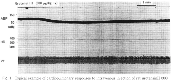
Typical examples of cardiopulmonary responses to the intravenous injection of rat urotensinII were shown in Fig. 1. The dose of 150 μg/kg of rat urotensinII showed almost no cardiopulmonary
Fig. 1 Typical example of cardiopulmonary responses to intravenous injection of rat urotensinII (300 μg/kg). Abbreviations: ABP: arterial blood pressure, bpm: beats per minute, HR: heart rate, mmHg: millimeter mercury, VT: tidal volume.
responses, but the dose of 300 μg/kg of the peptide showed clear cardiac responses of decreased ABP, and increased or decreased HR with the maximum level at around 2-3 minutes after the injection of rat urotensinII. The data in four experiments were summarized in Table I. There was, however, no
statistically significant difference in the ABP between before and 3 minutes after injection of urotensinII (see Table I). Respiratory response was slightly observed as shown in Fig. 1. All these responses recovered to the original level at around 12 to 15 minutes after the injection of rat urotensinII.
Table I:Cardiovascular responses to the intravenous injection of rat urotensinII (300 μg/kg)
It was found that intraperitoneal injection of urotensinII induced expression of c-Fos immunoreactive neurons in several nuclei in the brain. Table II showed number of c-Fos-ir neurons induced in four nuclei of the brain in the test experiment (i.p. injection of urotensinII) and control experiment (i.p. injection of 0.9% NaCl solution). c-Fos-ir neurons were found in the ventromedial nucleus of hypothalamus (VMH), lateral parabrachial nucleus (PBL), the complex of the solitary tract nucleus (NTS)/dorsal motor nucleus of the vagus nerve (DMX), and the ventrolateral medulla (VLM). Numbers of c-Fos-ir neurons these nuclei in the test experiment were found to be 2- to 3-times those in the control experiment.
DISCUSSION
It was found that intravenous injection of rat urotensinII induced decreases in the ABP, though there were no statistically significant differences in the ABP between before and 3 minutes after the i.v. injection of urotensinII. This present result was in agreement with the report by Gardiner et al, but contrary to the report by others who observed strong contraction of blood vessels by the injections of urotensinII8). It has been suggested that endogenous urotensinII and receptors of urotensinII exists in the rat arterial vessels. The depressor response may be induced via the receptors, though the mechanism remains to be clarified. In this experiment it was found that many c-Fos-ir neurons were expressed in the VLM. Thus, the depressor response may be induced via the depressor neurons located in the caudal ventrolateral medulla13). On the other hand, the i.v. injection of urotensinII did not induce constant responses of HR. Thus no definite mechanism might not be suggested on the response of HR, even though the increase of HR might be induced at least via the baroreceptor reflex HR14, 15). Actually, c-Fos-ir neurons were found to be expressed in the PBL and NTS/DMX. The PBL have been known to mutual connections with neurons in the hypothalamus and those in the brainstem such as NTS and DMX. The NTS is known to be the site receiving primary sensory information from the internal organs, and the neurons in the DMX to have parasympathetic efferent
projections to heart15, refs in there). Thus, as already described above, the depressor and tachy- or hypo-cardiac responses may be induced through neurons in these nuclei in the medulla oblongata, though further study is needed to clarify the mechanism of the responses of HR to the i.v. injection of urotensinII.
c-Fos-ir neurons were also found to be expressed in the VMH. It was reported that in the noradrenalin turn-over experiment, electrical stimulation of the VMH induced acceleration of sympathetic activity to wide-range peripheral organs. The VMH is also well known to be the site of satiety center16). Thus, the rat urotensinII may have some role in the regulation of sympathetic activity to the internal organs, as well as the regulation of feeding. Taking other physiological and behavioral functions into consideration, the neurons in the VMH may influence the sympathetic pathways.
In conclusion intravenous injection of rat urotensinII was found to induce depressor, tachy- or hypo-cardiac responses and slight respiratory response. Intraperiotneal injection of rat urotensinII induced stimulation of neurons in the VMH as well as neurons in the nuclei of cardiovascular center of the NTS/DMX and the VLM. The present results suggest that rat urotensinII might play some roles in the regulation of feeding as well as the regulation of cardiovascular function via central neuronal pathway as well as peripheral direct pathways.
REFERENCES
1) Handbook of biologically active peptides, Kastin, A. J., Academic Press, NY, 2006.
2) Yakabi K, Iwabuchi H, Nakamura T, Endo K., Fukunaga Y, Kumai I, Takayama K.. Neuronal expression of c-Fos protein in the brain after intravenous injection of gastrin in rats. Neurosci Lett. 317 (2002) 57-60.
3) Hayashi K., Takayama, K. Neuronal expression of c-Fos protein in the brain after intraperitoneal injection of galanin in Wistar rats. Ann Gunma Health Sci., 27 (2006) 27-31.
4) Takayama, K., Kawata, H., Takahara, R., Tomita, K., Neuronal expression of c-Fos protein in the brain after intraperitoneal injection of leptin in Wistar rats. Ann Gunma Health Sci., 28 (2007) 1-8.
5) Takayama, K., Johno Y., Hayashi K., Yakabi K., Tanaka T., Ro S. Expression of c-Fos protein in the brain after intravenous injection of ghrelin in rats. Neuroscience Letters, 417 (2007) 292-296.
6) Takayama, K., Iwazaki, H., Hirabayashi, M., Yakabi, K., Ro, S. Distribution of c-Fos immunoreactive neurons in the brain after intraperitoneal injection of apelin-12 in Wistar rats. Neuroscience Letters, 431 (2008) 247-250. 7) Peason,D., ShivelyJ.E., Clark, B.R., Geschwind, I.J.,
Barkley, M., Nishioka, R.S., et.al. urotensinII: a somatostatin-like peptide in the caudal neurosecretory system of fishes. Proc. Natl. Acad. Sci. USA 77 (1980) 5021-5024.
8) Ames, R.S., Sarau, H.M., Chambers, J.K., Willette, R.N., Aiyer, N.V., Romanie, A.M., et.al. Human urotensinII is a potent vasoconstrictor and agonist for the orphan receptor GPR14. Nature 401 (1999) 282-286.
9) Gardiner, S.M., March, J.E., Kemp, P.A., Davenport, A.P., Bennett, T. Depressor and regionally selective vasodilator effects of human and rat urotensinII in conscious rats. Br J Pharmacol. 132 (2001) 1625-1629.
10) M. Dragunow, R. Faull, The use of c-fos as metabolic marker in neuronal pathway tracing, J.
Neurosci. Methods 29 (1989) 261-265.
11) S.M. Sagar, F.R. Sharp, T. Curran, Expression of c-fos protein in brain: metabolic mapping at the cellular level, Science 240 (1988) 1328-1331.
12) L.W. Swanson. Brain Maps: Structure of the Rat Brain, Elsevier, Amsterdam, 1999.
13) Suzuki, T., Takayama, K., Miura, M. Distribution and projection of the mdullary cardiobascular control neurons containing glutamate, glutamic acid decarboxylase, tyrosine hydroxylase and phenylethanolamine N-methyltransferase in rats. Neurosci. Res., 27 (1997) 9-19.
14) Spyer, K. M., The central nervous organization of reflex circulatory control. In: A.D. Loewy and K.M. Spyer (Eds.) Central Regulation of Autonomic Functions, Oxford University Press, Oxford, 1990, pp.168-188.
15) Miura, M., Takayama, K., Okada, J., Neuronal expression of Fos protein in the rat brain after baroreceptor stimulation, J. Auto. Ner. Syst., 50 (1994) 31-43.
16) Nieuwenhuys, R., Voogt, J., van Huijzen C., Diencephalon: Hypothalamus in “The Human Central Nervous System”, Springer-Verlag, Berlin, pp. 289-323, 2007.