REVIEW
Strategy for improving the prognosis of patients with
intrahepatic cholangiocarcinoma by surgical treatment :
Considerations based on experience and a literature review
Seiki Tashiro, PhD1,2, Tatsuya Tsuji, PhD3, Hidenori Miyake, PhD4, Hitomi Kamo, MD1, Yuko Sumise, MD1, Shigeharu Takai, PhD1, and Kazuo Yoshioka, PhD1
1Taoka Hospital, Tokushima, Japan, 2The University of Tokushima (Professor Emeritus), Tokushima, Japan, 3Hasuda Clinic, Kumamoto, Japan, 4Tokushima Municipal Hospital, Tokushima, Japan
Abstract : The prognosis of patients with intrahepatic cholangiocarcinoma (ICC) is still poor, and the 5-year sur-vival rate in patients undergoing radical surgery (R0) is less than one-third. Since the prognosis depends main-ly on tumor factors, so earmain-ly diagnosis is necessary. To extend the survival time of these patients with a poor prognosis, cases of long-term survival were examined based on the results of our experiences and the literature. It was found that the hepatitis virus was highly involved in the carcinogenesis of ICC, and patients who were infected with hepatitis virus had rather good survival. J. Med. Invest. 68 : 15-21, February, 2021
Keywords : intrahepatic cholangiocarcinoma (ICC), hepatocellular carcinoma (HCC), hepatitis virus, nibolumab, lenvatinib
INTRODUCTION
The prognosis of patients with intrahepatic cholangiocarcino-ma (ICC) is less than one-third of patients undergoing curative resection (R0) surviving for more than 5 years. The prognosis de-pends on a high recurrence rate. So it is important to find more effective adjuvant therapy to prevent this recurrence. The aim of this review article is to develop a strategy to prolong the survival of these difficult groups of ICC patients.
THE HEPATITIS VIRUS IN ICC ; ROLE OF THE
HEP-ATITIS VIRUS
In the primary liver cancers, ICC developing from the bile duct epithelium was relatively rare, accounting for only 21 (7.2%) of the 292 cases of liver cancers in the 18 years (1980-98) at Kumamoto University Hospital (1). It was said that ICC and hepatitis virus were not related, but according to Shen et al. (2), 198 of 429 cases (46.4%) were positive for HBsAg, and the number of cases has been increasing in recent reports. Epide-miological studies in Japan and Asia have shown that chronic hepatitis, such as hepatitis B and hepatitis C, and progression to liver cirrhosis are also important factors in the development of ICC, and the reasons for this were shown by the RIKEN team. These studies were performed by a joint research group (including 37 other collaborators) with Hidetoshi Nakagawa as the leader and Akihiro Fujimoto as the vice leader of the Genome Research Team of the RIKEN Center for Biomedical Sciences (3, 4). To summarize these results, the collaborative research group used a next-generation sequencer and a supercomputer to decipher and identify all genomic mutations occurring in ICC. Next, they compared the genome mutations of 60 hepatocellular carcinoma (HCC) identified by the same genome-wide sequence
analysis and found an average of 4,300 mutations per tumor on the whole genome of ICC. The base substitution pattern of these mutations was similar to that of HCC. They also discovered that the genome sequence of hepatitis B virus has been inserted into the genomes of multiple ICCs, indicating a strong involvement in cancer. Furthermore, it was also found that several mutated genes in ICC were consistent with genes that mutated frequently in HCC. On the other hand, mutations in genes specifically found in cholangiocarcinoma outside the liver (extrahepatic cholangio-carcinoma) were also found in ICC. These cases have proven to have a very poor prognosis.
SURGICAL PROCEDURE FOR R0 RESECTION
First and second authors’ experiences from 1980 to 1998 at Kumamoto University Hospital.
In the beginning, full resection was the only means to cure ICC, and we applied the concepts, technologies of vascular surgery, and hepatic transplant surgery, so worked on how to perform surgery aimed at R0 resection. In three cases, we performed surgery aiming at R0 applying these techniques for advanced ICC patients. The three cases that underwent these operations are presented below.
Case 1 : A 51-year-old woman
The patient was admitted to our clinic on May 24, 1990, with jaundice as the chief complaint. Computed tomography (CT) showed a low-density mass (6 cm in diameter) with an irregular shape in segments IV and V and two small, low-density masses in segments VI and VIII.
Superior mesenteric arteriography showed encasement of the right hepatic artery, which branched off the superior mesenteric artery of approximately 5 cm in length. The left hepatic artery branched off from the left gastric artery, and the medial branch of the left hepatic artery was connected with the right anterior branch, making collateral vessels due to stenosis of the right anterior hepatic artery. On transarterial portal vein imaging, smooth encasement was shown approximately 2 cm in length on the trunk of the portal vein. On the basis of these imaging stud-ies, ICC with lymph node involvement in the hepatoduodenal
The Journal of Medical Investigation Vol. 68 2021
Received for publication November 13, 2020 ; accepted January 27, 2021.
Address correspondence and reprint requests to Dr. Seiki Tashiro, 1252 Nishiyama, Kamihachiman-chou, Tokushima, 770-8041, Tokushima, Japan and Fax : +81-88-644-3975.
ligament was diagnosed. Therefore, liver resection of the right 3 segments with hepatoduodenal ligamentectomy (combined en bloc resection of the extrahepatic bile duct, the hepatic artery, the portal vein, and lymph node dissection in the hepatoduode-nal ligament) and lymph node dissection around the abdomihepatoduode-nal aorta were carried out. The histological summary according to the Japanese Classification of Primary Liver Cancer was as fol-lows : mass forming type + periductal type, moderately differen-tiated tubular adenocarcinoma, Mt (4)-A(2), 5.6 × 3.9 cm2, 3 × 3 cm2, P (1) 3 × 3 cm2, L (1) 1 × 1 cm2, Hg, Fc (-), S1, N2, Vp0, Vv0, B1(RHD), PV2 (by metastatic lymph node), A2 (by metastatic lymph node), IM3, P0, Z0, EV(-). NL : n3 (No 3 1 / 6, 12p 1 / 1, 13a 2 / 5, 14 1 / 1, 16 (para-aortic lymph node) 4 / 11), pv2, a1, T4, N1, M1, stage 1VB, Hr 3 (PAML) + Hr0 (L), + D2+α (para-aortic lymph node dissection), SM (-), and Curability B (with no cancer at the surgical margin).
She survived 5 years and 10 months after surgery, but died on April 1, 1996. The cause of death was multiple metastases to the liver and lumbar and pelvic bones. It was thought that this patient should have been given adequate postoperative chemotherapy.
Fortunately, in this case, the right hepatic artery branched from the superior mesenteric artery, and the left hepatic artery branched from the left gastric artery ; therefore, the circulation of the remaining liver in the left lateral segment was main-tained, and there was thus no need to remove or reconstruct the left hepatic artery. It could be resected en bloc, including the bil-iary tract, the portal vein, right hepatic artery, and lymph nodes in the hepatoduodenal ligament, and each vessel could be easily rebuilt. Thus, surgery could be performed without cancer at the stump. However, this patient experienced recurrence and died 5 years and 10 months after the surgery, suggesting the need for postoperative adjuvant chemotherapy. The regimen and the administration period, etc., require further study.
Case 2. A 54-year-old man
The patient had experienced dull abdominal pain in the epi-gastrium since May 1992. A large tumor in the liver was seen on ultrasonography (US) ordered by his primary care doctor. The patient was referred and admitted to our clinic on July 3, 1992. US showed a large, low echoic mass (10 × 8 cm) in segments I and IV and obstruction of the middle hepatic vein. In addition, two small low-echoic masses (2.1 cm and 1.2 cm in diameter) were observed in segments V and VIII. Dynamic CT demonstrated a large mass in segments I ~ IV, and the inferior vena cava (IVC) was not detected in the hepatic portion. The tumor was 9 cm in diameter and had low intensity on T1-weighted imaging with a stricture at the origin of the right hepatic vein, and high inten-sity on T2-weighted imaging of magnetic resonance imaging (MRI). Arteriography of the common hepatic artery showed a large tumor with light staining in segment IV, with a few small light tumor stains surrounding the main tumor. The IVC was occluded on IVC venography, and the IVC blood flow was flowing to the intrahepatic vein with collateral veins through the right inferior hepatic vein, and it was drained from the right hepatic vein to the suprahepatic IVC. Based on the preoperative imaging diagnosis, an infiltrative obstruction in the hepatic portion of the IVC due to a large ICC from S I to S IV was diagnosed, but there were no clear extrahepatic lymph node metastases. Venography of the IVC showed obstruction of the hepatic IVC and many col-lateral veins in the liver through the right inferior hepatic vein. It is impossible to perform removal of liver cancer invading the IVC without total hepatic vein exclusion (THVE) (5-7). Although THVE is a simple exclusion, it has an acceptable range of only 40 to 60 minutes. Therefore, it was decided to perform liver resec-tion and IVC resecresec-tion and reconstrucresec-tion under hepatic cooling
reperfusion. Left and caudal lobectomy with resection of the IVC by in situ hypothermic-perfused liver surgery was carried out. First, in situ hypothermic-perfused liver surgery was started using the veno-venous bypass technique under total hepatic vascular exclusion. After IVC resection, an approximately 10-cm defect of the IVC was reconstructed with a 20-mm expanded EPTFE graft (ringed Gore-Tex) (5). Veno-venous time and total hepatic exclusion time were 187 and 181 minutes, respectively. Bypass flow was 1.3 - 2.1 L / min. Next, the two daughter tumors in segment V were extirpated.
The macroscopic findings were as follows : T4, N0, M0, Stage 1V, Curability 3, P3, Vv3. In the cut face of the resected speci-men, the IVC was surrounded and compressed by the S IV and S I tumors, and it was closed. The histology of the tumor was moderately differentiated adenocarcinoma. The patient had a good quality of life postoperatively. After postoperative adjuvant therapy with 5-fluorouracil (5-Fu) 250 mg / day + cis-diamine dichloroplatinum (CDDP) 125 mg for 2 weeks, he was discharged 7 weeks after surgery. However, he died of lung and liver metas-tases 10 months after surgery.
Hepatic resection under liver cooling reperfusion was per-formed in 3 cases including this case, and the postoperative course was good in all cases. THVE with a hepatic temperature of 23 - 25°C for 3 hours was temporary and transient to avoid postoperative liver injury, since it has been found that THVE under cooling reperfusion is possible for 3 hours.
Case 3:A 59-year-old man
The patient was admitted to our clinic with general fatigue and epigastric pain as chief complaints. He had no jaundice, ane-mia, or swollen superficial lymph nodes. The liver was palpable 10 cm below the xiphoid process, with an elastic hard consisten-cy, rounded edge, and uneven surface.
The spleen was not palpable, and ascites and edema were absent. Blood chemistry showed slightly elevated transaminase levels, and there were no abnormal coagulation findings. The indocyanine green (ICG) test was within normal limits. Hepatic US showed a large mass without a halo in segments II, III, IV, V, and VI of the liver. This mass clearly compressed the right hepatic vein and IVC. CT also demonstrated a large low-density mass, measuring 13 × 9 cm2 at the same site in the liver, which was observed to have compressed the right hepatic vein and IVC. Venography of the IVC showed compression, approximately 10 cm in length from the ventral side, by the tumor with collateral vessels. Based on the above imaging examination, the tumor was diagnosed as a large ICC, and infiltration of the IVC and right hepatic vein by the large tumor, located from the left hepatic lobe to the anterior of the right lobe, was suspected, which rendered the situation unresectable by conventional techniques. Then, two joint conferences with the Department of Anesthesiology, Central Surgery staff, Intensive Care Unit Department, and surgery staff were convened before the operation. After thor-ough discussion, it was decided to perform extracorporeal liver resection on the ex situ perfused liver (8). Ex vivo left trisegmen-tectomy with partial resection of the right hepatic vein by bench surgery after removing the whole liver was carried out. The summary of the surgery is as follows : the surgical procedure was extracorporeal surgery with left hepatic trisegmentectomy, lymphatic dissection, and partial resection of the right hepatic vein. An autologous transplant of the right posterior segment was then performed. Veno-venous bypass time was 5 hours and 30 minutes, and bench operation time was 4 hours and 9 min-utes. The duration of the anhepatic phase was 5 hours and 20 minutes. The bleeding volume was 6,500 ml, and transfusion volume was 5,000 ml. The operation time was 15 hours and 45 minutes. After extracorporeal hepatectomy, AST was 578 U / l,
and ALT was 480 U / l on the 2nd day, and they then gradually decreased. On the 9th day, AST and ALT were 46 U / l and 64 U / l, respectively, and they then continued in double digits. How-ever, total bilirubin increased to 13.0 mg / dl on the next day and decreased to 8.7 mg / dl on the third day, but it then increased for a while and reached 16 mg / dl on the 9th day after the operation. Therefore, bilirubin adsorption and plasma exchange therapy were started and continued, but the level did not improve. To identify the cause of cholestasis, blood flow of the portal vein and hepatic vein was confirmed by ultrasound Doppler examination, and on hepatic arteriography, the hepatic artery and portal vein were well imaged without any obstruction. Bleeding from gastric ulcers occurred from the 78th day after the operation, and liver function further deteriorated. Renal failure also developed, and he died of liver failure 3.5 months after surgery. Three factors were considered as the causes of liver failure : (1) hepatic resec-tion was too large, with 75% massive resecresec-tion of the liver (re-sidual liver ; 25%) ; (2) damage to the peripheral small bile duct epithelium due to prolonged low temperature ; and (3) since there was an intraperitoneal infection due to bile leakage from the cut surface of the liver, postoperative liver regeneration was delayed. It was thought that, due to the overlap of these causes, recovery of sufficient liver function could not be achieved.
In their first paper on extracorporeal hepatectomy, Pichlmayr
et al. (8) stated that “postoperative cholestasis could have
sig-nificant hazard in the postoperative course after an ex situ procedure.” At autopsy, the weight of the remaining liver was 1,180 g, which was twice as large as that at the time of surgery, and it was a jaundiced liver. However, dilatation of the intra-hepatic bile duct was not observed. There was no intraintra-hepatic metastatic lesion or recurrence. The anastomoses of the portal vein, hepatic artery, and inferior vena cava were also opened. Pichlmayr et al. (8) performed the world’s first extracorporeal
liver resection on the ex situ perfused liver for malignant tumors in the liver on February 1988, and 10 cases were performed by March 1990. The first 10 cases were performed by Pichlmayr
et al., and four (45%) of them developed liver failure ; one died
at 44 days, and three cases died of liver failure even after liver retransplantation. These four patients underwent a greater than 65% massive hepatectomy. The eleventh case succeeded in resec-tion of S IV and S VIII for metastatic liver cancer by Sugimachi
et al. on March 22, 1990 (9). The twelfth case was our case and
was performed on June 22, 1990 (10). In our case, 75% massive hepatectomy was performed, and only 25% remained. Therefore, it was thought that the surgical indication for extracorporeal liver resection should be stricter than for conventional liver re-section. Currently, adjuvant therapy such as anti-PD-1 antibody and lenvatinib should be performed before and / or after surgery (details described below). Also recently, as adjuvant therapy, LIU Xiaoliang et al. (11) performed programmed cell death protein1
(PD-1) blockade therapy and / or radiotherapy for three patients with high tumor mutation burden (TMB), high microsatellite instability (MSI-H), deficient mismatch repair (dMMR), and / or positive programmed cell death ligand 1 (PD-L1) expression. They provided the first report on the therapeutic responses of ICC patients treated with combined PD-1 blockade with stereo-tactic body radiotherapy (SBRT) (cyberknife) in the background of low TMB, MSS, pMMR, and negative PD-L1 expression. One stage 4A ICC patient and two postsurgical recurrent ICC patients were involved in this study, and the responses to the combined therapy of both locally irradiated tumor(s) and the abscopal tumors or metastases were assessed by magnetic res-onance imaging (MRI) and positron emission tomography-com-puted tomography (PET-CT). The stage IVA ICC patient (patient A) had a TMB of 1.2 muts / Mb with MSS, pMMR, and PD-L1 expression < 1%. Both the intrahepatic lesion and the lymph
node metastases were well controlled for 7 months, and partial response (PR) was achieved, with the sum of lesion diameters de-creased by 40.9%. Postsurgical recurrent ICC patients (Patient B) had a TMB of 3.8 muts / Mb with MSS, pMMR, and PD-L1 expression < 1%. Both the recurrent intrahepatic lesion and the lymph node metastases were well controlled by the combined therapy, and the sum of lesion diameter decreased by 86.3% (PR). The other postsurgical recurrent patient (Patient C) had a TMB of 0.98 muts / Mb with MSS, pMMR, and PD-L1 expression < 1%, and achieved complete response (CR) that was maintained for 11 months. Abscopal effects were observed in all three pa-tients. This study provided the first evidence for the effectiveness of combined SBRT and PD-1 blockade therapy in late-stage or re-current ICC patients with low TMB, MSS, pMMR, and negative PD-L1 expression, and potentially expanded the indications for the combined therapy to such patients who were previously not suitable for immunotherapy. Furthermore, PD-1 blockade with stereotactic body radiotherapy (SBRT) seems to be appropriate for cases where surgery is not possible due to reduced immunity. The progress of another case of anti-PD inhibitor treatment (12) that achieved a good response will be detailed later. In addition, drugs such as geranylgeranylacetone (13) (a non-toxic HSP70 inducer) to prevent postoperative liver failure are necessary, and it may be important to consider once again whether R0 surgery is possible with massive hepatectomy under low-temperature in situ perfusion of the liver in the body.
Liver resection and results of ICC patients at The University of Tokushima Hospital
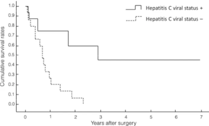
ICC surgeries (14 males and 9 females) were performed over the 9 years from April 1994 to July 2002 at The University of Tokushima Hospital, where the first author went after Kumamoto University Hospital. The patients’ average age was 65 years. In 11 cases of peripheral type and 12 cases of portal hilar type, the operation methods were : right, 3-segment resection, 4 cases ; left 3-segment resection, 1 case ; extended right hepatic lobectomy, 1 case ; extended left hepatic lobectomy, 7 cases ; right hepatic lobectomy, 1 case ; right anterior segmentectomy, 1 case ; right posterior segmentectomy, 2 cases ; and partial liver resection, 3 cases. In total, major hepatic resection was performed for 20 cases (87%), and resection of the extrahepatic bile duct was also carried out in 14 (60.1%) of these 23 cases, 8 (39.8%) were infect-ed with the hepatitis C virus (HCV). The cumulative survival rates of the HCV-related ICCs are shown in Figure 1. Survival curves of patients who were HCV-positive were significantly better (p = 0.0049) than of those who were HCV-negative, but the reason is not clear. However, an article (14) later revealed the reasons. Recently, lymph node metastasis was found to be significantly lower in hepatitis B virus (HBV)-associated ICC
Figure 1. Survival curves of patients who were HCV-positive were significantly better (p = 0.0049) than of those who were HCV-negative.
patients than in those without HBV infection. Seogsong et al. (14) found that HBV infection in cancer-associated lymphan-giogenesis / HBV infection could be involved in the suppression of cancer-associated fibroblasts adopting lymphatic endothelial morphology and function (mesenchymal-to-lymphatic endotheli-al transition), which resulted in the low extent of cancer-associ-ated lymphangiogenesis and led to a relatively low incidence (14). They also showed decreased lymph node metastases in HBV-as-sociated ICC. They did not discuss HCV infection, but all of our cases were HCV-infected, so that they had an improved survival rate compared with non HCV-infected cases. It was suggested that a similar mechanism might work to improve survival after resection of HCV-positive ICC. The case of a patient who sur-vived for a long time after resection is presented. The patient was a 67-year-old woman who had undergone subtotal gastrectomy with lymph node dissection 14 years earlier. After a periodic follow-up visit, ALP and γGPT were increased, and US and CT showed dilatation of the left intrahepatic bile duct and atrophy of the left lateral hepatic segment. She was referred to our depart-ment. After careful examination, she was diagnosed as having a small (2.5 cm in diameter), HCV-positive ICC with invasion to the hilar bile duct, and extended left lobectomy combined with resection of the caudate lobe and the extrahepatic bile duct, and lymphadenectomy and hepaticojejunostomy with the Roux-en-Y method for ICC, were performed. Lymph nodes along the lesser curvature of the stomach, left gastric artery, and the common hepatic artery had already been dissected in the operation for gastric cancer, so only the lymph nodes in the hepatoduodenal ligament with the extrahepatic bile duct were dissected in this procedure. She died of other disease at 8.5 years, and there was no recurrence at that time. Factors that contributed to long-term survival were : diagnosis of small tumors by periodic examina-tion after gastric cancer surgery ; hepatitis C virus infecexamina-tion ; no lymph node metastases ; easy flow of lymph in the ICC of the left lobe ; and lymph nodes were dissected during gastric cancer surgery. Thus, it was thought that this patient survived for a long time without recurrence because of the overlapping of these four conditions.
Recent experience with combined HCC and ICC in Taoka Hospital
An 88-year-old man with left back pain visited the orthopedic department on March, 2019. CT showed an abnormality in the liver, and he was referred to our department. Blood tests showed increases of AFP (777.7 ng / ml), PIVKA-II (241.0 ng / ml), CEA (21.7 ng / ml), and CA19-9 (5.9 U / ml). Contrast-enhanced CT showed a tumor in the left lateral segment. The tumor was di-agnosed as a combined tumor based on the contrast pattern and shape of the tumor, and this was proven by needle biopsy of the tumor. A left hepatic lateral segment resection was performed. The tumor was exposed on the serosa of the surface of the liver. The hepatoduodenal ligament was exposed. After taping of the portal vein, lymph nodes were dissected in the hepatoduodenal ligament. And left hepatic lateral segment resection was per-formed. The hepatectomy specimen was a 6 x 5.5-cm2 tumor. On pathological examinations, it was im (-), fc-inf (-), sf (-), s (0), vp (2), vv(0), va (0), b (0), sm (-), 12p (-).
The patient was discharged 11 days after surgery, and 2.8 months after surgery, multiple liver recurrences and right lung metastases developed, and he died 5.5 months after surgery. Even the combined tumor of ICC and HCC had a poor prognosis. Kim et al. (15) reported that patients with combined HCC and
ICC had poor postoperative survival rates. A high CA-19-9 level was associated with worse survival, suggesting that the chol-angiocarcinoma portion may be a major determinant of patient prognosis. In addition, Jung et al. (16) showed that combined
cancer carries a worse overall prognosis than either HCC or ICC
alone. Thus, it was considered necessary to treat it according to the same strategy used for ICC or a more radical strategy.
LYMPH NODE METASTASES AND THE
SIGNIFI-CANCE OF LYMPH NODES DISSECTION
There have been very few reports of the pattern of lymphatic spread of ICC. This pattern was elucidated to help define the extent of radical lymph node dissection. Thirty-nine consecu-tive patients who underwent hepatectomy with radical lymph node dissection were reviewed retrospectively (1). Lymph node metastases were detected in 24 (62%) of the 39 patients. The metastatic nodes were found in the hepatoduodenal ligament, along the common hepatic artery, around the abdominal aorta, on the posterior surface of the head of the pancreas, along the left gastric artery, along the superior mesenteric artery, around the celiac artery, along the lesser curvature of the stomach, and around the cardia only in the left peripheral and hilar types of cholangiocarcinoma, whereas all other sites included both the left or right peripheral type and the hilar type cholangiocarci-noma. ICCs, irrespective of their intrahepatic location, spread mainly to the nodes in the hepatoduodenal ligament, and then to the para-aortic nodes, retropancreatic nodes, or common hepatic artery nodes. In addition to these routes of spread, the left peripheral type or hilar type of cholangiocarcinoma tends to spread along the left gastric nodes through the lesser curvature. From our clinicopathological findings and the anatomical fea-tures, the efferent lymphatic flow from the liver is complicated and extensive (1).
A recent meta-analysis of 13 studies, including 1,337 pa-tients from 2000 to 2018 by Zho et al. (17) showed that there
were no significant differences in overall survival, disease-free survival, or recurrence between a lymph node dissection group and a non-lymphnode dissection group. They also found that postoperative morbidity was significantly higher in the lymph node dissection group. Therefore, they concluded that routine or prophylactic lymph node dissection could not be recommended because of the uncertain survival benefit. Furthermore, many multivariate analyses showed that the significant independent predictor of poor prognosis is not the omission of lymph node dissection, but the pathological lymph node metastasis itself. However, it is not clear whether there is metastasis without dissection. If there are cancer cells in the sentinel lymph node (18-21), the lymph nodes in the area of the tumor are dissected. If there are no cancer cells in the sentinel node, lymph node dis-section is not performed. Thus, it is more reasonable to search for the sentinel lymph node at the location of the tumor. Therefore, in the near future, we plan to inject indigo carmine into the liver tissue on the tumor side several times around the circumference during the operation to search for metastases to determine the need for dissection. If the sentinel nodes do not stain well, the lymph nodes of the hepatoduodenal ligament, which is the main flow of lymph from the liver, are dissected, and if frozen section examination is positive for cancer cells, radical dissection is performed, but if it is negative, radical dissection may not be considered.
ADJUVANT CHEMOTHERAPY FOR ICC
Despite R0 resection, the 5-year survival rate is low, at only 30%. Multifocal, node- or margin-positive disease is at a higher risk of recurrence after resection. A recent adjuvant therapy phase III trial from the Partenariat de Recherche en Oncologie Digestive-Actions Concertees dans les Cancers Colo-Rectaux
at Digestifs (PRODIGE) group reported no survival advantage with adjuvant Gemcitabine and Oxaliplatin therapy (22).
However, recently, immune therapy using programed cell death 1 (PD-1) was reported. Kubo et al. (23) showed that, in
addition to the main tumor, chronic bile duct injury and pre-cancerous lesions were observed at various sites of the large bile duct. Whole-exome analysis of occupational cholangiocarcinoma showed that an average of 44.8 somatic mutations per Mb was detected in the genome, approximately 30-fold higher than observed in typical cholangiocarcinoma. Moreover, a high num-ber of programmed death-1-positive T cells were found to have infiltrated the tumor, suggesting that PD-L1 may interact with the PD-1 expressed on T cells in the tumor microenvironment, thereby causing immune escape via the PD-1 / PD-L1 axis (24). The Ministry of Health and Welfare of Japan classified this type of cholangiocarcinoma “occupational ICC”, and a PD-1 inhibitor was approved. The treatment outcome with a PD-1 inhibitor in this case will be described later. Molecular profiling studies have indicated that about 30% to 40% of intrahepatic cholangiocarci-noma cases have actionable mutations. These include fibroblast growth factor receptor (FGFR) and BRAF genetic aberrations. Clinical trials targeting these mutations showed that inhibitors provided a promising early signal showing clinical efficacy, and patients with HCC expressing high vascular endothelial growth factor (VEGF) have a very poor prognosis. High expression of VEGF in patients with hepatocellular carcinoma (HCC) is asso-ciated with a poor prognosis. This seems to have led to the begin-ning of research with lenvatinib, with lenvatinib mesylate as the active ingredient. It is an oral multikinase inhibitor. Lenvatinib is a vascular endothelial growth factor. In addition to (VEGF) receptors VGFR1-3 and fibroblast growth factor (FGF) receptors (FGFR),1-4 tumor markers such as platelet-derived growth fac-tor recepfac-tor (PDGFR) PDGFRα, and KIT, this drug has potent and selective inhibitory activity against receptor tyrosine kinas-es involved in angiogenkinas-esis and malignancy (25).
Cancer tumor tissues are composed of a number of different types of cells, including cancer cells, vascular endothelial cells, and stromal cells. They express VGFR and their ligands VEGF and FGF, and they also express VEGF and FGFR induced by FGF (especially FGFR1). Both signals cause tumor angiogen-esis. Since hepatocellular carcinoma is a typical pluripotent tumor with a developed tumor vascular network, it is thought that suppressing both VEGFR and FGFR signals at the same time can suppress tumor angiogenesis very effectively. He-patocellular carcinoma expresses FGFR1-4, suggesting that increased FGFR signaling is associated with malignant trans-formation of cancer cells and a poor prognosis. Lenvatinib has a selective inhibitory activity against receptor tyrosine kinases such as VEGFR1-3 and FGFR1-4, and it inhibits tumor angio-genesis and malignant transformation by inhibition of VEGFR and FGFR, thus preventing hepatocellular carcinoma (26). It was expected to show a therapeutic effect. The global phase III study (Study 304, REFLECT) showed that overall survival was noninferior to that of sorafenib, so the drugs approved for “unresectable hepatocellular carcinoma” expanded for the first time in Japan. Tanaka and Kubo (12) have reported a case of relapse of ICC treated with a PD-1 inhibitor. A 39-year-old man underwent curative liver resection in October 2012, followed by adjuvant chemotherapy with S-1(Tegafur) or gemcitabine until postoperative 4 years because of a continuous increase in serum CA19-9. At postoperative 4 years 1 month, the serum CA19-9 concentration increased rapidly to 4,140 U / ml with lymph node metastasis around the superior mesenteric artery. Although the concentration decreased with gemcitabine and cisplatin chemotherapy, it increased again to 8,769 U / ml at postoperative 4 years 11 months. Positron emission tomography (PET) showed
enlarged para-aortic lymph nodes. They then started the admin-istration of a PD-1 inhibitor (nivolumab 3 mg / m2) every 2 weeks. After seven cycles of treatment, the serum CA19-9 concentration decreased to within the normal limit, and PET demonstrated diminished lymph nodes. The patient appeared healthy without recurrence 12 months after the complete response. Recently, a combination of a PD-1 inhibitor and lenvatinib has been report-ed to be effective for bone metastasis of ICC (26). A significant response to anti-PD-1-based immune therapy plus lenvatinib for recurrent intrahepatic cholangiocarcinoma with bone metasta-sis was previously reported.
To date, only 2 cases were reported in which nivolumab, an agent against (PD-1), combined with lenvatinib chemothera-py led to a complete response (26). The safety and efficacy of nivolumab-based immunotherapy combined with lenvatinib for ICC were not previously reported. A 40-year-old woman was identified as having a lesion of 7.0 cm in diameter in the right lobe of the liver. Furthermore, they started the administration of a PD-1 inhibitor (nivolumab, 3 mg / m2) every 2 weeks. The patient was diagnosed as having intrahepatic cholangiocarcino-ma. Surgery was performed, intraoperative ultrasound showed an 8 x 8 cm 2 mass in segment VIII of the liver, invasion of the diaphragm could be seen, and the mass was observed to jostle against the right and middle hepatic veins. The patient under-went resection of liver segment VIII, dissection of regional lymph nodes, and resection of lesions on the diaphragm. The patient developed recurrent lesions in the 5th month after surgery, and the cholangiocarcinoma expanded to the right thoracic vertebra (Th7-8) in the 6th month. The patient received nivolumab plus lenvatinib, and the lesions in the liver decreased in size and disappeared after this treatment. Additionally, the metastases in the right thoracic vertebral pedicle were stable after 9 months of therapy. Although the details of the operation and postopera-tive course are not described for the second case, both cases are described as having shown a complete response in combination with chemotherapy. Recently, based on basic research on immu-notherapy and molecular drug therapy, a small number of clini-cal trials has tried to determine whether it may also be effective for ICC. If this adjuvant therapy is used widely in patients with ICC, prognosis must be improved. This is a great pleasure for ICC patients with a poor prognosis and for the physicians who treat them.
PROPOSED STRATEGY OF SURGICAL
TREAT-MENT FOR ICC PATIENTS
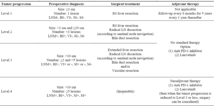
After summarizing the review of ICC, we developed a Pro-posed Strategy to improve the prognosis of ICC patients, as shown in Table 1. We decided to use the level because the tumor factors would be better expressed at the level than at the stage. Please see Table 1 for detail.
We expect that further investigations will prove the feasibility of our proposed strategy.
CONFLICTS OF INTEREST AND SOURCE OF FUNDING
The authors declare no conflicts of interest relevant to this review. This research received no specific grant from any fund-ing agency in the public, commercial, or not-for-profit sectors.REFERENCES
1. Tsuji T, Hiraoka T, Kanemitsu K, Takamori H, Tanabe D, Tashiro S : Lymphatic spreading pattern of intrahepatic cholangiocarcinoma. Surgery 129 : 401-407, 2001
2. Shen WF, Zhong W, Xu F, Kan T, Geng L, Xie F, Sui CJ, Yang JM : Clinicopathological and prognostic analysis of 429 patients with intrahepatic cholangiocarcinoma. World journal of gastroenterology 15 : 5976-5982, 2015
3. Fujimoto A, Furuta M, Shirashi Y, Gotoh K, Kawakami Y, Koji Arihiro K, Nakamura T, Ueno M, Ariizumi S, Nguyen HH, Shigemizu D, Abe T, Boroevich KA, Nakano K, Sasaki A, Kitada R, Maejima K, Yamamoto Y, Tanaka H, Shibuya T, Shibata T, Ojima H, Shimada K, Hayami S, Shigekawa Y, Aikata H, Ohdan H, Marubashi S, Yamada T, Kubo M, Hirano S, Ishikawa O, Yamamoto M, Yamaue H, Chayama K, Miyano S, Tsunoda T, Nakagawa H : Whole-genome mutational landscape of liver cancers displaying biliary phenotype reveals hepatitis impact and molecular diversity. Nat Commun 6 : 6120, 2015
4. Wardell CP, Fujita M, Yamada T, Simbolo M, Fassan M, Karlic R, Polak P, Kim J, Hatanaka Y, Maejima K, Lawlor RT, Nakanishi Y, Mitsuhashi T, Fujimoto A, Furuta M, Ruzzenente A, Conci S, Oosawa A, Sasaki-Oku A, Nakano K, Tanaka H, Yamamoto Y, Michiaki K, Kawakami Y, Aikata H, Ueno M, Hayami S, Gotoh K, Ariizumi S, Yamamoto M, Yamaue H, Chayama K, Miyano S, Getz G, Scarpa A, Hirano S, Nakamura T, Nakagawa H : Genomic character-ization of biliary tract cancers identifies driver genes and predisposing mutations. Hepatology 68 : 959-969, 2018 5. Kumada K, Shimahara Y, Fukui K, Itoh K, Morikawa S,
Ozawa K : Extended right hepatic lobectomy: combined resection of inferior vena cava and its reconstruction by EPTFE graft (Gore-Tex). Case report. Acta chirurgica scan-dinavica 154 : 481-483, 1988
6. Hemming AW, Reed AI, Langham MR Jr, Fujita S, Howard
RJ : Combined resection of the liver and inferior vena cava for hepatic malignancy. Annals of surgery 239 : 712-721, 2004
7. Grazi GL, Mazziotti A, Jovine E, Pierangeli F, Ercolani G, Gallucci A, Cavallari A : Total vascular exclusion of the liver during hepatic surgery : selective use, extensive use, or abuse?. Archives of Surgery 132 : 1104-1109, 1997
8. Pichlmayr R, Grosse H, Hauss J, Gubernatis G, Lamesch P, Bretschneider HJ : Technique and preliminary results of extracorporeal liver surgery (bench procedure) and of sur-gery on the in situ perfused liver. Br J Surg 77 : 21-26, 1990 9. Sugimachi K, Yanaga K, Shimada M : Extracoporeal liver resection-the first experience in Japan. Geka (in Japanese) 52 : 717-720, 1990
10. Tashiro S, Tsuji T, Inoue K, Sawada T, Kawamoto S, Kanemitsu K, Miyauchi K : Three segment resection of the left liver and partial resection of the right hepatic vein by extracorporeal liver surgery (in Japanese). Operation 45 : 507-514, 1991
11. Liu X, Yao J, Song L, Zhang S, Huang T, Li Y : Local and abscopal responses in advanced intrahepatic cholangiocar-cinoma with low TMB, MSS, pMMR and negative PD-L1 expression following combined therapy of SBRT with PD-1 blockade. J Immunotherapy Cancer 7 : 204, 2019
12. Tanaka S, Kubo S : Programmed death-1 inhibitor for occupant intrahepatic cholangiocarcinoma caused by chlo-rinated organic solvents. J Hepatobiliary Pancreat Sci 26 : 242-243, 2019
13. Tashiro S, Miyake H, Rokutan K : Role of geranylgeranylac-etone as non-toxic HSP70 inducer in liver surgery ; Clinical application. J Hepatobiliary Pancreat Sci 25 : 269-274, 2018 14. Jeong S, Tong Y, Sha M, Gu J, Xia Q : Hepatitis B virus-as-sociated intrahepatic cholangiocarcinoma : a malignancy of distinctive characteristics between hepatocellular car-cinoma and intrahepatic cholangiocarcar-cinoma. Oncotarget 8 : 17292-17300, 2017
15. Kim K, Lee S, Park E, Hwang S, Ahn C, Moon D, Ha T, Song G, Jung D, Kim K, Lim Y, Lee H, Chung Y, Lee Y, Suh D : Surgical treatment and prognosis of patients with combined hepatocellular carcinoma and cholangioma. Ann Surg Oncol 16 : 23-29, 2009
16. Jung DH, Hwang S, Kin KH, Hong SM, Lee YJ, Ahn CS, Moon DB, Ha TY, Song GW, Park GC, Yu ES, Lee SG : Clinico-pathological features and postresection progno-sis of double primary hepatocellular carcinoma and intrahe-patic cholangiocarcinoma. World J Surg 41 : 825-834, 2017 17. Zhou R, Lu D, Li W : Is lymph node dissection necessary
cholangiocarcinoma? A systematic review and meta-analy-sis. HPB (Oxford) 21 : 784-792, 2019
18. Wada H, Hyun H, Vargas C, Genega EM, Gravier J, Gioux S, Frangioni JV, Choi HS : Sentinel lymph node mapping of liver. Ann Surg Oncol 3 : S1147-S1155, 2015
19. Christophi C, Nguyen L, Muralidharan V, Nikfarjam M, Banting J : Lymphatics and colorectal liver metastases : the case for sentinel nodes mapping. HPB (Oxford) 16 : 124-130, 2014
20. Ceranic MS, Kecmanovic DM, Pavlov MJ, Nale DP, Micev MT, Kovacevic PA, Stamenkovic AB : Validation and fea-sibility of ex vivo sentinel lymph node “mapping” by meth-ylene blue in colorectal cancer. Hepato-gastroenterology 57 : 1113, 2010
21. Lupinacci RM, Paye F, Coelho FF, Kruger JAP, Herman P : Lymphatic drainage of the liver and its implications in the management of colorectal cancer liver metastases. Up-dates in surgery, 66 : 239-245, 2014
22. Edeline J, Benabdegharni M, Bertaut A, Watelet J, Hammel P, Joly JP, Boudjema K, Fartoux L, Bouhier-Leporrier K, Jouve JL, Faroux R, Guerin-Meyer V, Kurtz JE, Assenat
E, Seitz JF, Baumgaertner I, Tougeron D, Fouchardiere C, Lombard-Bohas C, Boucher E, Stanbury T, Louvet C, Malka D, Phelip JM : Gemcitabine and Oxaliplatin Chemo-therapy or Surveillance in Resected Biliary Tract cancer (PRODIGE 12-ACCORD 18-UNICANCER Gl) : A Rand-mized Phase III Study. J Clin Oncol 37 : 658-667, 2019 23. Kubo S, Nakamura Y, Takemura S, Sakata C, Urata Y,
Nozawa A, Nishioka T, Kinoshita M, Hamano G, Terajima H, Tachiyama G, Matsumura Y, Yamada T, Tanaka H, Nakamori S, Arimoto A, Kawada N, Fujikawa M, Fujishima H, Sugawara Y, Tanaka S, Toyokawa H, Kuwae Y, Ohsawa M, Uehara S, Sato KK, Hayashi T, Endo G : Case series of 17 patients with cholangiocarcinoma among adult workers of a printing company in Japan. J Hepatobiliary Pancreat Sci 21 : 479-488, 2014
24. Sato Y, Kinoshita M, Takemura S, Tanaka S, Hamano G, Nakamori S, Fujikawa M, Sugawara Y, Yamamoto T, Arimoto A, Yamamura M, Sasaki M, Harada K, Nakanuma Y, Kubo S : The PD-1 / PD-L1 axis may be aberrantly activated in occupational cholangiocarcinoma. Pathol Int 67 : 163-170, 2017
25. Matsuki M, Hoshi T, Yamamoto Y, Ikemori-Kawada M, Minoshima Y, Funahashi Y, Matsui J : Lenvatinib inhibits angiogenesis and tumor fibroblast growth factor signaling pathways in human hepatocellular carcinoma models. Can-cer medicine 7 : 2641-2653, 2018
26. Chen WX, Li GX, Hu ZN, Zhu P, Zhang BX, Ding ZY : Sig-nificant response to anti-PD-1 based immunotherapy plus lenvatinib for recurrent intrahepatic cholangiocarcinoma with bone metastasis : A case report and literature review. Medicine 98 : 45, 2019