Roles of IL-1α/β in Regeneration of
Cardiotoxin-Injured Muscle and Satellite Cell
Function
著者 CHAWEEWANNAKORN CHAYANIT
学位授与機関 Tohoku University
学位授与番号 11301甲第18885号
Doctoral Thesis
Roles of IL-1α/β in Regeneration of
Cardiotoxin-Injured Muscle and Satellite Cell Function
カルディオトキシン誘導性筋損傷における
IL-1α/β の役割と筋衛星細胞の機能
Chayanit Chaweewannakorn
(B5DD5042)
Graduate School of Dentistry
Tohoku University
ABSTRACT
Skeletal muscle undergoes tissue repair, which called regeneration or remodeling process throughout a life and these are specific adaptive functions of muscle tissue. The muscle regeneration after injury involving cascades of inflammatory responses and myogenic program of muscle stem cells, the satellite cells. Recent studies have shown a close relevant between inflammatory process and myogenesis, but the involved cellular and molecular components that link these processes are diverse and not completely elucidated. Interleukin (IL)-1 is a novel cytokine of inflammatory responses and exerts pleiotropic impacts on various cell types. During injury, IL-1 is secreted from activated immune cells and majorly acts on pro-inflammatory function which may directly drives the subsequent tissue healing process. Thus, the aim of this thesis is to investigate the role of IL-1 during skeletal muscle regeneration. A series of in-vivo and in-vitro experiments were performed in interleukin-1α/β-double knockout (IL-1KO) mice and their isolated skeletal muscle satellite cell culture to examine the functions of IL-1. Firstly, the IL-1KO mice exhibit delayed muscle regeneration after cardiotoxin (CTX) injection, characterized by delayed infiltration of immune cells accompanied with suppressed local production of pro-inflammatory factors including IL-6 and delayed increase of PAX7-positive satellite cells post-injury, along with the reduced myogenic gene expression as compared with those of wild-type mice. For the in-vitro examination, the satellite cells cultures obtained from the IL-1KO mice unexpectedly revealed that IL-1KO myoblasts have impaired myogenic properties in terms of both proliferation and differentiation, indicating by myotube formation. However, the delayed proliferation and differentiation were reversed by exogenous IL-1 administration to the cultures. Interestingly, the biological analysis revealed the delay in myogenesis was not attributable to the myogenic transcriptional program since MyoD and myogenin were highly upregulated in IL-1KO cells. Furthermore, the cellular response of myoblast to IL-1 observed via live-image technique unveiled that the delayed myogenic characteristics of IL-1KO are at least in part, to be due to dysregulation of cellular fusion events, possibly follow from aberrant actin regulatory systems. Data from this thesis suggests IL-1 plays a positive role in muscle regeneration by coordinating the initial inflammatory interactions between various cells in the regenerating tissue niche. Moreover, findings also provide compelling evidence that IL-1 is intimately engaged in regulating the fundamental function of myocytes, partly involves with cellular actin remodeling which subsequently accounted for myogenic functions like myoblast fusion.
CONTENTS
Abstract……….... ii List of Abbreviations……….2 List of Figures………...3 Chapter 1: Introduction 1.1 Backgrounds………....…...4 1.2 Objective……….5 1.3 Outline of dissertation……….6Chapter 2: Review of literatures 2.1 Skeletal muscle repair……….7
2.2 The skeletal muscle satellite cells………...8
2.3 Involvement of inflammatory process to the muscle regeneration………….9
2.4 Cellular and Molecular pathways of myogenesis………10
2.5 Interleukin-1 cytokines………11
Chapter 3: Materials and methods 3.1 Experimental designs………13
3.2 Experimental procedures……….….……….……14
Chapter 4: Study results 4.1 Results……….……….……….21 4.2 Discussion……….………27 4.3 Figures……….…..31 Chapter 5: Conclusion 5.1 Conclusion………47 5.2 Future perspectives………48
List of Scientific publications and presentations………50
Acknowledgements………51
List of Abbreviations
BSA, Bovine serum albumin
Cdc42, Cell division control protein 42 homolog CTX, Cardiotoxin
CXCL1, Mouse chemokine (C-X-C motif) ligand 1 DAPI, 4',6-Diamidino-2-phenylindole, dihydrochloride DM, differentiation medium
DMEM, Dulbecco's Modified Eagle Medium EDTA, Ethylenediaminetetraacetic acid FACS, Fluorescent-activated cell sorting
GAPDH, Glyceraldehyde 3-phosphate dehydrogenase GM, Growth medium
H&E, hematoxylin and eosin IFN-γ, Interferon γ
IL, Interleukin
IL-1α, Interleukin-1α IL-1β, Interleukin-1β
IL-1KO, Interleukin-1α/β-double knockout MAP kinase, mitogen-activated protein kinase MRFs, Myogenic regulatory factors
MRF4, Myogenic regulatory factor 4 Myf5, Myogenic factor 5
MyHC, skeletal myosin heavy chain NF-κB, nuclear factor-κB
N-WASP, neural Wiskott-Aldrich syndrome protein PAK, p21-activated kinase
PAX3, Paired box 3 PAX7, Paired box 7
PBS, phosphate-buffered saline PFA, Paraformaldehyde
qPCR, Quantitative real-time polymerase chain reaction Rac1, Ras-related C3 botulinum toxin substrate 1
Rho A, Ras homolog gene family, member A TA, Tibialis anterior muscle
TNF-α, tumor necrosis factor α WT, Wild-type
List of Figures
Fig. 1 Geneticschemes of IL-1α/β-double-knockout (IL-1KO) mice Fig. 2 Genetic phenotyping of IL-1α/β-double-knockout (IL-1KO) mice Fig. 3 Hematoxylin and eosin stain of cardiotoxin-injured muscle cyosections Fig. 4 Histological analysis of regenerating muscles cryosections
Fig. 5 Histological analysis of PAX7-expressing cells in muscle cryosections Fig. 6 Biological analysis of cardiotoxin-injured muscle tissues.
Fig. 7 Fluorescent activated cell sorting of satellite cells Fig. 8 Cell proliferation assay of myocyte cultures
Fig. 9 Myogenic differentiation analysis of myocyte cultures (I) Fig. 10 Myogenic differentiation analysis of myocyte cultures (II)
Fig. 11Responsiveness of myocyte cultures to stimulation with IL-1β (Ι) Fig. 12 Responsiveness of myocyte cultures to stimulation with IL-1β (ΙΙ) Fig. 13 Live cell imaging analysis of wild type myocyte cultures
Fig. 14 Live cell imaging analysis of IL-1KO myocyte cultures (I) Fig. 15 Live cell imaging analysis of IL-1KO myocyte cultures (II) Fig. 16 Schematic illustration from the in-vivo results
Chapter 1: Introduction
1.1 Background
Skeletal muscle function represents a major determinant of physical performance, apart from their recognized role to body locomotive, this organ involves with whole body metabolic activities and directly influences the quality of life. Altering of muscle tissue structure and function due to various kind of stressor, including environmental factors, macro/microtrauma and degenerative condition could increase susceptibility to rapid onset or deterioration of adverse health conditions issues with chronic diseases and aging (Wolfe, 2006). Especially for the aging and aged population, a parallel decline in muscle volume and strength occurred simultaneously from intrinsic regulation and environmental influences like nutritional and physical activity. However, the skeletal muscles are known for a remarkable ability to adapt their properties, such as energy expenditure and even their total mass in order to meet the changes. The topic of muscle mass maintenance attracts a great scientific interest because it apparently reflects both structural and reparative functions of tissue. The maintenance of muscle mass is resulted from the balance between myogenesis or degrading cellular signals which are triggered by mechanical loads, nutrients, hormones, cytokines or environmental status.
Recently the role of inflammatory signals in muscle homeostasis has become a central focus in order to develop new therapeutic targets against muscle morbid conditions. Firstly, local and systemic inflammatory programs are potentially subjected to the pathological muscle wasting. Chronic diseases such as chronic renal failure, diabetes and cancer are involved with increased proinflammatory chemical molecules dominating by tumor necrosis factor α (TNF-α), IL-1 and interferon γ (IFN-γ) (Forrester & Bick-Forrester, 2005). These cytokines chronically activate the inflammatory signaling and push the balance toward the overall protein breakdown systems, include the ubiquitin proteasome, the lysosomal and the calcium ion dependent system, that latterly lead to the loss of muscle mass (Costamagna, Costelli, Sampaolesi, & Penna, 2015; Yang & Hu, 2018). On the contrary to conventional inflammation-triggered protein catalytic effect, the local immune response in muscle tissues was found positively involved to muscle functions. The roles of neutrophils in vivo during muscle contraction in both general exercise and masticatory activity pointed their beneficial regulation for the fatigue alleviation effect (Chiba et al., 2015; Tsuchiya et al., 2018). Currently, for the field of tissue regeneration, the critical roles of immune cells
during muscle repair has been studied concurrently with the fast-moving stem cell studies.
In addition to the contribution from protein synthesis or breakdown regulation, the alteration of myogenic programs is also essential for quantitative and qualitative maintenance of muscles. Normal regenerative myogenesis requires a well-tailored immune cascade that mediated by various pro and anti-inflammatory cytokines (Karalaki, Fili, Philippou, & Koutsilieris, 2009; Yang & Hu, 2018). Thus, both an aberrant inflammatory action in pathologic health conditions or the use of immuno-modulating approach in current medical practice could someway affect the muscle homeostasis. Among several cytokines, IL-1 has been studied extensively, partly due to its pleiotropic functions. Besides, during inflammation, IL-1 could be produced by multiple cellular sources like neutrophils and macrophages (Sims & Smith, 2010). However, knowledges regarding of how the immunomodulatory triggered by IL-1 interact to the myogenic program is still limited. Therefore, this study mainly questions on the roles of IL-1 cytokine during regeneration of injured muscle, including the impact on regenerating tissue environment, and also specify on the stem cell myogenic process.
1.2 Objectives
This study hypothesized IL-1 to be constitutively involved in the muscular regeneration process, and therefore took advantage of the IL-1KO mouse model as a principal method.
The aims of the study are pointed out as follows;
1.2.1 To directly investigate the role of IL-1 in the process of muscle
regeneration after CTX-induced skeletal muscle injury.
1.2.2 To examine whether IL-1 deficiency was directly responsible for
1.3 Outline of dissertation
The outline of dissertation is showed as below:
Chapter 1: Introduction
This chapter gives the general background and concepts for conducting this study. The research question and hypothesis regarding to the main idea are described, along with research objectives and overall contents of this thesis.
Chapter 2: Review of literatures
This chapter explained the involved information and previous research in the related research topics including the skeletal muscle functions, skeletal muscle remodeling process and tissue immune response to stressors within skeletal muscle.
Chapter 3: Material and methods
The detailed information on the methodology, consists of the experimental design and experimental procedure are explained in this chapter. The analysis methods used for each experimental procedures and interpretation are also described.
Chapter 4: Results
This chapter provides the results from each experiment, along with their interpretations and figures. The discussed details on each part of results, also the limitation and suggestion for improving the experimental designs are mentioned in this section.
Chapter 5: Conclusion
In the last chapter, the summarized information from all studies are presented and discussed with their practical implication. This section also added further consideration for future research development.
Chapter 2: Review of literatures
2.1 Skeletal muscle repair
Skeletal muscle is a markedly abundant tissue in the body which important for movement and stability during activities. Also, this tissue has specific ability which is the tissue plasticity that support the potential for adaptation of the tissue to altered environments. Another impressive qualification of muscle tissue is a postnatal repair of muscle fibers throughout life (Karalaki et al., 2009; Francesco Saverio Tedesco, Arianna Dellavalle, Jordi Diaz-Manera, Graziella Messina, & Giulio Cossu, 2010). In terms of muscle repair, this process constitutes of regeneration of damaged muscle fibers, constant remodeling of muscle proteins and cellular contents, or the hypertrophic remodeling in excessive used muscles. Thus, the muscle repair is a crucial component to promote skeletal muscle health and physical performances. Skeletal muscle tissue consists of diverse cell types with different physiologic properties that incorporate to muscle fiber itself and nearby components, including myocytes, myogenic stem cells, immune cells, endothelium and others. Each of muscle tissue contains a large number of single unit myocyte fusing to each other to form a unit of myofiber. Skeletal muscle constantly undergoes structural and functional remodeling with self-repair in response to damages cause by excessive physical loads, degenerative diseases, nutritional alterations, traumatic injuries and even constantly remodel under normal conditions (Charge & Rudnicki, 2004). Historically, studies provided the observation that myofiber could increase their size and nuclei contents in the absence of apparent nuclear division activities. The nuclei of myocytes within mature myofiber themselves are post-mitotic, they cannot re-proliferate and incorporate to the repair the primitive myofiber. Therefore, the roles of muscle plasticity during postnatal muscle growth and repair depends mainly from the function of muscle stem cells, called the satellite cells. The regeneration process after injury involves various cellular activities within skeletal muscle tissue, mainly begins with a necrotic and inflammatory stage of injured tissue, then followed by a regeneration phase (Charge & Rudnicki, 2004; Tidball & Villalta, 2010). Thus, the highlights are given to the involvement of inflammatory cell coordination to muscle tissue niches, and the activation of muscle progenitor cells, myoblasts, that would proliferate and differentiate to give rise for the new myofibers.
Several experimental models were used to help investigating the muscle regeneration process. The myotoxin (venom toxin), chemical (barium chloride)
and mechanical (tissue freeze or crush) induced injury are commonly used in mice muscle regeneration studies. Even though all models lead to similar sequential steps of inflammatory and regenerative phase, each model could display different kinetics of muscle regeneration such as the interaction of each cell types in the regenerative tissue niches and importance of particular stages during tissue repair (Hardy et al., 2016; Mahdy, Lei, Wakamatsu, Hosaka, & Nishimura, 2015). Moreover, the morphologic changes in repaired tissues compare to the non-injured tissues could be the effect from injury model selection. Among various injury models, this study selected the cardiotoxin (CTX) induced injury model to observe the stem cells and other tissue components response during muscle regeneration. The CTX is a simple, reproducible and the most common used model referring to muscle injury publications. The comparative study revealed this model exhibited a time restriction inflammatory response, indicating by the cytokine expression returned to the control level as the tissues are histologically appeared regenerated (Hardy et al., 2016). In addition, the morphological changes of the repaired microvasculature or tissue fibrosis were only marginally changed, suggesting the CTX-induced injury could be an appropriate method for studying the muscle stem cell and local immune behavior during muscle regeneration.
2.2 The skeletal muscle satellite cells
The skeletal muscle satellite cells have a distinct relationship and exert multifunctional roles to give the adaptation plasticity to their companion muscle fibers. Exceptionally, it has been a clear understanding that muscle regeneration relies mainly on the function of a skeletal muscle stem cell, a satellite cell (F. S. Tedesco, A. Dellavalle, J. Diaz-Manera, G. Messina, & G. Cossu, 2010). In 1961, the study by Alexander Mauro observe the mononuclear cell that located between the basal lamina and the sarcolemma and later named the satellite cells (Mauro, 1961). The allocation of satellite cell presence was particularly found at high numbers nearby neuromuscular junctions and vasculature areas, but the explanation for these discoveries are not clarified. Interestingly, even though the satellite cells population presents in all skeletal muscle tissue, the percentage and myogenic properties could differ among each group of muscles. Studies compared the intrinsic properties between somite or branchiomeric derived muscles satellite cells and found the branchiomeric muscle satellite cell group exhibited longer differentiation time, confirming the different source and age of satellite cells could impact the myogenesis (Carvajal Monroy, Grefte, Kuijpers-Jagtman, Von den Hoff, & Wagener, 2017; Ono, Boldrin, Knopp, Morgan, &
Zammit, 2010). The satellite cells are normally maintained in quiescent state and are activated post-injury by stimuli including inflammation-related factors, which are produced in a coordinated fashion by recruited/activated leukocytes and skeletal muscle fibers (Munoz-Canoves, Scheele, Pedersen, & Serrano, 2013; Serrano, Baeza-Raja, Perdiguero, Jardi, & Munoz-Canoves, 2008). The activation signals to satellite cells could be derived from the muscle niche, proximal microvasculature, and through the activated inflammatory signals. Later to their activation, the satellite cells undergo a process called asymmetric division, in which they give rise to either the myoblasts that are committed to the differentiation, or a few divided cells still remain their quiescence state to maintain the stem cell pool (Karalaki et al., 2009; Francesco Saverio Tedesco et al., 2010). The roles of satellite cells are not limited only as the main cellular source for muscle formation, these cells also convey the improved phenotype of new muscle fibers, serves as a tracking marker in muscle responses to stimuli and also communicate within myogenic networks such as with the stromal cells that control extracellular matrix environment (Fry, Kirby, Kosmac, McCarthy, & Peterson, 2017). Despite its pivotal role that has been distinctly clarified, the cellular and molecular regulation of satellite activation and myoblast fusion is diverse and not fully understood so far, then the developed ability to isolate and culture the satellite cells support the advancement in the study of myogenesis.
2.3 Involvement of inflammatory process to the muscle
regeneration
Inflammatory process is considered the first step of damaged/injured muscle repair. At this stage, the injured muscles are necrotized and release the damaged cellular contents to tissue areas (Hardy et al., 2016). The CTX is a peptide that acts as protein kinase C inhibitor and induce local myo-necrotizing. The necrosis of muscle tissue consists of calcium influx, loss of sarcolemma, contractile content and cellular organelles. These cellular substances and cell debris act as the chemoattractant molecules that recruit various immune cells into the damaged areas and multiple events of immune cells infiltration occurred according to time and stage of injury. During the first event of cell infiltration, neutrophil which is accounted for the first line defense of innate immunity is majorly recruited. The muscle injury can directly stimulate tissue resident neutrophils, results in the release of pro-inflammatory cytokines such as TNF-α and IL-1β which further recruit circulating neutrophils to the injury site (Teixeira et al., 2003; Yang & Hu, 2018). The cellular components of inflammatory
response characteristics of CTX injured tissues are contributed by neutrophils and macrophages respectively. Neutrophil is also well-known for their rapid accumulation to the injured fibers, occurring within few hours. Apart from the cytokine release, neutrophil works via different mode of actions such as phagocytosis and production of chemical molecules including granule enzyme and oxidative factors (Mayadas, Cullere, & Lowell, 2014; Rosales, 2018). These specific and nonspecific actions facilitate the removal of damaged particles and induce the pro-inflammatory status. In the later phase principally plays by the adaptive immunity, the recruited immune cells are dominant by macrophages which also secrete substantial amount of cytokine, mainly TNF-α and IL-1β. These chemical molecules not only promote pro-inflammatory condition to provide debris clearance, but also lead to environmental modification for the activation, expansion, and differentiation stem cells during muscle regeneration. Many animal studies used gain-and-loss of functions models of inflammatory molecules showed the impaired inflammatory response negatively affected the developmental and regenerating myogenesis (Costamagna et al., 2015; Teixeira et al., 2003), yet the effects from aberrant IL-1 regulation have not been elucidated. Lastly, the switching of immune cells phenotypes from pro-inflammatory to anti-inflammatory comes up with the microenvironment modification that support the late stage of myogenesis, the maturation of newly formed myofibers. Conclusively, the systematic activation of innate immune responses is crucial for inducing infiltration of neutrophils and macrophages, which in turn contributes to activating satellite cells, leading to coordinated and efficient muscle regeneration (Tidball & Villalta, 2010; Zhang et al., 2013)
2.4 Cellular and Molecular pathways of myogenesis
The regulation process of skeletal muscle regeneration relatively resembles that of the muscle development, thus review of cellular and molecular pathway during embryogenic myogenesis would be crucial for the study of muscle regeneration. The cellular and molecular events during myogenesis consists of three main parts; progenitor cell activation, expression pattern of transcriptional myogenic program and myoblast fusion. Firstly, the satellite cells specification during embryogenic development partly acquire from the roles of paired box 3 (Pax3) and paired box 7 (Pax7) genes, thus the satellite cell lineage in quiescent stage are indicated as Pax7 expressing cells. After the myofiber injury, the satellite cells receive signals from neighboring tissue, become activated and then proliferate into myogenic commitment cell population, regulated by the myogenic
regulatory factos (MRFs), a group of muscle-specific, basic helix-loop transcription factors consisting of MyoD, myogenic factor 5 (Myf5), myogenin and myogenic regulatory factor 4 (MRF4). The primary myogenic regulatory factors, MyoD and Myf5 are expressed in the activated satellite cell and the divided myogenic cells called myoblasts. Then, the shift from proliferation to differentiation of myogenic cells activates the late stage myogenic regulatory markers, dominated by myogenin, MRF4 and myosin heavy chain (MyHC)that are expressed during the cell transformation into mature myocyte and the terminal differentiation which is myoblast fusion (Karalaki et al., 2009). In addition, many new skeletal muscles govern proteins were discovered, one of them is myomaker which is a muscle specific protein necessary for fusogenic activity of myoblast membranes (Millay et al., 2013). An important aspect of myogenic differentiation is myoblast fusion by means of the cell membrane dynamics with actin cytoskeleton remodeling, which is regulated by the Rho family of small GTPases (Rho A; Ras homolog gene family-member A, Rac1; Ras-related C3 botulinum toxin substrate 1, and Cdc42; Cell division control protein 42 homolog) in association with their downstream effectors such as p21-activated kinase (PAK) and neural Wiskott-Aldrich syndrome protein (N-WASP) (Charrasse, Causeret, Comunale, Bonet-Kerrache, & Gauthier-Rouviere, 2003; Gruenbaum-Cohen et al., 2012; Joseph et al., 2017; Vasyutina, Martarelli, Brakebusch, Wende, & Birchmeier, 2009). Although skeletal myogenesis is a highly sophisticated process driven by several key myogenic transcription factors including MyoD and myogenin (Yin, Price, & Rudnicki, 2013), several lines of evidence have indicated that these transcriptional myogenic programs and the aforementioned actual cellular fusion events are executed somewhat independently (Gruenbaum-Cohen et al., 2012; Millay et al., 2013).
2.5 Interleukin-1 cytokines
One of the pivotal mediators of the initial inflammatory response is IL-1, which is highly upregulated in damaged muscle tissues, especially in neutrophils and macrophages recruited around the site of injury (Kohno et al., 2012; Mackiewicz et al., 2003; Oliveira Cde et al., 2009). The IL-1 consists of two isoforms, IL-1α and IL-1β, in which both isoforms function as proinflammatory cytokines, hence the functional different especially on the roles of IL-1α are controversial and needed further investigation. Traditionally, the IL-1 are well-known for mediating the inflammatory response locally and systemically, but in some cases the presence of IL-1 is still indispensable for physiologic function
(Holmes, 2017). Given, however, that IL-1 has pleiotropic function including pro-inflammatory, cellular differentiation and/or proliferation-inducing effects on various cell types (Strle et al., 2004), surprisingly little is known about the precise role of IL-1 in the process of muscle regeneration; i.e. whether IL-1 positively or negatively contributes to the regeneration process. Moreover, the existence of two IL1 isoforms creates further complexity adding to the challenge of understanding the precise role(s) of IL-1 in regenerating skeletal muscles. Several myokines including interleukin-6 (IL-6) have been implicated in supporting the differentiation and proliferative function of satellite cells (Baeza-Raja & Munoz-Canoves, 2004; Otis et al., 2014; Tierney et al., 2014), skeletal muscles are capable of producing IL-6 due to several circumstances such as exercise, inflammation, or local injury. The IL-6 is considered as a multifaceted cytokine with both pro-inflammatory and anti-inflammatory functions. Several cellular pathways draw the relationship that IL-1 could lead to IL-6 production via its classical signaling like nuclear factor-κB (NF-κB)and mitogen-activated protein (MAP) kinase. Therefore, in the muscle microenvironment level, IL-1 cytokine possibly acts as an intermediate mediator for spontaneous cascades of immune responses, then contributes to a conserved inflammatory-regenerative phase transition of muscle repair. However, the potential direct involvement of IL-1s in the particular regulation of satellite cell function remains to be clarified.
Chapter 3: Materials and methods
3.1 Experimental designs
In this study, two main parts of quantitative experiments were performed, including in vivo and in vitro experiments. Firstly, the in vivo experiment was performed on the group of wild type or IL-1α/β-double knockout (IL-1KO) mice to compare the tissue regenerating property. The measurements consisted of changes in myofiber size, satellite cells activation pattern, protein and gene expression during muscle regeneration were done at different representative phase. All variables include mice phenotype (WT or IL-1KO), CTX injection, and time after injury.
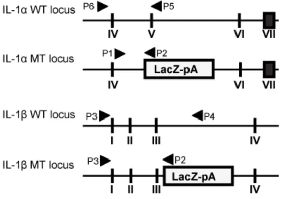
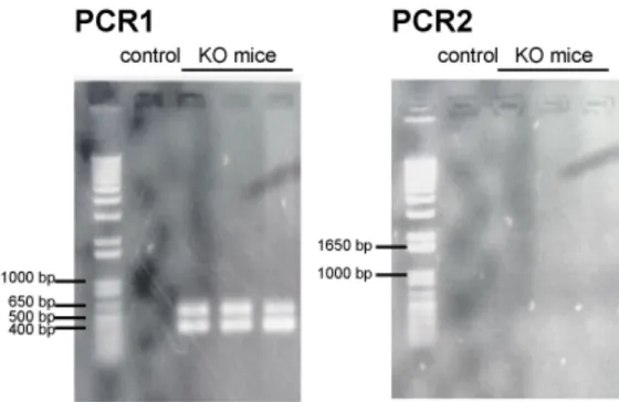
Male 6-7-week old Balb/c and IL-1KO mice weighing 20-25 g were used in this study. WT mice were purchased from CLEA-Japan (Tokyo, Japan). IL-1KO mice, homozygous for deficiencies of both IL-1α and IL-1β were bred, maintained with routine genotyping (Fig. 1 and 2) and obtained from animal facilities of Tohoku University, Graduate School of Dentistry (Chiba et al., 2015; Horai et al., 1998). The experimental design, as well as the care and use of the mice, followed the guidelines for animal experiments of Tohoku University and the Ethics Committee for Animal Experiments, Tohoku University, approved these studies (Ethic approval #2015MdA-335). The mice were kept in standard cages in an air-conditioned room at 23±1°C and with a 12-hour light-dark cycle. They were allowed access to standard food pellets and tap water ad libitum.
In the following in vitro experiment, myogenic properties of specific cell type, the satellite cells, were examined on the isolated primary cells culture from both mice types. Cell culture model was used to compare myoblast proliferation and differentiation properties, myogenic gene and protein expression and also the cellular responses to exogenous treatment with IL-1β solution. The variables include satellite cell phenotype (WT or IL-1KO), exogenous IL-1β treatment, and differentiation time. All details of measurement and analysis procedures were described as follows.
3.2 Experimental procedures
3.2.1 Cardiotoxin-induced mouse skeletal injury model
The mice were anesthetized with medetomidine hydrochloride (0.75 mg/kg) (Zenoaq, Fukushima, Japan), midazolam (4 mg/kg) (Sandoz, Tokyo, Japan) and butorphanol (5 mg/kg) (Meiji Seika, Tokyo, Japan). Cardiotoxin-induced muscle injury was performed by unilaterally injected 10 µl of 100 µM CTX into a Tibialis Anterior muscle (TA), to serve as the injured side, while the contralateral TA was injected with the same volume of saline to serve as the non-injured side (Garry, Antony, & Garry, 2016). The muscles were harvested on
Days 3, 7 and 14 after injection. The mice were euthanized by cervical dislocation
under anesthesia by isoflurane (MSD Animal Health, Summit, NJ, USA) inhalation. The TA from both sides were dissected, kept at -80°C and then subjected to quantitative real-time polymerase chain reaction (qPCR) and Bio-Plex assay, or immediately immersed in 4% paraformaldehyde (PFA) in phosphate-buffered saline (PBS) solution and then prepared for histological analysis (n=6 per group for either qPCR, Bio-plex assays or histological analysis).
3.2.2 Histological Analyses
The TA cryosections were stained with hematoxylin and eosin (H&E) solution and photographed employing an inverted microscope, the Olympus CKX41 with Olympus UPlanFL 20X/0.75 objective lens (200 folds magnification) equipped with DP Controller Software (Olympus, Tokyo, Japan). For immunostaining procedures, the cryosections were washed, blocked with 5% calf serum in PBS, and then incubated with the primary antibodies at 4°C overnight, all from Abcam, Cambridge, UK and all antibodies were used at 1:200 dilutions in 1% bovine serum albumin (BSA) in PBS as follows; rabbit anti-laminin IgG (#ab11575) for muscle cross-sectional area measurement or rabbit anti-PAX7 IgG (#ab34360) for satellite cell staining. The sections were washed with PBS, and then incubated for one hour at room temperature with Alexa Fluor 488 donkey-anti rabbit IgG (#A21206, Thermo Fisher Scientific) and 4',6-Diamidino-2-phenylindole, dihydrochloride (DAPI) (Dojindo, Kumamoto, Japan) (1:1000 dilution) diluted in 1% BSA in PBS and then mounted and examined using an Olympus FV1000 confocal microscope with an oil-immersion objective lens (UPlanApo 20X NA 0.80 and UApo/340 40X NA 1.35) equipped with FV10-ASW1.7 software (Olympus). Images were taken at five random fields with approximately 120-150 myofibers. The muscle cross-sectional area was
measured in µm2 in which myofibers with centrally-positioned nuclei were considered to be regenerating fibers. The PAX7+/DAPI+ cells were counted as satellite cell numbers per field (Liu et al., 2017). Then images were analyzed by Fiji ImageJ software (National Institutes of Health, Bethesda, MD, USA).
3.2.3 Skeletal Muscle Satellite Cell Isolation and Culture
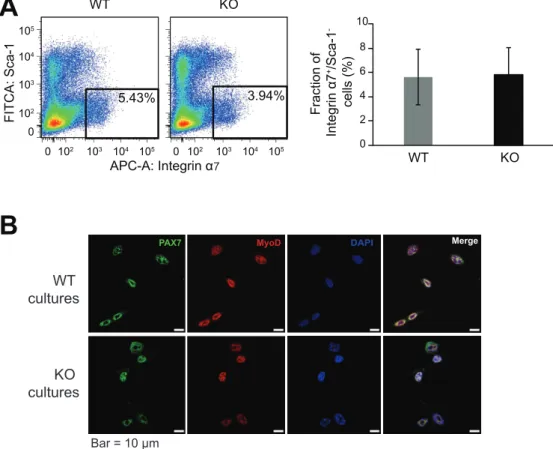
Mouse satellite cells derived from hind limbs of WT and IL-1KO mice, 6-10 weeks old (five and six independent isolation experiments, for a total of n=6-10 IL-1KO and n=16 WT mice, respectively). Cells were isolated by the fluorescent-activated cell sorting (FACS) method as previously described (Yi & Rossi, 2011). Briefly, the skeletal muscle tissues resected from hind limbs were minced and digested with 0.2% collagenase (Wako, Tokyo, Japan) and 0.01% DNase I (Sigma-Aldrich, St. Louis, MO, USA) for 1 hour at 37°C. They were then filtered through a 70 µm cell strainer (Becton, Dickinson and Company, Franklin Lakes, NJ, USA) and centrifuged at 700 g for 20 min. The pellets were resuspended in 1 ml of staining solutions, which had been treated with Fc receptor blocking solution (TruStain fcX, 1:20 in FACS buffer; Biolegend, San Diego, CA, USA) for 10 min. The cells were labeled with the following monoclonal antibodies (all used at 1:20 dilution): Pacific Blue-conjugated anti-CD45 (clone 30-F11; BioLegend), FITC-conjugated anti-Sca-1 (clone D7; BioLegend), PE/Cy7-conjugated anti-CD31 (clone 1A8; BioLegend), PE-PE/Cy7-conjugated anti-CD34 (clone MEC14.7; BioLegend), and APC-conjugated anti-integrin-α7 (clone 334908; R&D systems, Minneapolis, MN, USA) antibodies. Following 45 min incubation on ice, the cell suspension was washed with staining solution and centrifuged for 5 minutes at 700 x g twice. Satellite cells were defined as single live mononuclear CD45-Sca-1-CD31-CD34-Integrin-α7+ cells. These cells were sorted and analyzed with a BD FACS ARIA II flow cytometer (Becton, Dickinson & Co.). The cells were grown on Matrigel (Becton, Dickinson & Co.) coated plates at 37°C with 5% CO2 and were supplemented with growth medium (GM); Dulbecco's
Modified Eagle Medium (DMEM) with 20% fetal bovine serum, 10 ng/mL basic fibroblast growth factor (ReproCELL, Kanagawa, Japan), 1% chicken embryo extract (United States Biological, Salem, MA, USA), 30 µg/ml penicillin, and 100 µg/ml streptomycin (Wako). After expanding the satellite cells for 3-4 passages, the obtained myoblasts were stocked in CellBanker (Zenoaq) and the experimental myoblast stocks were used for cell culture experiments. In vitro experiments were performed on replicate cultures of the same cell isolation stocks in three independent experiments. For differentiation experiments, the myoblasts
were grown to confluence for 3-4 days and then myogenic differentiation was induced (Day 0) by changing to differentiation medium (DM); DMEM with 5% horse serum, 30 µg/ml penicillin, and 100 µg/ml streptomycin. Recombinant IL-1α (#400-ML) and IL-1β (#401-ML) were obtained from R&D Systems.
3.2.4 Quantitative real-time polymerase chain reaction (qPCR)
Total RNA was extracted by Tri reagent (Molecular Research Center Inc., Cincinnati, OH, USA) from cell cultures or TA tissues with homogenization using Micro Smash MS-100R (Takara Tomy, Tokyo, Japan). cDNA was synthesized using a Transcriptor First Strand cDNA Synthesis Kit with oligo-dT primers (Roche, Basel, Switzerland). Then, qPCR was performed with Lightcycler 480 SYBR Green reagent mixed with the desired primers and measured with a Lightcycler 480 II instrument. The relative mRNA expression levels of the target genes were calculated using Glyceraldehyde 3-phosphate dehydrogenase (GAPDH) as a reference gene. Primer sequences were as follows;
Primers Sequences
Myogenin Forward 5’AGCGGCTGCCTAAAGTGGAGA Reverse 5’GGAGGCGCTGTGGGAGTTG MyoD Forward 5’GCCGCCTGAGCAAAGTGAAT
Reverse 5’GCGGTCCAGGTGCGTAGAAG Myomaker Forward 5’ATCGCTACCAAGAGGCGTT
Reverse 5’CACAGCACAGACAAACCAGG IL-1α Forward 5’ GACCGACCTTTTCTTCTG
Reverse 5’ AGGTGCACCCGACTTTGTTCTT IL-1β Forward 5’ GCACCTTCTTTTCCTTCATCTTTG
Reverse 5’ GTTGTTCATCTCGGAGCCTGT IL-1R1 Forward 5’-TGGAGGCAAAGCAAACTGTAAG
Reverse 5’-TCGGGGCACTGGGAACTC IL-1R2 Forward 5’-GCGCTGGCACCTCTGTCTCT
Reverse 5’-TAGTTTGGGCTGGAAGGGAAGTC GAPDH Forward 5’GGAGAAACCTGCCAAGTATGA
3.2.5 Cytokine measurement by Bio-Plex assay
Cell culture media and tissue extracts from TA homogenized with lysis buffer were subjected to this cytokine measurement assay. The cytokine concentrations were determined with a Bio-Plex 200 system and a Bio-Plex pro wash station (Bio-Rad, Hercules, CA, USA) according to the manufacturer’s guidelines. Briefly, assay beads conjugated with capture antibodies were incubated on a 96-well plate (20 folds bead stocks dilution for 50 µl/well) and then washed with washing buffer (containing 0.1% BSA, 0.1% Tween 20 and 0.2% sodium azidein PBS). Next, the standard dilution series and cell lysate or culture medium samples (100-150 µl/well) were incubated for two hours, then washed three times. Following incubation with the detection antibody (IL-6; 150 ng/ml and CXCL1 200 ng/ml at 25 µl/well) for 30 minutes and fluorescent conjugated Streptavidin-phycoerythrin (500 folds stock for 50 µl/well) for 10 minutes, respectively, the samples were separated and then washed three times. Finally, 70µl/well of the assay buffer (containing 0.1% BSA and 0.2% sodium azide in PBS) was added prior to cytokine level measurements. Data were obtained employing Bio-Plex Manager Software 5.0 (Bio-Rad). The antibodies used for measuring cytokines were as follows; mouse chemokine (C-X-C motif) ligand 1 (CXCL1) capture antibody (#840325) and detection antibody (#840326), and mouse IL-6 capture antibody (#840171) and detection antibody (#840172) (R&D Systems).
3.2.6 Western Blot Analysis
Lysis buffer (30 mM Tris, 100 mM sodium chloride, 1 mM ethylenediaminetetraacetic acid (EDTA), 1% TritonX-100, 2.5 mM sodium fluoride, 2 mM sodium polyphosphate, 1 mM sodium orthovanadate, 1 mM phenylmethylsulfonyl fluoride, 10 µg/ml aprotinin, 1 µg/ml pepstatin, and 5 µg/ml leupeptin) was added to the cultured myoblasts to extract whole cell lysates. The total protein concentration was measured using a BCA Protein Assay Kit (Thermo Fisher Scientific). Then, 30 µg of protein per well were loaded for 10% SDS polyacrylamide gel electrophoresis and transferred to Immobilon-P polyvinylidene difluoride membranes (Merck, Kenilworth, NJ, USA). The protein-transferred membranes were blocked with 5% BSA in Tris-buffered saline with 0.1% Tween-20 buffer, then incubated at 4°C overnight with 1:1000 diluted primary antibodies, as follows; mouse monoclonal anti-skeletal myosin heavy chain (MyHC) fast (#M4276, Sigma-Aldrich, St. Louis, MO, USA), mouse monoclonal MyHC slow (#M8421, Sigma-Aldrich), mouse monoclonal
anti-CDC42 (#610928, Becton, Dickinson & Co.), rabbit polyclonal anti-N-WASP (H-100) (#sc20770, Santa Cruz Biotechnologies, Dallas, TX, USA), rabbit monoclonal anti-GAPDH (#2118), rabbit polyclonal anti-Phospho-PAK1 (Ser144)/PAK2 (Ser141) (#2606), rabbit polyclonal anti-Phospho-PAK1 (Thr423)/PAK2 (Thr402) (#2601), rabbit polyclonal anti-PAK1/2/3 (#2604), rabbit monoclonal anti-Phospho-NF-κB p65 (#3033), rabbit monoclonal anti-NF-κB p65 (#8242), rabbit polyclonal anti-Phospho-NF-anti-NF-κB2 p100 (#4810), and rabbit polyclonal anti-NF-κB2 p100/p52 (#4882) antibodies (Cell Signaling, Beverly, MA, USA). The membranes were washed with Tris-buffered saline with 0.1% Tween-20 buffer, followed by incubation at room temperature for one hour with 1: 10,000 dilutions of secondary antibodies, as follows; stabilized peroxidase conjugated goat anti-mouse (#32430) or stabilized peroxidase conjugated goat anti-rabbit (#32460) (Thermo Fisher Scientific) antibodies. Then, a signal was produced employing chemiluminescence reagents, SuperSignal West Femto Maximum Sensitivity Substrate (Thermo Fisher Scientific), and detected the relative density of chemiluminescent signals for further analysis with Image Quant TL systems (GE Healthcare, Chalfont St. Giles, UK).
3.2.7 Myoblast Proliferation assay
Myoblasts obtained from WT and IL-1KO mice were seeded at 1.0 × 10⁴ cells per well on 96-well plates at four replications and supplemented with growth medium. All wells were replaced with growth medium with or without 2-2000 pg/ml of recombinant IL-1α or IL-1β. Cell proliferation was measured at Day0, Day1, and Day3 by colorimetric WST-1 assay, as follows (Alexsandra da Silva Neto Trajano et al., 2016). The WST-1 solution at 5 mg/ml (Dojindo) was added to cell cultures at a ratio of 1:10, followed by a 4-hour incubation at 37°C with 5% CO2. The absorbance of samples against a control
blank (medium only) was then measured at 450 nm with the reference wavelength at 650 nm using a Spectramax M5-LV (Molecular Devices, Sunnyvale, CA, USA).
3.2.8 Cytological Analyses
Cells seeded onto glass on cover-slips were collected at 8 hours after seeding (for satellite cell staining) and 2 days after plating, as well as the day begin differentiation (replacement of GM with DM), 2 and 4 days after differentiation, and then fixed with 1% PFA and 0.1% Triton X-100 in PBS. These cells were blocked with 5% calf serum in PBS and then incubated with
rabbit anti-PAX7 IgG (#ab34360) and mouse anti-MyoD IgG (#sc377460, Santa Cruz Biotechnologies) for satellite cell staining or rabbit polyclonal anti-desmin IgG (#4024) (1:200 dilutions; Cell Signaling) diluted in 1% BSA in PBS at 4°C overnight. The cells were washed with PBS and counterstained with Alexa Fluor 488 donkey-anti rabbit IgG (#A21206, Thermo Fisher Scientific), Alexa Fluor 555 donkey-anti mouse IgG (#A31570, Thermo Fisher Scientific) and DAPI (Dojindo) (1:1000 dilution) diluted in 1% BSA in PBS at room temperature for an hour and then mounted in Vectashield (Vector Laboratories, Burlingame, CA, USA). All samples were examined with a confocal microscope (Olympus FV1000; Olympus) equipped with an oil-immersion objective lens (UApo/340 40X NA 1.35) and five random fields per sample were photographed using FV10-ASW 1.7 viewer software (Olympus, Japan). Fiji ImageJ analysis of the fusion index was conducted to determine the percentage of nuclei contained in desmin-positive myotubes (Millay et al., 2013; Teng et al., 2015) with three independent non-blinded observations. For observing actin cytoskeletal structures, filamentous (F)-actin staining was performed with TRITC-phalloidin at RT for 1 hr. Twelve-bit images of five random fields were taken with the FV1000 confocal microscope and FV10-ASW1.7 software (Olympus) equipped with the aforementioned oil-immersion objective lens or the SP8 confocal microscope and LAS X software (Leica) equipped with another oil-immersion objective lens (HC PL APO CS2 63x, NA 1.40), and processed with Fiji ImageJ. Approximately 50-80 cells were observed in each experiment, and representative images are shown in Figure 15B as maximum projections of z-stack images from three independent experiments.
3.2.9 Live-cell imaging of actin dynamics
WT and IL-1KO myoblasts were seeded in matrigel–coated glass-bottom dishes (No. 1S, thickness: 0.16–0.19 mm; Matsunami Glass) and then transfected with Lifeact-Venus expression plasmid using Lipofectamine 3000 (Thermo Fisher Scientific) according to the manufacturer’s instructions. On the following day, the cells were immersed in imaging buffer consisting of 150 mM NaCl, 5 mM KCl, 2 mM CaCl2, 1 mM MgCl2, 10 mM HEPES-NaOH (pH 7.4) and 5.5
mM D-glucose. IL-1β for acute stimulation was diluted in the imaging buffer at 2,000 pg/ml and added to the cells by perfusion. Images were acquired with an SP8 confocal microscope (Leica) equipped with a white-light pulsed laser and an oil-immersion objective lens (HC PL APO CS2 63x, NA 1.40) at ~30°C with a
lens heater (TOKAI HIT). Excitation was at 498 nm and fluorescence was acquired at 503–650 nm. Twelve-bit images were acquired with LAS X software (Leica) as a t-series at intervals of 10 s with the same acquisition settings. Image analysis and processing were performed with Fiji ImageJ with StackReg plug-in for image alignment. Images shown in figures are maximum projections of six successive frames and were median-filtered. For quantification, mean fluorescent intensities of LifeAct-venus within a cell was obtained, followed by calculation of the difference in intensities from a define period (as indicated in the Figure Legends). Linear fitting and statistical analyses were performed using ORIGIN (Origin Lab).
3.2.10 Statistical analysis
The statistical analyses were performed using SPSS statistics software version 21 (IBM, Chicago, IL, USA). All experimental data are presented as means ± SE (standard error of the mean). The statistically significant of differences was determined by multi-factor ANOVA with a Bonferroni post-hoc comparison test, Tukey’s multiple comparison and a chi-square analysis was used to determine the difference of the distribution. For live-cell imaging analysis, F-test was used. Differences were considered to be statistically significant at a P-value less than 0.05.
Chapter 4: Study results
4.1 Results
IL-1KO mice exhibited delayed muscle regeneration after cardiotoxin injection-induced injury
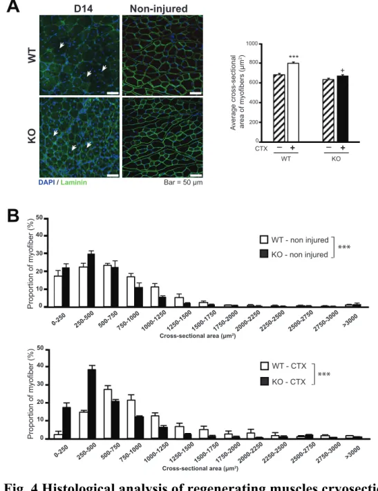
The TA muscles of WT and IL-1KO mice injected with CTX at 3, 7 and 14 day-post-injury were histochemically analyzed (Fig. 3-5). The H&E sections revealed myofiber necrosis on Day 3 after CTX injection in both WT and IL-1KO mice. The necrotizing muscle fibers were characterized by a loss of the usual polygonal shape and an increase in the interstitial space between fibers due to the destructive action of CTX. The major distinction between WT and IL-1KO mice on Day 3 after CTX injection was inflammatory cell infiltration. In WT mice, numerous mononuclear cells had been recruited to the interstitial spaces, while there was less cell infiltration among necrotic myofibers in IL-1KO mice. On Day 7 after CTX-injection, the damaged areas had mostly been replaced with newly-formed myofibers characterized by centrally-localized nuclei in the TA of WT mice, but necrotic fibers remained in those of IL-1KO mice. Consistent with these qualitative observations, morphometrical analysis on Day 14 after CTX injury clearly demonstrated that the TA in IL-1KO mice exhibited significantly smaller average cross-sectional areas of regenerating myofibers, along with a different distribution (towards a shift to smaller) of myofiber size compared with those in WT mice (Fig. 4)
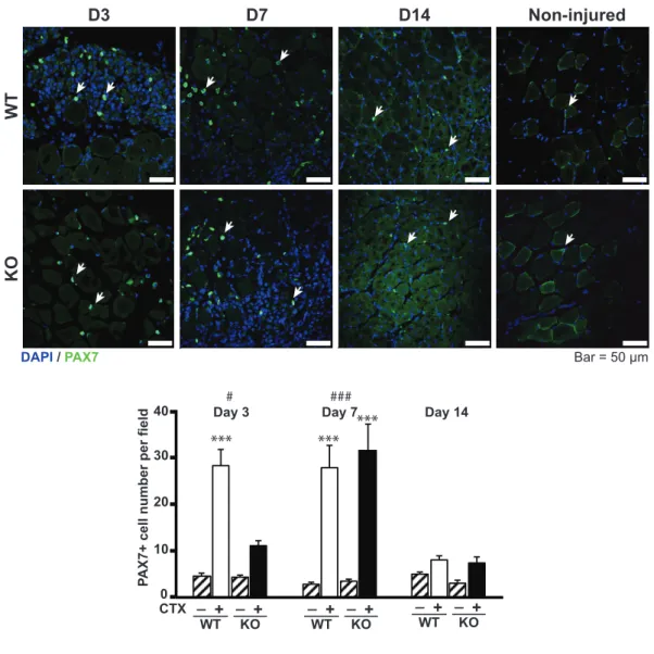
To assess the regeneration competence of CTX-injected TA in both mouse models, I investigated myogenic indicators within the TA. Since satellite cells are well recognized for their skeletal muscle regenerating capacity, I performed immunofluorescent staining for PAX7, a satellite cell marker, to evaluate the temporal pattern of PAX7-positive cell numbers (Fig. 5) within TA during the muscle regeneration process. On Day 3 after CTX-injection, significantly greater numbers of PAX7-positive cells were detected in the TA of WT mice (Fig. 5, upper panels) than in those of IL-1KO mice (Fig. 5, lower
panels), while PAX7-positive cell numbers were similar on Day 7 (Fig. 5). On Day 14, in both WT and IL-1KO mice, numbers of PAX7-positive cells in the
CTX-injected TA decreased to counts similar to those in non-injected control TA (Figs. 5). The satellite cell activation pattern was obtained by immunostaining skeletal muscle sections from WT and IL-1KO mice at Day3 post-injection.
Although both strain had a different number of satellite cells (Fig.5), most PAX7-positive cells co-expressed MyoD, which was indicative of them being activated satellite cells. It should be noted that the number of satellite cells per an equivalent amount of muscle mass obtained from IL-1KO mice under intact conditions were comparable to those from WT mice, as determined by FACS analysis described in detail below.
The real time PCR analysis for myogenic differentiation markers further demonstrated the delayed regeneration status in the TA of IL-1KO mice (Fig.
6A). Namely, mRNA upregulations of myogenin, MyoD and myomaker, a
muscle-specific fusogenic factor, were detected in the TA from the CTX-injected side on Day 3, particularly in WT mice, while CTX-induced upregulations of these mRNAs in the TA of IL-1KO mice were low.
Furthermore, the Bio-Plex analysis for pro-inflammatory chemokines/interleukins in the homogenates of TA muscles indicated that both IL-6 and CXCL1 were significantly higher on the CTX-injured side than in TA from the control side, but these CTX-induced productions of pro-inflammatory factors in the TA of IL-1KO mice were significantly blunted (Fig. 6B).
IL-1KO myoblasts displayed delayed proliferation and differentiation, both of which were normalized by exogenous IL-1β administration
As described above, IL-1KO mice exhibited clearly delayed muscle regeneration after CTX injection, which was characterized by i) delayed infiltration of immune cells accompanied by inadequate production of pro-inflammatory factors (CXCL1 and IL-6), and ii) delayed appearance of PAX7-positive cells along with low mRNA expressions of myogenic differentiation markers in the regenerating muscles.
Given that regeneration of damaged muscle in vivo is a highly complex process involving a wide array of cytokines, chemokines and interleukins being secreted by various residential and recruited cell types as well as injured myofibers within the regenerative niche, relatively little is known about the direct role(s) of IL-1s in myoblast function. To determine whether IL-1 deficiency is directly responsible for fundamental properties of satellite cells and myoblasts, a series of cell culture experiments was performed using myoblasts expanded from primary satellite cells obtained from IL-1KO and WT mice by the FACS technique. After isolating satellite cells from hind limbs of WT and IL-1KO mice
by FACS, these satellite cells were cultured and expanded for 3-4 passages to obtain adequate numbers of myoblasts, which could be used for in vitro experiments. The WT and IL1-KO myoblasts both expressed PAX7 and MyoD by immunofluorescent staining (Fig. 7B). Given that the number of satellite cells per an equivalent amount of skeletal muscle obtained from IL-1KO mice under intact conditions were comparable to those from WT mice (Figs. 7A), these results imply that the in vivo behaviors of IL-1KO satellite cells under normal intact conditions to be similar, whereas their expansion after CTX-induced injury was significantly delayed (Fig. 5).
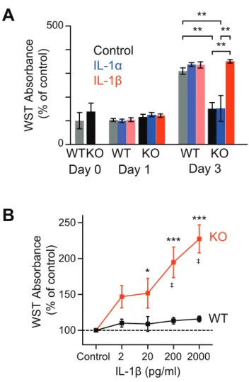
As shown in Figure 8, the cell proliferation assay using WST-1 demonstrated that myoblasts obtained from IL-1KO mice (IL-1KO myoblasts) showed significantly slower growth than those from WT-mice (WT myoblasts) during the time-course to Day3 (Fig. 8A, black bars). However, exogenous administration of 1β, but not α, restored the delayed proliferation of IL-1KO myoblasts to levels similar to those observed in WT myoblasts at Day 3 (Fig. 8A, red bar) and the restorative effect of IL-1β was exerted in a dose-dependent manner (Fig. 8B, red line). In contrast, the exogenous addition of either IL-1β or IL-1α failed to evoke further augmentation of the proliferation of WT myoblasts (Figs. 8A, WT and Fig. 8B, black line). It should be noted that WT myoblasts reached confluence at Day3 and WST-1 absorbance had not further increased by Day 5, whereas IL-1KO myoblasts eventually reached confluence on Day 5 even in the absence of exogenous IL-1β.
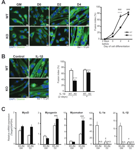
The IL-1KO myoblasts also manifested their delayed myogenesis in terms of myotube formation as assessed by fusion index quantification based on immunofluorescent staining of desmin, a myogenic differentiation marker protein (Fig. 9A and 9B), even though IL-1KO myoblasts eventually forming myotubes reached a fusion status similar to that in WT myoblasts (Fig. 9A, right graph). Despite an apparently delayed myogenic fusion process on Day 2 after the start of differentiation (Fig.9A), RT-PCR analysis revealed that IL-1KO cells adequately, even highly, expressed key myogenic transcription factors (myogenin and MyoD) as well as Myomaker (Millay et al., 2013) (Fig. 9C). Also, both IL-1α and IL-1β were detected in WT myoblasts, and their expression levels were decreased on Day2 of differentiation (Fig. 9C). It should be noted that in this set of myogenic differentiation experiments, inoculation with 1.5 times the number of IL-1KO myoblasts (compared to WT) was required to compensate for their poor growth and achieve 80-90% confluence on Day 0. Interestingly, exogenous
administration experiments revealed that the delayed myotube formation of IL-1KO cells was restored, showing essentially the same fusion index as that of WT cells, on Day2 in the continuous presence of a relatively high concentration of IL-1β (Fig. 9B).
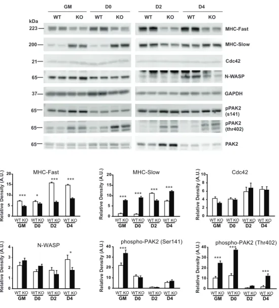
To explore possible derangements in the intracellular signaling intermediates in IL-1KO cells, I performed western blot analysis using the culture cells and found that the phosphorylation status of group I PAK proteins, direct effectors of Cdc42 and Rac1 involving myogenic regulation (Joseph et al., 2017), differed markedly between IL-1KO and WT cells. Namely, in IL-1KO cells, persistently higher phosphorylation of PAK2 (especially at Thr402) than in WT cells was observed (Fig. 10). Moreover, the gradual decrease of N-WASP levels in IL-1KO cells correlated with a tendency for delayed differentiation, which was obvious on Days 2 and 4 after the start of differentiation, as compared to WT cells, while Cdc42 protein levels were similar (Fig. 10). Amounts of fast skeletal myosin heavy chain (MyHC) were highly upregulated upon myogenic differentiation, especially in WT cells, while those in IL-1KO cells were markedly compromised. Interestingly, IL-1KO cells expressed slight but noticeable levels of slow MyHC even on Day 0 but their amounts in both WT and IL-1KO cells had become similar by Day 2 and remained so on Day 4.
IL-1KO myocytes displayed impaired intracellular signaling and CXCL1/IL6 secretory ability in response to IL-1β along with low expression of IL-1 receptors
Given that IL-1β normalized the delayed growth and myogenic fusion of cellular phenotypes of IL-1KO myoblasts, I further examined the cellular responses of myoblasts to 1β stimulation. While a high concentration of IL-1β (2,000 pg/ml) induced phosphorylation of NF-κB involving the canonical (p65) and the non-canonical (p100) pathway in both IL-1KO and WT myoblasts, these IL-1β responses were more obviously detected in the WT myoblasts (Fig.
11). On the other hand, IL-1β failed to induce further phosphorylation of PAK2
(Thr402) in either of WT and IL-1KO myoblasts under these experimental conditions (Fig. 11). Consistent with the compromised IL-1β-dependent NF-κB phosphorylation in IL-1KO myoblasts, RT-PCR analysis revealed that IL-1KO myoblasts expressed significantly lower levels of both IL-1R1 (a positive mediator of IL-1 signaling cascades) and IL-1R2 (a decoy receptor for IL-1) than
those in WT myoblasts (Fig. 12A). On the other hand, WT myoblasts highly expressed both IL-1R1 and IL-1R2 mRNAs, but they were both markedly decreased upon differentiation at Day2 to levels comparable to those in IL-1KO cells. Interestingly, the differentiation-dependent diminishment of IL-1R1expression in WT cells was not induced when the cells were cultured in the presence of IL-1β (2000 pg/mL), while IL-1KO cells failed to restore expression of either IL-1R1 and IL-1R2 following IL-1β treatment (Fig. 12A).
Then, I examined the effects of exogenous IL-1β on secretion of CXCL1 and IL-6 in IL-1KO and WT myotubes on Day 6 of differentiation. As shown in Fig. 12B, exogenous administration of a relatively low concentration of IL-1β (20 pg/mL) remarkably stimulated not only CXCL1 but also IL-6 secretion. Importantly, IL-1KO myotubes exhibited a significantly reduced capacity to secrete these myokines in response to IL-1β.
IL-1KO myoblasts displayed impaired actin remodeling activity and its restoration in response to IL-1β
Finally, the actin remodeling activity of myoblasts in response to IL-1β stimulation was assessed by live-imaging of F-actin with LifeAct-Venus (Hatakeyama, Nakahata, Yarimizu, & Kanzaki, 2017), since multiple regulators of the actin cytoskeleton including PAK 2 and N-WASP appeared to be altered in the IL-1KO myoblasts (Figs. 10 and 11). As shown in the representative time lapse images under basal conditions, WT myoblasts progressively potentiated actin intensity with typical ruffling formation (Fig. 13A) while IL-1KO myoblasts exhibited relatively stable formation of micro-spikes with a steady state of actin remodeling activity (Figs. 14A). These dissimilarities presumably reflected differences in the amounts and/or phosphorylation status of PAK2 and N-WASP between WT and IL-1KO myoblasts. Importantly, administration of IL-1β markedly restored actin dynamics in IL-1KO myoblasts within several minutes as evidenced by vigorous remodeling of actin filaments especially underneath the plasma membrane, promoting membrane ruffling (Figs. 14B). Interestingly, such IL-1β-dependent actin remodeling activities in IL-1KO myoblasts persisted even after chronic (overnight) treatment with a relatively high concentration of IL-1β, and additional IL-1β further enhanced the remodeling activities along with membrane ruffling (Figs. 15A). With F-actin staining by phalloidin, I observed not only membrane ruffling (Fig. 15B, right panel, arrowheads) but also
intercellular communications of F-actin (arrows) after overnight treatment with IL-1β in IL-1KO myoblasts, supporting the observations that exogenous administration of IL-1β effectively restores intercellular fusion events.
4.2 Discussion
Because of the potent pro-inflammatory effects exerted by IL1α/β in other tissues (Rider et al., 2011), it is reasonable to speculate that the delayed infiltration of immune cells into damaged muscle tissues displayed by IL-1KO mice may have played a role in the delayed muscle regeneration (Figs. 3 – 6). These data are consistent with the concept that an appropriate inflammatory response is a prerequisite for efficient regeneration of damaged tissues (Tidball & Villalta, 2010; Zhang et al., 2013). On the other hand, another key finding reported herein is the unexpected role of endogenous IL-1α/β to promote fundamental properties of satellite cells and/or myoblasts in vitro (Figs. 8 – 15). This effect would result in delayed regeneration as seen in the IL-1KO mice after CTX-induced injury in vivo (Figs. 4 and 5). While caution must be exercised when reconciling the in vivo data with the in vitro data regarding impaired phenotypes of IL-1KO cells, the present results directly confirm the conclusion that IL-1α/β are positively involved in the regeneration process of injured skeletal muscles. They appear to coordinate the initial inflammatory environments required to stem cell activation. These studies provide insights into the specific roles of autocrine and/or intracrine IL-1α/β in determining the properties of satellite cells after injury.
Muscle regeneration includes an initial inflammatory phase and a regenerative phase. After injury has occurred, M1-type macrophages are promptly recruited into the injured muscles and produce IL-1, IL-6 and tumor necrosis factor α (TNF-α), which contribute to stimulating the proliferation of satellite cells in a coordinated fashion as evidenced by an increase in PAX7-positive cells (Heredia et al., 2013; Tidball & Villalta, 2010; Zhang et al., 2013). Based on the in vivo data presented in Figure 3 to Figure 6, IL-1 molecules appear to be involved in the initial inflammatory responses including local productions of IL-6 and CXCL1, which are highly likely to contribute to triggering the stimulation of satellite cells to undergo proliferation and migration within the regenerating muscle tissues (Griffin, Apponi, Long, & Pavlath, 2010; Nedachi, Hatakeyama, Kono, Sato, & Kanzaki, 2009; Sachidanandan, Sambasivan, & Dhawan, 2002; Serrano et al., 2008). Consistent with this notion, other models of skeletal muscle injury have also revealed the importance of early phase inflammatory cytokine secretion in supporting regeneration. For example, crush injury reportedly increased the expression of IL-1β and IL-6 within a few hours of injury (Voss et al., 2017), and barium chloride injury increased these
inflammatory factors within a day and peaked at 5 days after injury (Otis et al., 2014). While the subsequent regenerative phase, characterized by the switching of macrophages from the M1- to M2-types, supports myogenesis of the activated myoblasts (Arnold et al., 2007; Ruffell et al., 2009), the in vivo data on this state do not reveal whether the IL-1-deficiency also influenced this later phase of the muscle regeneration process. However, it is noteworthy that another pro-inflammatory factor, as shown in TNF-α-deficient mice, reportedly did not differ in terms of either the initial inflammatory responses or patterns of muscle regeneration (i.e., leukocyte recruitment, activation and fusion of myoblasts) (Collins & Grounds, 2001). Thus, these findings demonstrated IL-1 to be an important factor serving as the initial pro-inflammatory trigger for accomplishing efficient regeneration of the damaged muscle. Future studies are warranted to precisely elucidate the in vivo roles of IL-1α/β as the key coordinators of the highly complex regenerative niche in injured muscle tissues.
The in vitro data presented in this study strongly imply that myoblasts, as well as perhaps satellite cells, are intrinsically regulated by endogenous IL-1α/β that can serve not only as autocrine factors exerting actions mediated through cell surface receptors (Weber, Wasiliew, & Kracht, 2010) but also as intracrine/intracellular factors, especially in the case of IL-1α (Di Paolo & Shayakhmetov, 2016; Werman et al., 2004). While several lines of evidence have demonstrated that IL-1 exerts pleiotropic effects on proliferation, differentiation and apoptosis of myoblasts including C2C12 cells and human myogenic cells (Authier et al., 1999; Broussard et al., 2004; Grabiec et al., 2013; Otis et al., 2014), the findings in this study obtained from IL-1KO satellite cells provide direct evidence that IL-1 molecules are intimately engaged in the regulation of myoblast competency. An important observation in the experiments using IL-1KO and WT myoblasts was that neither the proliferative nor the differentiating potency of WT myoblasts was additionally enhanced by exogenous 1 In contrast, IL-1-deficiency resulted in a delayed growth and differentiation phenotype, but these impairments were efficiently reversed by IL-1β, which appeared to be at least in part mediated via IL-1β-dependent stimulation of actin remodeling activities (Fig.
14 and 15). Given that WT myoblasts expressed both IL-1α and IL-1β mRNA,
though they both decreased upon differentiation, endogenous IL-1 molecules apparently contribute to maintaining myoblast properties. Future studies are warranted to clarify the precise mechanism of how endogenous IL-1 molecules participate in the regulation of myoblast growth and differentiation. Meanwhile 1β, but not 1α, effectively normalized these impaired phenotypes of
IL-1KO myoblasts, observations similar to those in other cell types (Horai et al., 1998). Although the functional discrepancies between IL-1β and IL-1α await further clarification, alterations in their binding properties for other functional proteins including adhesion molecules (Sahni, Guo, Sahni, & Francis, 2004) might be involved in the differences in their potencies for inducing dynamic actin remodeling. In this regard, several lines of evidence demonstrate the importance of extracellular matrix on myogenesis (Grabiec et al., 2013; Osses & Brandan, 2002).
A puzzling aspect of the exogenous IL-1 actions observed herein was that despite the highly potent restorative effects of exogenous IL-1β on the delayed phenotypes of IL-1KO myoblasts even with a low expression of IL-1R1 (Fig.
12A). On the other hand, the IL-1KO myotubes exhibited significantly lower
responses in terms of 6 and CXCL1 secretions induced by exogenous IL-1β administration than WT myotubes (Fig. 12B). These results strongly suggest that endogenous IL-1 molecules have a complex involvement in the maintenance of fundamental myocyte function, including the maintenance of susceptibility and cellular responses to IL-1 that could only be revealed when both IL-1α/β were genetically deleted. This notion is further supported by biochemical data showing that IL-1KO myoblasts exhibited clearly different profiles of muscle protein abundance (MyHC types) and the phosphorylation status of the key myogenic signaling intermediates (PAK2) (Joseph et al., 2017), as compared with those in the WT-myoblasts, even prior to differentiation (Fig. 10). Moreover, the highly phosphorylated state of PAK2 observed in the IL-1KO myoblasts displaying apparent delayed myogenic fusion (Fig. 10), suggest IL-1 molecules to be essential for maintaining the intact PAK-related signaling cascades and myogenic differentiation. These results thus support those of a recent study showing that group I PAKs are involved in muscle regeneration (Joseph et al., 2017) but also extend this notion further, indicating that proper regulation, not just enhanced phosphorylation of PAK2, is required for myoblast function and muscle regeneration.
The results also provide hypothetical mechanistic insights into the impaired myogenic phenotypes caused by IL-1-deficiency. Namely, the failure of myogenesis is not attributable to the myogenic transcriptional program since MyoD and myogenin were adequately upregulated in IL-1KO cells (Fig. 9C), instead appearing to be due to the dysregulation of cellular fusion events most likely resulting from malfunction of actin dynamics (Fig. 14 and 15) in accordance with derangements in Rac1 and/or Cdc42-related function involving
their downstream effectors, PAK2 and N-WASP (Fig. 10). Consistent with this hypothesis, N-WASP-deficient myoblasts reportedly exhibited discernible defects in their fusion events with no obvious interference with the myogenic transcriptional program (Gruenbaum-Cohen et al., 2012). Furthermore, IL-1 has been shown to exert pleiotropic actions mediated through the Rho-family of small GTPases including Rac1 and Cdc42 in other cell types (Lee & Heur, 2014; Windheim & Hansen, 2014). Thus, although the precise molecular mechanisms involved in the delayed myotube formation observed in IL-1KO myocytes remain to be further investigated, the results from this study strongly suggest that the aforementioned derangements in regulatory systems relating to actin dynamics are involved in the defects of IL-1-KO myocytes since exogenous IL-1β effectively restored dynamic actin remodeling properties (Fig. 14 and 15). Given that various intracellular signaling intermediates stimulated by IL-1R1, including TNFR-associated factors (TRAFs) and NFκB, have been reportedly involved in regulating the proliferation and differentiation of myoblasts (Mueck et al., 2011), the compromised phenotype observed in the IL-1KO cells is highly likely to have resulted from combinatorial defects among these signaling cascades and aberrant actin regulatory systems. Since myomaker, another key fusogenic factor (Millay et al., 2013), was even highly expressed in the IL-1KO myoblasts, there is presumably a further layer of complexity that must be unraveled in a future study. In addition, the detailed mechanisms underlying the disturbances in the signaling cascades involving the Rho-family small GTPases resulting from IL-1-deficiency await further clarification. Considering the variance of satellite cell properties (Deasy et al., 2007), as well as of immune responses stemming from sex and age (Klein & Flanagan, 2016), it should be noted that relatively young male mice (6-10 weeks) were used in this study, and the possible influence of IL-1α/β on these mice needs to be addressed in a future study.
In summary, I have clearly demonstrated that endogenous IL-1 molecules are positively involved in the muscle regeneration process by experimentally showing that IL-1-deficiency resulted in delayed infiltration of immune cells along with inadequate local production of pro-inflammatory factors and delayed appearance of satellite cells within the damaged muscle tissues. This study also found that IL-1-deficient myoblasts displayed delayed growth and myogenic fusion capabilities in vitro, both of which were efficiently restored to normal by exogenous IL-1β.