Title
Proteasome Activity and Biological Properties of Normal Prion
Protein: A Comparison between Young and Aged Cattle( 本文
(Fulltext) )
Author(s)
YOSHIOKA, Yumi; ISHIGURO, Naotaka; INOSHIMA, Yasuo
Citation
[Journal of Veterinary Medical Science] vol.[72] no.[12]
p.[1583]-[1587]
Issue Date
2010
Rights
The Japanese Society of Veterinary Science (社団法人日本獣医
学会)
Version
出版社版 (publisher version) postprint
URL
http://hdl.handle.net/20.500.12099/39513
Proteasome Activity and Biological Properties of Normal Prion Protein:
A Comparison between Young and Aged Cattle
Yumi YOSHIOKA1), Naotaka ISHIGURO1)* and Yasuo INOSHIMA1)
1)Laboratory of Food and Environmental Hygiene, Department of Veterinary Medicine, Faculty of Applied Biological Sciences, Gifu
University, 1–1 Yanagido, Gifu 501–1193, Japan
(Received 10 April 2010/Accepted 18 July 2010/Published online in J-STAGE 2 August 2010)
ABSTRACT. Atypical bovine spongiform encephalopathy (atypical BSE) has recently been identified in several countries including Japan.
Most cases of atypical BSE have been reported in cattle older than 8 years of age. To clarify the association between age and occurrence of atypical BSE, we investigated both the physiological properties and amount of cellular prion protein (PrPC) in brain homogenates from
young and aged cattle by enzyme-linked immunosorbent assay and immunoblotting. The amount of PrPC in the brain homogenates was
not significantly different between young and aged cattle, but the amount in the detergent-insoluble fraction in the aged cattle was sig-nificantly higher than that of young cattle. Significant differences were observed in neither of the glycosylation forms nor in proteinase K sensitivity in young and aged cattle. Age-related changes included deposition of lipofuscin pigment and a decrease of 33% in pro-teasome activity in the brains of aged cattle compared to that of young cattle.
KEYWORDS: atypical BSE, brain homogenate, cattle, prion, proteasome.
J. Vet. Med. Sci. 72(12): 1583–1587, 2010
Prion diseases, also known as transmissible spongiform encephalopathies (TSE), are a group of fatal neurodegener-ative disorders caused by accumulation of the proteinase K (PK)-resistant pathogenic isoform (PrPres) in the host
cellu-lar prion protein (PrPC) [17]. TSE, which have been found
in humans and other mammalian species, include bovine spongiform encephalopathies (BSE) in cattle and scrapie in sheep and goats. Only one strain of BSE prion was origi-nally thought to exist, in contrast to the many different scrapie strains found around the world [3]. Recently, how-ever, a strain of atypical BSE, which is different from the classical BSE, was independently reported in Italy [5], France [1], and Japan [21]. Atypical BSE is classified into two different types: H-type, characterized by a higher molecular mass associated with the unglycosylated protein band, and L-type or a bovine amyloidotic spongiform encephalopathy (BASE), characterized by an unglycosy-lated protein band with a lower molecular mass and the pre-dominance of a monoglycosylated band [1, 5].
To date, more than 40 cases of atypical BSE, including 2 cases in Japan [13, 21], have been documented globally through active surveillance systems. Most atypical BSE cases have occurred in cattle over 8 years of age, although classical BSE cases occur mostly in animals between 4 and 6 years of age. This indicates that atypical BSE might be associated with cattle age or age-related retrogressions such as proteasome activity. However, the biochemical proper-ties and expression of PrPC in the brain of aged cattle remain
largely unclear.
To clarify the existence of any relationship between cattle
age and occurrence of atypical BSE, the physiological prop-erties and amount of PrPC expression in brain homogenates
were compared between young and aged cattle. Relative expressions of PrPC in brain homogenates were not
signifi-cantly different between the young and aged cattle, but the amount of detergent-insoluble PrPC fraction in the aged
cat-tle was slightly higher than that of the young catcat-tle. The activity of proteasomes in the brain of aged cattle decreased by about 33% compared to that of young cattle, and deposi-tion of lipofuscin pigment increased with advancing age. MATERIALS AND METHODS
Brain samples: The medulla oblongatae of bovine brains
from 52 young healthy cattle (age: 20–35 months, Holstein) were obtained from the Gifu Prefectural Office of Meat Inspection in Japan. The medulla oblongatae from 63 aged healthy cattle (age: 120 months, Holstein) were obtained from the Matsumoto Meat Inspection Center in Japan. All medulla oblongatae samples were tested negative for BSE according to enzyme-linked immunosorbent assay (ELISA) kit. Medulla oblongatae from three young and three aged cattle were fixed in 10% formalin for histological analysis. The remaining samples were kept frozen at 80C until use in ELISA and immunoblotting analyses.
Preparation of brain homogenate and fractionation: Ten
percent (w/v) of brain homogenates from obex regions were prepared in 5 ml of lysis buffer [10 mM Tris-HCl pH 7.5, 150 mM NaCl, 0.5% nonidet P-40 (NP-40), 1.25 mM EDTA pH 8.0 and 0.5% sodium deoxycholate] on ice by means of a pestle, as described by Yuan et al. [23]. The brain homogenates were centrifuged at 2,500 rpm (570 × g) for 10 min at 4C to collect supernatant, designated as the S1 fractions. The S1 fractions were further centrifuged at
* CoRRESPONDENCETO: ISHIGURO, N., Laboratory of Food and
Envi-ronmental Hygiene, Department of Veterinary Medicine, Faculty of Applied Biological Sciences, Gifu University, 1–1 Yanagido, Gifu 501–1193, Japan.
Y. YOSHIOKA, N. ISHIGURO AND Y. INOSHIMA
1584
50,000 rpm (100,000 × g) in an Optima TLX Ultracentri-fuge with a TLA100.3 rotor (Beckman Coulter, Fullerton, CA, U.S.A.) for 1 hr at 4C. After ultracentrifugation, the detergent-soluble (S2) fraction and the detergent-insoluble (P2) fraction were separated. The S2 fractions were concen-trated with a 4-fold volume of methanol at 20C for 30 min, and then the S2 and P2 fractions were resuspended in the buffer (10 mM Tris-HCl pH7.5 and 150 mM NaCl). Protein concentrations in the S1, S2 and P2 fractions were determined by spectrophotometer and a DC protein assay kit (Bio-Rad Laboratories, Hercules, CA, U.S.A.).
20S proteasome activity assay: The proteasome activity
in the S1 fractions of brain homogenates from both young and aged cattle were measured with a 20S proteasome activ-ity assay kit (Chemicon International, Temecula, CA, U.S.A.) according to the manufacturer’s instructions. The assay kit works by detecting fluorophore 7-amino-4-methyl-coumarin (AMC) after it is cleaved from the labeled sub-strate LLVY-AMC by the hymotrypsin-like activity of cellular proteasome. Free AMC was subjected to quantified fluorescence measured with a 380/460 nm filter set with a Wallac 1420 ARVOsx Multilabel Reader (PerkinElmer, Waltham, MA, U.S.A.). To measure the proteasome activ-ity with the inhibitors, the S1 fractions were incubated with proteasome inhibitors such as 0.1 μg/μl of lactacystin.
PK digestion and deglycosylation of PrPC: For PK
(Roche Diagnostics, Mannheim, Germany) digestion of PrPC, 25 samples were incubated with several PK
concentra-tions ranging from 0 to 5 μg/ml for 30 min at 37C, and then the reaction was terminated by addition of 0.1 mM Pefabloc (Roche Diagnostics).
For deglycosylation of the PrPC, 9 μl amounts from six
samples were denatured with 1 μl of 10 × glycoprotein dena-turing buffer [6% 2-Mercaptoethanol and 10% Sodium Dodecyl Sulfate (SDS)] and boiled for 10 min. Next, 2 μl of 500 mM sodium phosphate (pH 7.5), 2 μl of 10% NP-40, 4
μl of water and 2 μl of recombinant Peptide N-glycosidase F
(PNGase F; Roche Diagnostics) were added and they were then incubated for 1 hr at 37C. The PNGase F reaction was stopped by addition of 20 μl of 2 × sample buffer (25 mM Tris-HCl pH 6.8, 2% Glycerol, 1.2 mM EDTA pH 8.0, 0.016% 2-Mercaptoethanol, 0.016% Bromo phenol blue, 2% SDS and 0.096% Urea) before subjecting samples to SDS polyacrylamide gel electrophoresis (SDS-PAGE).
SDS-PAGE and immunoblotting: Samples were mixed
with an equal volume of 2 × sample buffer and boiled for 10 min. Ten micrograms of proteins were separated by Tris-Glycine SDS-PAGE with 12% NuPAGE polyacrylamide gel (Invitrogen, Carlsbad, CA, U.S.A.). The size fraction-ated proteins on the gels were electrotransferred onto immo-bilon-P polyvinylidene fluoride (PVDF; Millipore, Billerica, MA, U.S.A.) membranes at 65 V for 2 hr at 4C. Membranes were blocked with 5% nonfat milk in phosphate buffered saline plus 0.1% Tween 20 (PBST) and reacted with anti-PrP 44B1 monoclonal antibody (mAb; [12]). The samples were also reacted with two anti-PrP mAbs 110 [12] or SAF32 (Bertin, Paris, France) recognizing N-terminal
octa-repeat region. The PVDF membranes were washed in PBST and then incubated with the appropriate secondary antibody conjugated with horseradish peroxidase (GE Healthcare, Buckingham, U.K.). The bands that reacted with antibodies were then visualized by enhanced chemilu-minescence (ECL kit, GE Healthcare), and detected by LAS-4000UVmini lumino image analyzer using Multi-Gauge software (Fujifilm, Tokyo, Japan).
After immunodetection with anti-PrP mAb 44B1, the antibodies bound to membranes were completely stripped, and reprobed with anti-synapsin I mAb (Sigma-Aldrich, St. Louis, MO, U.S.A.) by the same procedure as described above for cellular protein.
ELISA: To estimate the quantity of glial fibrillary acidic
protein (GFAP) in the brain homogenates, S1 fractions (young cattle, n=10; aged cattle, n=8) were analyzed with a RIDA SCREEN® Risk Material 10/5 ELISA (R-Biopharm
AG, Darmstadt, Germany) according to the manufacturer’s instructions. To determine the quantity of PrPC in the brain
homogenates from young and aged cattle, the S1 (young cat-tle, n=10; aged catcat-tle, n=8) and, S2 and P2 (young catcat-tle, n=20; aged cattle, n=45) were analyzed by FRELISA® BSE
(FUJIREBIO, Tokyo, Japan). Results were examined for statistical significance with the Student’s unpaired t-test.
Histological analysis: Formalin-fixed medulla
oblonga-tae at the obex were defatted with alcohol and embedded in paraffin. The paraffin-embedded blocks were cut into 4-μm sections, and routinely deparaffinized, rehydrated, and then stained with hematoxylin and eosin (HE).
RESULTS
Physiological comparison of obex samples from young and aged cattle: To examine whether the proteasome
activ-ity in bovine brain is influenced by advancing age, 20S pro-teasome activity in brain homogenates from young and aged cattle was determined in the S1 fractions with a 20S protea-some activity assay kit. The 20S proteaprotea-some activity of the aged cattle was about 33% lower than that of the young cat-tle: this represents a significant difference (P0.01, Fig. 1). When the samples were incubated with the lactacystin inhibitor, the proteasome activity decreased 24% in the young cattle and 26% in the aged cattle.
Histological analysis revealed deposition of lipofuscin pigment in the obex region in both the young and aged cat-tle, with more deposition in the aged cattle (data not shown).
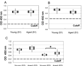
Expression of PrPC in brains of young and aged cattle:
To determine the expression level of PrPC in the brains of
the aged cattle, amounts of PrPC in the S1 fraction were
examined with a FRELISA® BSE kit. The amount of PrPC
in the S1 fraction was not significantly different between the young and aged cattle (Fig. 2A). The amount of glial marker GFAP in the S1 fraction was also not significantly different between the young and aged cattle (Fig. 2B). The amount of PrPC in the P2 fraction was significantly higher in
the aged cattle than in young cattle (P0.05), while the amount of PrPC in the S2 fraction showed no significant
dif-ference (Fig. 2C).
The PrPC immunoblot profile was examined with
anti-PrP mAb 44B1(Fig. 3). In the S2 fraction, one major band migrating at approximately 35 kDa (lanes 2–6) is shown in Fig. 3. In the P2 fraction, one major band migrating at 26– 17 KDa was detected with mAb 44B1, but not with N-termi-nal recognizing mAbs 110 and SAF32, suggesting that the 26–17 KDa band corresponds to the N-terminally truncated form (Fig. 3). The quantities of synapsin I in the S2 and P2 fractions were not significantly different between the young and aged cattle, although the bands reacted with anti-syn-apsin I were slightly different between S2 and P2 fractions.
Treatment with PNGase F and PK-sensitivity of PrPC:
After treatment with PNGase F, the PrPC in the S2 and P2
fractions shifted mainly to the 30-kDa band corresponding to the full-length PrPC and the 18-kDa band corresponding
to an N-terminally truncated PrPC fragments (Fig. 4). There
was no significant difference in glycosylation forms between the young and aged cattle.
To examine PK sensitivity, the S2 and P2 fractions from different aged cattle were treated in either the absence or presence of 1, 1.25, 1.6, 2.5 and 5 μg/μl of PK for 30 min, after which the PrPC molecules were studied by immunoblot
analysis (Fig. 5). After incubation with 5 μg/μl of PK, most PrPC in the S2 fraction was relatively digested in contrast to
PrPC in the P2 fraction. These results indicate that the S2
fraction was relatively sensitive to PK concentration in both the young and aged cattle, whereas the P2 fraction was rela-tively resistant to PK treatment in both the young and aged cattle.
DISCUSSION
To clarify the relationship between aged cattle and the occurrence of atypical BSE, we focused on the physiologi-cal properties and amount of PrPC in brain homogenates
from young and aged cattle. In this study, the brains of aged cattle (>120 months) exhibited a significant decrease in the
Fig. 1. Proteasome activity in brain homogenates from young and aged cattle. The 20S proteasome activity in the S1 fraction of brain homogenates from young (8 cattle: 17– 24 months) and aged (8 cattle: 60–100 months) cattle was determined by a 20S proteasome activity assay kit. The 20S proteasome activity was measured with or without the lac-tacystin inhibitor. Histograms were obtained from 8 sam-ples and show the mean SD. RFU, relative fluorescent unit, was determined by a 380/460 nm filter set with a Mul-tilabel Reader Wallac 1420 ARVOsx. *, P0.01.
Fig. 2. Comparison of the amounts of PrPC expression and
quan-tity of GFAP in brain homogenates. A) The amounts of PrPC in
the S1 were detected by a FRELISA® BSE. The cutoff value
was determined by the value at 0.150 plus the mean value of negative controls according to the manufacturer’s instructions. Young cattle, 10 samples; Aged cattle, 8 samples. B) The quan-tity of GFAP was determined by a RIDASCREEN® Risk
Mate-rial 10/5. Young cattle, 10 samples; Aged cattle, 8 samples. C) The amounts of PrPC in the S2 and P2 were detected by a
FRELISA® BSE. Young cattle, 20 samples; Aged cattle, 45
samples. *, P0.05.
Fig. 3. The relative amounts of S2 and P2 of PrPC in brain
homogenates from young and aged cattle. The samples were separated by SDS-PAGE, transferred onto PVDF mem-branes and reactd with mAb 44B1. Lane 1, Scrapie (Obihiro strain) affected mouse brain; lanes 2, 4, 6, 7, 9 and 11, young cattle (20-month old); lanes 3, 5, 8 and 10, aged cattle (lanes 3 and 8, 175-month old; lanes 5 and 10, 129-month old).
Y. YOSHIOKA, N. ISHIGURO AND Y. INOSHIMA
1586
20S proteasome activity, and deposition of lipofuscin pig-ment served as a sign of aging. Oxidative metabolism is essential for neurons to generate energy, because the high energy demands in neurons lead to aging vulnerability [7]. Additionally, lipofuscin contains some materials derived from lysosomal degradation; furthermore, it increases with aging [7].
We believe our study is support for the age-related decreases of protein turnover in cells. In our study, the amount of PrPC in the detergent-insoluble (P2) fraction was
significantly higher in the young than in the aged cattle (Fig. 2C), which agrees well with a decrease in protein turnover including 20S proteasome activity with aging. In vitro evi-dence has shown that the 20S proteasome selectively
degrades damaged proteins following an oxidative insult [8], and is a marker of the primary mechanism for the degrading oxidized proteins [6]. Age-related decline in pro-teasome activity is also reported in the brain [4] and spinal cord [11].
To analyze more details of the physiological properties of PrPC, the S1 fraction was then fractionated into
detergent-soluble (S2) and detergent-indetergent-soluble (P2) proteins. PrPC
amounts in the S2 and P2 fractions of the aged cattle was slightly higher than that of young cattle, while PrPC amount
of the S1 fraction was almost same in young and aged cattle (Fig. 2A). Several experiments using proteasomal inhibi-tors such as MG132 and N-acetyl-leucinal-leucinal-nor-leucinal (ALLN) revealed that wild type PrPC is
accumulated in both detergent-soluble and -insoluble spe-cies, when cells are incubated with proteasomal inhibitors [14, 22]. In particular, the insoluble fraction includes an unglycosylated 26 kDa PrPC molecules with a
protease-resistant core [14, 22]. Thus in our study, the remarkable accumulation of PrPC in the P2 fraction of the aged cattle
may be caused by the significant decreases in the 20S pro-teasome activity (Figs. 1 and 2). In our study, the P2 sam-ples from young and aged cattle were relatively resistant to PK treatement. However, it is not clear how the physiolog-ical properties of PrPC such as PK resistance and
accumula-tion of PrPC in the P2 fraction of the aged cattle are
associated with occurrence of atypical BSE. In the future, to elucidate the relationship between expression of PrPC and
occurrence of atypical BSE, brain samples from atypical BSE need to be characterized directly.
We found influences of aging on neither of the glycosyla-tion forms nor on PK sensitivity in the bovine PrPC.
How-ever, Goh et al. described both an increasing prevalence in the complex oligosaccharides in PrPC from aged mice and
an absence of any relationship between aging and PK sensi-tivity of mouse PrPC [10]. We conclude that age-related
physiological changes in PrPC are not the same in cattle and
mice. Likewise, Salès et al. reported that PrPC expression
increase with age in the brain of hamsters [18], while in the present study, the amount of PrPC in the S1 fractions were
similar in young and aged cattle, suggesting that PrPC
expression may not change drastically with age in the brains of cattle. Again, we conclude interspecies differences.
The present study provides insight into possible explana-tions for the correlation between cattle age and occurrence of atypical BSE. Perhaps aging should not be considered strictly as an influence on PrPC, but also as an influence on
variable structures and functions of the brain due through genetic and environmental mechanisms. Despite the pleth-ora of research on neurodegenerative diseases associated with aging in humans, such as Alzheimer’s and Parkinson’s diseases, questions concerning aging still remain [7]. Mito-chondrial dysfunction is thought to be a key factor in age-related diseases of humans [19] and should therefore be investigated in the cattle. Biological relationships between aging and PrPC, including antioxidant-like activity [2],
syn-aptic transmission [15], and the expression of PrPC exposure
Fig. 4. Immunoblot analysis of PrPC in brain homogenates
after treatment with PNGaseF. The samples were separated by SDS-PAGE, transferred onto PVDF membranes and reacted with mAb 44B1. Molecular mass markers are indi-cated on the left. Lane 1, Scrapie (Obihiro strain) affected mouse brain; lanes 2, 3, 5, 6, 8, 9, 11 and 12, young cattle (20-month old); lanes 4, 7, 10 and 13, aged cattle (175-month old).
Fig. 5. Immunoblot analysis of PrPC in brain homogenates
after PK treatment. The samples after PK treatment were separated by SDS-PAGE, transferred onto PVDF mem-branes and reacted with mAb 44B1. Lane 1, Scrapie (Obi-hiro strain) affected mouse brain; S2, lanes 2–7; P2, lanes 9–13; lanes 2 and 8, non PK control.
to oxidative stress in the aged mice [20], have also been reported. Thus, it is conceivable that PrPC plays a role in
protecting neurons from the effects of cellular stress. The origin and mechanisms of the occurrence atypical BSE remains unknown, as with classical BSE. Only one cases of atypical BSE have been reported in young cattle, while classical BSE occurred in aged cattle in Japan [13, 21]. It is very difficult to precisely estimate the influences of age on atypical and classical BSE cases. Atypical BSE and clas-sical BSE display unique incubation periods, PrPres
deposi-tion patterns, and histological lesions [9, 16]. However, because the onset of BSE is affected by exposure dose of BSE agent and host susceptibility, it is difficult to estimate the infection time for both classical and atypical BSE cases. There is a possibility that atypical BSE agents could infect young cattle and reside within the body for long periods before symptoms of BSE appears in aged cattle. In addition, the low prevalence of atypical BSE identified through mainly active surveillance means that whole brains are not often available for examination. Thus the need remains for detailed sampling of the brain, with the exception the obex region, to determine the etiology of atypical BSE.
ACKNOWLEDGMENTS. We would like to thank the staff of the local Meat Inspection Centers in Gifu and Nagano Prefectures for providing cattle brain samples. This study was supported in part by a Grant-in-Aid for Scientific Research (B) (No.17380180) from the Ministry of Educa-tion, Cultures, Sports, Science and Technology of Japan, and by a grant from the Ministry of Health, Labour and Wel-fare of Japan.
REFERENCES
1. Biacabe, A. G., Laplanche, J. L., Ryder, S. and Baron, T. 2004. Distinct molecular phenotypes in bovine prion diseases. EMBO Rep. 5: 110–115.
2. Brown, D. R., Wong, B. S., Hafiz, F., Clive, C., Haswell, S. J. and Jones, I. M. 1999. Normal prion protein has an activity like that of superoxide dismutase. Biochem. J. 344: 1–5.
3. Bruce, M. E. and Dickinson, A. G. 1987. Biological evidence that scrapie agent has an independent genome. J. Gen. Virol. 68: 79–89.
4. Carney, J. M., Starke-Reed, P. E., Oliver, C. N., Landum, R. W., Cheng, M. S., Wu, J. F. and Floyd, R. A. 1991. Reversal of age-related increase in brain protein oxidation, decrease in enzyme activity, and loss in temporal and spatial memory by chronic administration of the spin-trapping compound N-tert-butyl-alpha-phenylnitrone. Proc. Natl. Acad. Sci. U.S.A. 88: 3633– 3636.
5. Casalone, C., Zanusso, G., Acutis, P., Ferrari, S., Capucci, L., Tagliavini, F., Monaco, S. and Caramelli, M. 2004. Identifica-tion of a second bovine amyloidotic spongiform encephalopa-thy: molecular similarities with sporadic Creutzfeldt-Jakob disease. Proc. Natl. Acad. Sci. U.S.A. 101: 3065–3070. 6. Davies, K. J. 2001. Degradation of oxidized proteins by the 20S
proteasome. Biochemical 83: 301–310.
7. Esiri, M. M. 2007. Aging and brain. J. Pathol. 211: 181–187. 8. Ferrington, D. A., Sun, H., Murray, K. K., Costa, J., Williams,
T. D., Bigelow, D. J. and Squier, T. C. 2001. Selective degrada-tion of oxidized calmodulin by the 20S proteasome. J. Biol. Chem. 276: 937–943.
9. Fukuda, S., Iwamaru, Y., Imamura, M., Masujin, K., Shimizu, Y., Matsuura, Y., Shu, Y., Kurachi, M., Kasai, K., Murayama, Y., Onoe, S., Hagiwara, K., Sata, T., Mohri, S., Yokoyama, T. and Okada, H. 2009. Intraspecies tramsmission of L-type-like bovine spongiform encephalopathy detected in Japan. Micro-biol. Immunol. 53:704–707.
10. Goh, A. X., Li, C., Sy, M. S. and Wong, B. S. 2007. Altered prion protein glycosylation in the aging mouse brain. J. Neuro-chem. 100: 841–854.
11. Keller, J. N., Huang, F. F. and Markesbery, W. R. 2000. Decreased levels of proteasome activity and proteasome expres-sion in aging spinal cord. Neuroscience 98: 149–156. 12. Kim, C. L., Umetani, A., Matsui, T., Ishiguro, N., Shinagawa,
M. and Horiuchi, M. 2004. Antigenic characterization of an abnormal isoform of prion protein using a new diverse panel of monoclonal antibodies. Virology 320: 40–51.
13. Hagiwara, K., Yamakawa, Y., Sato, Y., Nakamura, Y., Tobi-ume, M., Shinagawa, M. and Sata, M. 2007. Accumulation of mono-glycosylated form-rich, plaque-forming PrPSc in the
sec-ond atypical bovine spongiform encephalopathy case in Japan. Jpn. J. Infect. Dis. 60: 305–308.
14. Ma, J. and Lindquist, S. 2001. Wild-type PrP and a mutant asso-ciated with prion disease are subject to retrograde transport and proteasome degaradation. Proc. Natl. Acad. Sci. U.S.A. 98: 14955–14960.
15. Maglio, L. E., Martins, V. R., Izquierdo, I. and Ramirez, O. A. 2006. Role of cellular prion protein on LTP expression in aged mice. Brain Res. 1097: 11–18.
16. Masujin, K., Shu, Y., Yamakawa, Y., Hagiwara, K., Sata, T., Matsuura, Y., Iwamaru, Y., Imamura, M., Okada, H., Mohri, S. and Yokoyama, T. 2008. Biological and biochemical character-ization of L-type-like bovine spongiform encephalopathy (BSE) detected in Japanese black beef cattle. Prion 2: 123–128. 17. Prusiner, S. B. 1982. Novel proteinaceous infectious particles
cause scrapie. Science 216: 136–144.
18. Salés, N., Hässing, R., Rodolfo, K., Di Giamberardino, L., Traiffort, E., Ruat, M., Frétier, P. and Moya, K. L. 2002. Devel-opmental expression of the cellular prion protein in elongating axons. Eur. J. Neurosci. 15: 1163–1177.
19. Vanitallie, T. B. 2008. Parkinson disease: primacy of age as a risk factor for mitochondrial dysfunction. Metabolism 57: 50– 55.
20. Williams, W. M., Stadtman, E. R. and Moskovitz, J. 2004. Age-ing and exposure to oxidative stress in vivo differentially affect cellular levels of PrPc in mouse cerebral microvessels and brain
parenchyma. Neuropathol. Appl. Neurobiol. 30: 161–168. 21. Yamakawa, Y., Hagiwara, K., Nohtomi, K., Nakamura, Y.,
Nishijima, M., Higuchi, Y., Sato, Y. and Sata, T. 2003. Atypical proteinase K-resistant prion protein (PrPres) observed in an
apparently healthy 23-month-old Holstein steer. Jpn. J. Infect. Dis. 56: 221–222.
22. Yedidia, Y., Horonchik, L., Tzaban, S., Yanai, A. and Tarabou-los, A. 2001. Proteasomes and ubiquitin are involved in the turn-over of the wild-type prion protein. EMBO J. 20: 5383–5391. 23. Yuan, J., Xiao, X., Mcgeehan, J., Dong, Z., Cali, I., Fujioka, H.,
Kong, Q., Kneale, G., Gambetti, P. and Zou, W.-Q. 2006. Insol-uble aggregates and protease-resistant conformers of prion pro-tein in uninfected human brains. J. Biol. Chem. 281: 34848– 34858.