SUMMARY
Keywords
Synchronous occurrence of breast cancer and refractory diffuse
large B-cell abdominal lymphoma: Management and review of
the literature
Nikolaos S. Salemis
1,2,*
1 Breast Cancer Surgery Unit, Army General Hospital, Athens, Greece; 2 IASO Women's Hospital, Athens, Greece.
synchronous, breast, carcinoma, diffuse lymphoma
The synchronous occurrence of primary breast cancer and lymphoid tissue malignant tumors has been rarely reported in the literature. We present an exceedingly rare case of synchronous breast invasive ductal carcinoma with an abdominal diffuse large B-cell lymphoma (DLBCL). A 78-year-old woman who was diagnosed with a luminal A invasive breast cancer on core biopsy, and complaint of progressively worsening low back pain. An abdominal computed tomography (CT) scan that was performed as part of the preoperative staging showed a large abdominal mass measuring 10.5 × 4.8 × 9.5 cm surrounding the lower part of the abdominal aorta, the right common iliac, right external, right internal iliac, and the left internal iliac arteries. A CT-guided fine-needle aspiration biopsy (FNAB) of the abdominal mass was then performed, to exclude the possibility of being an abdominal tumor metastasis of the known primary breast cancer. Histopathological findings were suggestive of DLBCL. Following a multidisciplinary team discussion, chemotherapy was initiated for DLBCL. The tumor however was refractory to multiple chemotherapy regimens and exhibited a highly aggressive clinical course. The diagnostic evaluation and management of the patient are discussed, along with a review of the relevant literature. This case underscores the fact that the presence of synchronous malignancies may pose both diagnostic and treatment challenges. Accurate staging of both malignancies and multidisciplinary team discussion is of utmost importance to guide an optimal therapeutic approach. Histopathological evaluation is essential for both tumors, for the second malignancy not to be misinterpreted as a secondary deposit of the primary one.
1. Introduction
Multiple primary malignancies (MPMs) are defined as the presence of two or more primary malignant tumors in an individual patient. Based on the timing of their diagnosis, MPMs are classified as either synchronous or metachronous. Synchronous MPMs are diagnosed at the same time or within a two month-interval, whereas in metachronous MPMs, the second tumor is found more than six months after the diagnosis of the first tumor (1). The incidence of MPMs ranges from 0.52% to 11.2% in various series (2-4). In a retrospective review of 8,428 autopsies, 1,870 which were performed in cancer patients, Lee et al. (5) found that 0.8% of all autopsies and 3.6% of all cancer autopsies revealed multiple primary tumors. In another retrospective clinical study, Lv et al. (2) found a 1% incidence of MPMs among 15,683 patients diagnosed with malignant tumors in
seven years. Metachronous tumors are more frequent than synchronous tumors (3), with a ratio of 2.7:1 (6). MPMs may occur at any age, but 75% of the patients are older than 50 years (2). The incidence of MPMs has increased in recent years (3). Synchronous occurrence of breast cancer with a lymphoid tissue malignancy is a very rare occurrence. Herein, a case of synchronous invasive ductal breast cancer with a refractory diffuse large B-cell abdominal lymphoma (DLBCL) is described, along with a review of the relevant literature.
2. Case Report
Α 78-year-old woman presented to our Breast Clinic with a 2-month history of a left breast mass. Her past medical history was unremarkable, and she had no family history of breast or ovarian cancer.
Physical examination revealed a non-tender
Intractable & Rare Diseases Research. 2021; 10(2):131-135. defined firm mass measuring approximately 2.5cm at
the 9-o'clock position, 3 cm from the nipple of the left breast. Mammography showed a spiculated left breast mass in the left upper inner quadrant (Figure 1), whereas ultrasonography revealed a hypoechoic irregular mass, highly suspicious for malignancy. A core needle biopsy revealed a luminal A invasive ductal breast carcinoma. During the preoperative staging investigations, the patient had a complaint of progressively worsening low back pain radiating to the left leg. A computed tomography (CT) scan of the abdomen revealed a
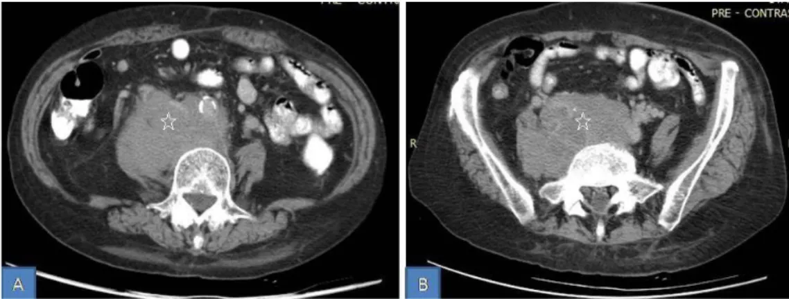
large soft tissue mass, measuring 10.5 × 4.8 × 9.5 cm surrounding the lower part of the abdominal aorta, the right common iliac, right external, right internal iliac, and the left internal iliac arteries with extension at L4-L5 spinal segment compressing the L4 and L5 nerve roots (Figure 2). The computed tomography scan of the chest and the remainder of the staging investigations were unremarkable.
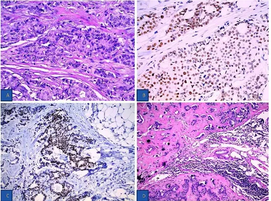
A CT-guided fine-needle aspiration biopsy (FNAB) of the abdominal mass was then performed, which showed a diffuse large B-cell lymphoma (Figure 3). The patient underwent wide local excision of the breast mass with sentinel node biopsy. The two sentinel nodes contained macrometastases, and an axillary lymph node dissection was then performed. The histological examination revealed an invasive grade II ductal breast carcinoma measuring 2.6 × 2.1 × 1 cm according to the modified Bloom-Richardson grading system (Figure 4). On immunohistochemical analysis, the tumor cells were positive for Estrogen and Progesterone receptors, whereas the expression of Her2 was negative, and the ki-67 proliferation index was 8-10%. Two of the thirteen removed axillary lymph nodes contained metastases. Staging investigations showed that the DLBCL was confined exclusively to the abdominal cavity. The patient was categorized into the high-intermediate risk group according to the International Prognostic Index (IPI) and revised IPI (R-IPI) categories. Soon after, treatment was initiated with the R-CHOP regimen (rituximab 375 132
Figure 1. Left mediolateral oblique (A) and craniocaudal (B) mammograms showing a spiculated mass with slight nipple retraction (arrows).
Figure 2. Abdomen CT scan showing a large mass measuring 10.5 × 4.8 × 9.5 cm extending to the pelvis (stars).
Figure 3. Histopathological findings of DLBCL. (A,B) Tumor cells are of medium and large size with irregular or oval intensely colored nuclei
and one or more prominent nucleoli. A. (H-E ×100), B (H-E ×400). (C) On immunohistochemical analysis, the tumor cells were strongly and diffusely positive for CD 20 (×200).
compared with the general population, however, the synchronous occurrence of a non-breast malignancy in breast cancer patients is exceedingly rare (9).
Breast cancer is the most frequently diagnosed malignancy after treatment for Hodgkin's Lymphoma, especially in young patients who have undergone radiation therapy. On the other hand, the development of non-Hodgkin lymphoma (NHL) after breast cancer treatment is a very rare occurrence (10).
SPMs in patients with breast cancer are rare. In a retrospective review, Sas-Korczyńska et al. (11) found synchronous malignancies in only 112 (0.009%) patients among 118,952 patients with breast cancer who were treated between 1965 and 2014. The most common type of synchronous primary malignancy was contralateral breast cancer (63.4%), whereas the most frequently observed non-breast synchronous malignancy derived from female genital organs (36.6%). Synchronous lymphatic tissue tumors were found in only 3.6% of the cases (11).
In a literature review, Woo et al. (12) found only 37 cases of synchronous breast cancer with NHL of all types. DLBCL was synchronous with breast cancer in 29.7% of the cases. Other types of concomitant NHL included mantle cell lymphoma (MCL): 8.1%, follicular lymphoma (FL): 27%, marginal zone B-cell lymphoma (MZBL): 5.4%, chronic lymphoid leukemia (CLL): 18.9%, and mucosa-associated lymphoid tissue lymphoma (MALT): 5.4%. The diagnosis of the mg/m2, vincristine 1.4 mg/m2, doxorubicin 50 mg/m2,
cyclophosphamide 750 mg/m2, and prednisolone 100
mg orally). Despite a partial remission due to the initial therapy, the tumor rapidly relapsed, and the patient received rituximab in combination with gemcitabine and vinorelbine chemotherapy (rituximab 375 mg/ m2, gemcitabine 880 mg/m2, vinorelbine 25 mg/m2).
Despite chemotherapy, the disease progressed causing severe neurological symptoms that led to the inability to walk. Radiotherapy was then administered and resulted in improvement in neurological symptoms. Unfortunately, the intraabdominal disease continued to progress, and the patient started salvage therapy with the DICE (dexamethasone, ifosfamide, cisplatin, etoposide) regimen but again with no response. She died of progressive disease 11 months after the initial diagnosis.
3. Discussion
MPMs were first described as a clinical entity by Billroth in 1889 (7). However, Warren and Gates in 1962 (8) proposed the following specific criteria for the diagnosis of MPMs. First, the presence of clear histological evidence for malignancy in both tumors, second the tumors must be histologically distinct and third, the second tumor cannot be a metastatic tumor of the first (8).
Although there is an increased risk of developing a second malignancy in women treated for breast cancer,
Figure 4. Histopathological findings of invasive ductal breast carcinoma. (A) Tumor cells are medium in size with weakly eosinophilic
cytoplasm and include oval slightly irregular nuclei with granular chromatin small nucleus and moderate pleomorphism and atypia (H-E ×400). (B,C) On immunohistochemical analysis, the tumor cells stained positive for ER (H-E ×400) and PR (H-E ×200). (D) Lymph node with
Intractable & Rare Diseases Research. 2021; 10(2):131-135. 134 concomitant DLBCL was established in all patients
after an axillary lymph node biopsy. It is noteworthy that not even a single case of abdominal DLCBL was mentioned in that review. The authors stated that it is not well known if synchronous tumors share a common etiology or each tumor arises independently. In the vast majority of cases (88.9%), the second malignancy had not been detected until initiation of treatment for the first tumor (12).
DLBCL is the most common type of Non-Hodgkin Lymphomas (13,14). It usually runs an aggressive clinical course characterized by rapidly enlarging lymphadenopathy and constitutional symptoms (13). DLBCL can involve extranodal sites such as kidneys, adrenal gland, brain, and bones. The definitive diagnosis is established by excisional lymph node biopsy. Chemoimmunotherapy with R-CHOP (rituximab, cyclophosphamide, doxorubicin, vincristine, prednisolone) is the most common upfront treatment, resulting in a durable remission in 50-60% of the patients. However, the prognosis is poor for patients who develop disease refractory to up-front treatment (13). Papajik et al. (15) studied 209 NHL patients with F-18FDG PET/CT and found 6 (2.9%) suspicious cases for a second tumor. Only one of these patients had breast cancer. In 83% of the patients, the second malignancy was asymptomatic. The authors suggested that in patients with NHL, the additional imaging of lesions detected at F-18FDG PET/CT may reveal synchronous malignancies (15).
In a retrospective analysis of 809 DLBCL patients, Tanba et al. (16) found 123 (15.2%) cases of multiple malignancies. The majority (76.4%) developed metachronous malignancies, whereas only (23.6%) were diagnosed with synchronous tumors. Stomach cancer was the most frequent second malignancy followed by colorectal cancer, lung cancer, and breast cancer. The overall survival and progression-free survival were significantly shorter in patients having a second malignancy than in those without (16).
Synchronous breast cancer and malignant lymphoma is an exceedingly rare occurrence. In most cases, the malignant lymphoma was diagnosed either in the breast or in the ipsilateral axillary lymph nodes (17-22). Synchronous breast cancer with abdominal DLBCL is an exceedingly rare clinical entity. We were able to find only one case reported in the English literature (9). The exact pathogenesis of multiple primary malignancies has not been clearly defined (1,23). However, it is suggested, that several potential causative factors may contribute, including intrinsic, environmental, genetic, or therapeutic factors (24). Intrinsic factors may include susceptibility, immune status, endocrine, and embryonic development. Environmental factors may include lifestyle, prolonged exposure to radiation or pollution. Genetic factors may include genetic mutations, whereas therapeutic factors
mainly include chemotherapy and radiation therapy (23,24). Subramanian et al. (25) suggested that the oncogenic Epstein-Barr virus may be the causative factor for the dissemination of both invasive breast carcinoma and lymphomas through the EBNA-3C, which interacting with the Nm23-H1 metastatic suppressor protein reverses its ability to stop breast and lymphoma cell migration. Wiernik et al. (20) suggested that some patients with both breast cancer and lymphoma may share a viral etiology as a causative factor.
Our patient had synchronous breast cancer with a refractory DLBCL that showed a highly aggressive clinical course refractory to chemotherapy. It has been reported that 50% of DLBCL patients become refractory to or relapse after treatment (14).
In 636 patients with refractory DLBCL who were included in the international, multicohort retrospective SCHOLAR-1 study, the objective response rate was 26%, whereas the median overall survival was 6.3 months. Only 20% of patients with refractory DLBCL were alive at two years (14).
There is no consensus of prognostic factors for MPMs (24). Patients with synchronous malignancies are associated with shorter survival than patients with metachronous tumors (2). The presence of synchronous primary malignancies may pose a diagnostic and therapeutic dilemma (11,12,21). Axillary lymphadenopathy secondary to NHL can be attributed to metastases of the known breast cancer (12,19), whereas the second tumor should not be confused with a progression of the known primary (17). Especially in patients with synchronous breast cancer and NHL, the B symptoms of NHL may be mistaken for perimenopausal symptoms (12). A biopsy is of utmost importance to obtain a definitive diagnosis of NHL (10). Operable synchronous primary malignancies can be surgically treated in a single setting with less morbidity and better survival rates (1). The optimal treatment should be discussed in a multidisciplinary team, taking into consideration the prognosis and possibility of a curative or palliative treatment (11).
In conclusion, we present an exceedingly rare case of synchronous breast cancer with a refractory abdominal DLBCL and briefly review the relevant literature. The presence of multiple primary malignancies may pose a diagnostic and therapeutic dilemma. A thorough staging investigation of both malignancies and multidisciplinary team discussion is necessary to determine the optimal treatment plan. In our case, which appears to be the second reported in the literature, DLBCL was confined to the abdominal cavity and exhibited a highly aggressive clinical course.
Acknowledgements
The author would like to thank Dr. Poulakidas E. from the Hematology Department for providing the treatment
details of DLBCL and Dr. Papadopoulou G. and Fragia K. from the Pathology Department for providing the histology slides.
Funding: None.
Conflict of Interest: The authors have no conflicts of
interest to disclose.
References
1. Das S. Synchronous and metachronous cancers: an update. Annals of Clinical Case Reports. 2017; 2:1388.
2. Lv M, Zhang X, Shen Y, Wang F, Yang J, Wang B, Chen Z, Li P, Zhang X, Li S, Yang J. Clinical analysis and prognosis of synchronous and metachronous multiple primary malignant tumors. Medicine (Baltimore). 2017; 96:e6799.
3. Lee J, Park S, Kim S, Kim J, Ryu J, Park HS, Kim SI, Park BW. Characteristics and survival of breast cancer patients with multiple synchronous or metachronous primary cancers. Yonsei Med J. 2015; 56:1213-1220. 4. Liu Z, Liu C, Guo W, Li S, Bai O. Clinical analysis
of 152 cases of multiple primary malignant tumors in 15,398 patients with malignant tumors. PLoS One. 2015; 10:e0125754.
5. Lee TK, Myers RT, Scharyj M, Marshall RB. Multiple primary malignant tumors (MPMT): study of 68 autopsy cases (1963-1980). J Am Geriatr Soc. 1982; 30:744-753. 6. Testori A, Cioffi U, De Simone M, Bini F, Vaghi A,
Lemos AA, CiullaMM, Alloisio M. Multiple primary synchronous malignant tumors. BMC Res Notes. 2015; 8:730.
7. Billroth T. Die Allgemeine chirurgischepathologie und therapie. In: G. Reimer (ed.). 51 Vorlesungen-Ein Handbuch fur Studierende und Artze, 14. Auflage, Berlin, 1889, pp. 908.
8. Warren S, Gates O. Multiple malignant tumors. A survey of the literature and statistical study. Am J Cancer. 1932; 16:1358-1414.
9. Alonso-Ruano M, López-Bonet E, Huerta-Anaya MV, Vila-Camps E, Bernado L, Tuca-Rodriquez F, Suarez-Pumariega P, Menendez JA. Synchronous solid neuroendocrine breast carcinoma and abdominal lymphoma: A case report and review of the literature. Oncol Lett. 2013; 5:459-462.
10. Tamaoki M, Nio Y, Tsuboi K, Nio M, Tamaoki M, Maruyama R. A rare case of non-invasive ductal carcinoma of the breast coexisting with follicular lymphoma: A case report with a review of the literature. Oncol Lett. 2014; 7:1001-1006.
11. Sas-Korczyńska B, Mituś JW, Kamzol W, Rzepa MK, Jakubowicz J, Wysocki WM. Synchronous malignancies in patients with breast cancer. Nowotwory Journal of Oncology. 2017; 67: 336-341.
12. Woo EJ, Baugh AD, Ching K. Synchronous presentation of invasive ductal carcinoma and mantle cell lymphoma: a diagnostic challenge in menopausal patients. J Surg Case Rep. 2016; 2016:rjv153.
13. Liu Y, Barta SK. Diffuse large B cell lymphoma: 2019 update on diagnosis, risk straticication, and treatment. Am J Hematol. 2019; 94:604-616.
14. Crump M, Neelapu SS, Farooq U, et al. Outcomes in refractory diffuse large B-cell lymphoma: results from the international SCHOLAR-1 study. Blood. 2017; 130:1800-1808.
15. Papajík T, Mysliveček M, Sedová Z, Buriankova E, Prochazka V, Raida L, Kubova Z, Neopal C, Starostka D, Mikula P, Melichar B, Kucerova L, Tichy M, Indrak K. Synchronous second primary neoplasms detected by initial staging F-18 FDG PET/CT examination in patients with non-Hodgkin lymphoma. Clin Nucl Med. 2011; 36:509-512.
16. Tanba K, Chinen Y, Uchiyama H, et al. Prognostic impact of a past or synchronous second cancer in diffuse large B cell lymphoma. Blood Cancer J. 2018; 8:1.
17. Frey BM, Morant R, Senn HJ, Fisch T, Schmid U. Simultaneous occurrence of breast carcinoma and malignant lymphoma. Case observations and literature review. Schweiz Med Wochenschr. 1994; 124:1010-1016. (in German)
18. Hołody Ł, Kunikowska J, Braziewicz J. Simultaneous breast cancer and DLBCL lymphoma - role of PET/CT examination with 18F-FDG and 18F-FES. Nucl Med Rev Cent East Eur. 2018; 21:113-114.
19. Hahm MH, Kim HJ, Shin KM, Cho SH, Park JY, Jung JH, Jeong JY, Bae JH. Concurrent invasive ductal carcinoma of the breast and malignant follicular lymphoma, initially suspected to be metastatic breast cancer: a case report. J Breast Cancer. 2014;17:91-97.
20. Wiernik PH, Hu X, Ratech H, Fineberg S, Marino P, Schleider MA, Etkind P, Walewski JA. Non-Hodgkin's lymphoma in women with breast cancer. Cancer J. 2000; 6:336-342.
21. Cuff KE, Dettrick AJ, Chern B. Synchronous breast cancer and lymphoma: a case series and a review of the literature. J Clin Pathol. 2010; 63:555-557.
22. Liu W, Zhu H, Zhou X. Synchronous bilateral non-Hodgkin's diffuse large B-cell lymphoma of the breast and left breast invasive ductal carcinoma: a case report and review of literature. Int J Clin Exp Pathol. 2014; 7:7042-7048.
23. De Luca A, Frusone F, Vergine M,Cocchiara R, LaTore G, Ballesio L, Monti M, Amabile MI. Breast cancer and multiple primary malignant tumors: case report and review of the literature. In Vivo. 2019; 33:1313-1324. 24. Xu LL, Gu KS. Clinical retrospective analysis of cases
with multiple primary malignant neoplasms. Genet Mol Res. 2014; 13:9271-9284.
25. Subramanian C, Cotter MA 2nd, Robertson ES. Epstein-Barr virus nuclear protein EBNA-3C interacts with the human metastatic suppressor Nm23-H1: a molecular link to cancer metastasis. Nat Med. 2001; 7:350-355.
Received January 24, 2021; Revised March 23, 2021; Accepted March 31, 2021.
*Address correspondence to:
Nikolaos S. Salemis, Breast Cancer Surgery Unit, Army General Hospital, 19 Taxiarhon Street, 19014 Kapandriti, Athens, Greece.
E-mail: nikos.salemis@gmail.com
Released online in J-STAGE as advance publication April 8, 2021.