Title
restriction in vivo due to spleen extract and diluted antiserum
Author(s)
Kinjo, Kiyokatsu; Kosugi, Tadayoshi; Yamaguchi, Hideyuki;
Rokushima, Yoshikazu; Miyawaki, Keiko
Citation
琉球大学保健学医学雑誌=Ryukyu University Journal of
Health Sciences and Medicine, 5(2): 92-103
Issue Date
1982
URL
http://hdl.handle.net/20.500.12001/4135
琉大保医誌5(2) : 92-103, 1982.
Specific immunosuppression without genetic restriction in
vivo due to spleen extract and diluted antiserum
Kiyokatsu Kinjo, Tadayoshi Kosugi
Hideyuki Yamaguchi; Yoshikazu Rokushima and Keiko Miyawaki
Department of Physiology, School of Medicine, University of the Ryukyus
'Department of Physiology, Kobe University School of Medicine
The effect of spleen extract and diluted antiserum to antibody production was studied in order
to clarify the role of spleen extract in re如Iation ofっntibody production. Extracts of normal or
pertussis-vaccine-stimulated mouse spleen did not by themselves suppress the primary immune response of mice to sheep or horse red blood cells. Similarly, anti-sheep-red-blood-cell mouse serum did not suppress if administered after 20- to 50イold dilution. However, when the spleen extract and the diluted antiserum were administered ei也er together with, or a day before the antigen, a very strong suppression of the primary response was observed. Antibody production was not suppressed by a combination of kidney or liver extracts and diluted mouse antisera.
The suppressive action observed was specific, and the specificity was determined solely by the specificity of也e diluted antiserum used. On the other hand, the stimulatory action of the spleen extract appeared non-specific. Indeed not only mouse spleen extracts but also rat spleen extracts or concentrated bovine spleen extracts exerted a suppressive action on antibody production in mice. The secondary response was suppressed very effectively by the administration of spleen extracts alone. From the above-mentioned results, it was clarified that a specific immunosuppression without genetic restriction was due to spleen extract and diluted antiserum m vivo.
INTRODUCTION
There are many reports that primed spleen extracts or primed thymus extracts exhibit specific immunosuppression in vivo (6-8, 10-18). For example, Takemori et al. (12, 13) found that primed spleen or thymus extract specifically suppressed IgG antibody production and that this phenomenon occurred only when the donor and the recipient contained the same I region of H-2 locus.
In contrast, comparatively few reports exist on the suppression of antibody production by normal spleen extracts. Gevorkyan et al. (3) observed that suppression by normal spleen extracts was nonspecific when the extracts had a high concentration of protein, and that at low concentrations, they enhanced, rather than suppressed, antibody production. Reinertsen and Steinberg (9) and Egan et al. (1) reported that supematants from concanavalin A-activated spleen cells had a nonspecific immunosuppressive effect. From the above-mentioned literatures, it was clarified that the active
Abbreviations used in this paper : HRC, hor監red blood cell ; PFC, plaque forming cells ; PV, pertussis vaccine ; SRC, sheep red blood cells.
spleen cell and extract played an important role in regulation of antibody production. However, until present time it was not clarified whether the normal spleen extract suppressed the antibody production in vivo or did not.
In the present paper, the role of normal spleen extract was studied in detail concerning with the regulation of antibody production in vivo.
That is to say, in the present study, extracts from normal spleen or pertussis vaccine (PV) enlarged spleen were shown to act as an immunosuppressant, but in a manner very different from the studies described above. Thus in our system, the spleen extracts by themselves were inactive. However, when the extracts were combined with diluted antisera, which again by themselves extert-ed little suppressive action, and administerextert-ed to mice either together with or just before the antigen,
a remarkable suppression of the primary response was observed- Under certain conditions the number of plaque forming cells (PFC) in the experimental mice could be made as small as those in normal, unimmunized mice. This suppressive action was specific to antigen to which the diluted antiserum was directed. The effect of the spleen extracts, on the other hand, was not limited by the histocompatibility barrier.
MATERIALS AND METHODS
Animals : Outbred female ICR mice (6-12 wks old) were purchased from Nihon Clea Co. and Oriental C0., and maintained on pellets and water ad a lib. Wistar rats and outbred dogs were purchased from Fujii Co., and bovine spleen was obtained from the Kobe Food Center.
Antigens : Sheep red blood cells (SRC) and horse red blood cells (HRC) purchased from the Japan Blood Co. were washed twice with saline. An immunizing dose or 109 erythrocytes was used. Antisera : ICR mice were immunized with a single intraperitoneal (i.p.) injection of 109 SRC or HRC and bled 8 days later. The antisera were inactivated at 56。C for 30 mm and diluted 20-, 25-, or 50 -fold (anti-SRC sera) or 12.5-fold (antトHRC sera) with saline and stored in small aliquots at -200
C until required. The hemolysin titers of the antLSRC sera before dilution were 320-, 640-, or 1024 -fold, and that of the antトHRC serum was 80-fold according to the conventional antiserum dilution
method. Preparation of spleen extracts : PV spleens were obtained from mice injected l.p. with a dose of PV containing 1010 cells 8 daysbefore. Normal or PV spleens (10.0g) were added to ll.0 ml of cold saline. The mixture was homogenized for 2 min with an Ultra Turrax Homogenizer and then disrupted with a Sonifier Cell Disruptor for 10-20 sec. All these procedures were carried out at O-C. The spleen suspensions were centrifuged at 15,000 rpm for 60 or 30 min at 0-C and the supernatant solutions were used as the spleen extracts. These were either used immediately or stored at -80。C until required.
Preparation of kidney and liver extracts : Kidney and liver of PV-stimulated mice were extracted by the same procedure as that used for the preparation of spleen extracts.
Preparation of concentrated bovine spleen extracts : Fresh bovine spleen (lO.0g) was added to 6.0 ml of cold saline. Subsequent procedures were the same as those used for the preparation of mouse spleen extracts.
Immunization : For the study of primary response, mice were injected i.p- with 0.5 ml of SRC (109 cells), 0.5 ml of normal or PV spleen extracts and 0.5 ml of diluted anti-SRC serum (1 : 20, 1 :25 or 1 :50 or anti-HRC serum (1:12.5). For the study of secondary response, mice were injected i. p. with
94 Kiyokatsu Kmjo, et al.
0.5 ml of SRC(109cells), and after 20 days 0.5 ml of SRC (109 cells) was administered together with spleen extracts and diluted antisera. The number of mice per group was 4 t0 9.
Hemolytic plaque technique : PFC to SRC or HRC were assayed by a modification of the Jerne plaque assay (16). The mice were sacrificed by anestesia. Spleen cell suspensions were prepared by dissociating the spleens with forceps in cold saline, and agitating gently with a pipette. Large cell clumps sedimented after standing for 10 seconds, and they were removed from the cell suspensions. Appropriately diluted cell suspension (0.1 ml) was added to 1.0 ml of 0.85% solution of Difco agarose containing 199 culture medium and 4 x 10⁸ SRC or HRC at 50-C. The mixture was immediately poured onto a supporting bottom layer cor止aining 1.4 % Difco agar-agar in a petridish- After incubation at 37-C for 30 min (or as soon as the agarose solidified), this layer was covered with 1.Oml of guinea pig complement (1 : 2.5 dilution), and the numbers of direct PFC (IgM PFC) were determined after a further incubation at 37-C for 2 hrs. The numbers of indirect PFC (IgG PFC) were determined by adding 0.1 ml of 1 : 40 dilution of goat antiserum mouse 7S-globulins (Hyland Co.) to the agarose solutions. The numbers of IgG PFC were obtained from the difference in values between the assays performed in the presence and absence of this anti-mouse IgG serum. The PFC
assay were carried out 4 days after antigen injection, unless indicated otherwise.
Statistical analysis : Arithmetic means and standard e汀ors (S. E.) were calculated, and the P value was determined by Student's t-test.
RESULTS
Suppression of the primary re坤onse by the simultaneous administration of spleen extracts and diluted mouse antisera.
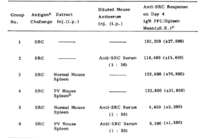
In confirmation of earlier results (2, 4, 19, 20), we could also show that the primary immune response was suppressed by the presence of high concentrations of antibody. Thus when undiluted mou監 anti-SRC sera (0.5 ml) were administered to mice together with the immunizing antigen, SRC, the primary response measured by the increase of PFC in spleen was moderately depressed (data not shown). However, when the anti-SRC sera were diluted 20-to 50-fold before administra-tion, significant suppression was not observed (compare Group 2 with 1 in Table 1). In the study, we discovered that very strong suppression occurred when extracts of normal mouse spleen were administered together with the diluted antiserum (Group 5 in Table 1). It should be emphasized that the administration of spleen extracts alone (Groups 3)did not produce a statistically significant su-ppression; thus the suppression apparently requires the presence of bo也spleen extracts and
antisera.
Extracts of spleen enlarged by the pretreatment of mice with pertussis vaccine (PV spleen extracts) showed a behavior identical to the extracts of normal spleen (cf. Groups 4 and 6 in Table 1). Since the treatment significantly improved the yield of the spleen extracts, they were always prepared from PV spleen in subsequent experiments.
The amounts of antisera and spleen extracts required for suppression were apparently rather critical. Thus in the experiment of Table 1, no or little suppression was observed if the anti-SRC sera were diluted 100-fold or if the dosage of the spleen extracts was reduced to one-half (data not shown).
Table 1. Suppression of IgM PFC formation by mouse spleen extracts and diluted mouse anti-SRC sera assayed on day 4 after antigen challenge, a :.anti-SRC(109) given intraperitoneally. b : Spleen extracts from PV-stimulated mice. C : Arithmetic mean ± S,E.; 4 mice j光r group. The hemolysin titer to anti-SRC serum was 1024-fold. P values for groups 1 vs. 2, N.S.;
1vs.3, N.S.; 1vs.4, N.S.; 1vs.5, < 0.001 ; 1vs.6, <0.001.
Diluted Mouse Group Antigena Extract Antiserum No. Challenge lnj.d.p.) (. (. } Anti-SRC Response on Day 4 IgM PFC!Spleen Mean(士:S.E. )C 1 SRC 2 SRC SRC Normal Mouse Spleen 4 SRC PV Mouse s pleen" 183,200 (士27,300) Anti-SRC Serum 114,400 (士15,400) (1 : 50) 132,600 (土70,800) 122,600 (土31,800)
sRC Normal Mouse Anti-SRC Serum 4,400 {土3, 200) S pleen PV Mouse Spleen (1 : 50) Anti-SRC Serum 5, 100 (土1,500) (1 : 50)
Table 2. Suppression of IgM PFC formation by extracts of various organs and diluted mouse anti-SRC sera assayed on day 4 after antigen challenge, a : anti-SRC (109) given intraperitoneally. b : Various organ extracts from PV-stimulated mice, c : Arithmetic mean ± S且; 4 to 8
mice 】光r group. The hemolysin titer to anti-SRC serum was 320-fold. P values for groups l vs.6, N.S.; 1 vs. 7, N.S.; 1vs. 8,く0.01.
Group Antigen8 ExtnaCtb No. Challenge Inj.(i.p.) Diluted Mouse Antiserum ln.(i.p.) AntトSRC Response on Day 4 IgM PFC/Spleen Mean (士S.E.)C SRC SRC SRC Kidn<∋y S RC Liver S R C Spleen SRC Kidney sRC Liver S RC S pleen 238,600 (土73,100) Anti-SRC Serum 184,600 (土37,300) (1 : 25 388,000 (土80,900) 153,000 (土29,100) 136,000 (士26,900) Anti-SRC Serum 172,000 (士66,100) (1 : 25) Anti-SRC Serum 81,000 (±42,800) (1 : 25) Anti-SRC Scrum 17,600 (士16,200) (1 : 25
96 Kiyokatsu Kinio, et al.
Table 3. Antigen-sr治cific suppression by mouse PV spleen extracts and diluted mouse antisera. a : SRC (109) and HRC (10s) given intraperitoneally. b : Spleen extracts from PV-stimulated mice, c : Arithmetic mean ± S.E.; 4 to 9 mice per group. The hemolysin titer to anti-SRC serum was 640-fold; that of anti-HRC serum was 80-fold. P values for groups IgM PFC toSRC :groups lvs.6, N.S.; 1 vs7, N.S.; 1 vs. 8, < 0.001 IgM PFCto HRC:group
lvs.4,N.S.; 1vs.6,く0.02;1vs.7,く0.001; 1vs.8,N.S.
Group Antigen" Extract Diluted Mouse Anti-SRC Response on Day 4 Anti-HRC Response on Day 4 IgM PFC/Spleen lgM PFC/Spleen
challenge InJ.d.p.) In,.(1 , ± Mean(アS.E.)c
1 SRC+HRC 2 SRC 3 HRC 4 SRC+HRC 5 SRC+HRC AntトHRC Serum (1 :12.51 Anti-SRC Serum (1 :20) sRC+HEC PV Spleenb
SRC+HRC PV Spleen AntトHRC Serum (1 :12.5) SRC+HRC PV Spleen Anti-SRC Serum
(1 :20) 246,600 (士75,200) 255,600 (土22,600) 1,000 (土600) 274,600 (土56.600) 27(),600 (±82,700) 129.600 (土34,000) 95,800 (i.42,000) 24,000 (土14,900 31,400 (土8,000) 650 (土330) 30.200 (土7.900) 14,400 (士6,600) 29,500 (士3,900) 8.900 (士1.200) 630 (土510) 15,900 (上8,100)
active (Table 2). No immunosuppressive action was observed in the groups (6 and 7) in which a combination of kidney or liver extracts and diluted anti-SRC sera was administered. In some experiments, suppression by lung extracts and diluted anti-SRC sera was seen, but the effect was not reproducible (data not shown).
Speaficiかof the immunosuppressive action
The immunosuppression in our system was apparently antigen specific, and the specificity was determined by the antigen specificity of the diluted antisera administered. Thus in an experiment in which SRC and HRC were used as antigens (Table 3), the mounting of the primary response to SRC and HRC was strongly suppressed only when spleen extracts plus diluted antトSRC and antLHRC sera, respectively, were administered (Group 7 and 8). Neither cross reaction nor antigen competト tion was observed. These results indicated that suppression of antibody production by spleen ext-racts and diluted antisera is specific to the antigen, to which the diluted antiserum was directed.
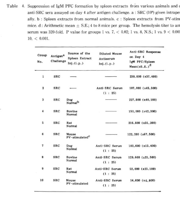
The activity of the spleen extracts was not hindered by the histocompatibility or even the species barrier. This is illustrated in Table 4, in which spleen extracts of xenogenetic animals were tested.
Table 4. Suppression of IgM PFC formation by spleen extracts from various animals and diluted anti-SRC sera assayed on day 4 after antigen challenge, a : SRC (109) given intraperitone-ally, b : Spleen extracts from normal animals, c : Spleen extracts from PV-stimulated mice, d : Arithmetic mean ± S.E.; 4 to 8 mice per group. The hemolysin titer to anti-SRC serumwas 320-fold. Pvalueforgroups 1 vs. 7, < 0.02; 1 vs. 8, N.S.; 1 vs. 9 < 0.005; 1 vs.
10,<0.001.
Group Anti&en且 No. Challenge
Source of the Diluted Mouse Spleen Extra Antiserum
Inj.d.p.) Ini.(i.p.) Antl-SRC Response on Day 4 IgM PFC/Spleen Mean(アS.E.)d SRC SRC こII sRC 冒。rmalb SRC Bovine Normal SRC Rat Normal SRC Mouse PV - stiraulatedc SRC Dog Normal SRC Bovine N orm al SRC Rat Norma1 10 SRC Mouse PV- stimulated Anti-SRC Serum (1 : 25) Anti-SRC Serum (1 : 25) Anti-SRC Serum (1 : 25) Anti-SRC Serum (1 : 25) Anti-SRC Serum (1 : 25) 230,600 (土37,400) 187,800 (士45,300) 227,600 (土40, 100) 150,000 (士42,300) 216,600 (士31,200) 122, 200 (土67,500) 102,600 (土13,400) 129,600 土21,500) 52,000 (士23, 100) 14,600 (土6,800)
Thus immune response in mice was suppressed quite effectively by a rat spleen extract and diluted mouse anti-SRC serum (Group 9).A bovine spleen extract and diluted mouse anti-SRC serum did not display statistically significant suppressive action (Group 8). However, when a concentrated bovine spleen extract was administered with diluted mouse anti-SRC sera, the suppressive action was as strong as in the case where a combination of mouse spleen extract and diluted mouse anti -SRC serum was administered (Table 5 ).
Time course of lgM PFC production
Fig. 1 shows the time course of the suppression of antibody production by combined spleen extracts and diluted anti-SRC sera. Statistically significant suppression was observed on the 3rd, 4th and 5th day after immunization, but on the 6th day the difference between the
ex-penmental and the control groups became rather small. Effect of the adminねtration scheme
98 Kiyokatsu Kinjo, et al.
Table 5. Suppression of IgM PFC formation by cone, bovine spleen extracts and diluted mouse anti-SRC sera measured on day 4 after antigen challenge, a : anti-SRC (109) given intraperitoneally. b : Concentrated bovine spleen extract (1.0ml) given intraperitoneally・ C : Arithmetic mean ± S.E.; 4 mice per group. The hemolysin titer to anti-SRC serum was 320-fold. P values for groups 1 vs. 2, N.S.; 1 vs. 3, N.S.; 1 vs. 4 < 0.005.
Group Antigena Nature of the Diluted Mouse E xtract Antiserum No. Challenge Inj.(, , Inj.(i.p.)
Anti-SRC Response on Day 4 IgM PFC/Sple<∋n Mean(土S.E. )-SRC SRC SRC SRC B ovin e Cone. Spleen" B ovin e Cone. Spleen 308,000 (士110,000) Anti-SRC Serum 284.000 (土35,200) (1 : 25) Anti-SRC Serum (1 : 25) 221,000 (± 48,300) 14,600 (土6,500)
Fig. 1. Numbers of IgM PFC to SRC in spleen of mice at various times after intraperitoneal injection of 109 SRC or 109 SRC, PV mouse spleen extracts and diluted mouse anti-SRC sera (1 : 25). The hemolysin titer of the anti-SRC serum was 320-fold. Arithmetic means
of results obtained from 4 mice are shown : the vertical bars represent the S.E.*--・mice
administered SRC on day 0. --- mice administered SRC, mouse spleen extracts and diluted mouse anti-SRC sera on day 0.
Table 6. The day of administration of mouse spleen extracts and diluted mouse anti-SRC sera and the suppression of IgM primary response, a : SRC (109) given intraperitoneally. b : Spleen extracts Pv-stimulated mice, c : Arithmetic mean ± S.E.; 4 mice per group. The hemolysin titer to anti-SRC serum was 320-fold. P values for groups 1 vs. 2,く0.05 ; 1 vs. 3,く0.002, 1vs.4, < 0.001,1vs.5,く0.001;1 vs.6,N.S.
Group
Anti-SRC Si汚p°n88 °n Day 4 IBM PFC!Spleen N。 Day-3 Day-2 Day- Day 0 線n (iS.E.) 1 2 Spl-ExtP Anti-SRC Serum (1:25) 3 4 5 6 SpトExt. Anti-SRC Serum (1:25) Spl-Ext. Anti-SRC Serum <1:25) SRC SRC SKI SRC S氏c SpトExt. Anti-SRC Serum (1:25) SRC SpトExt. Anti-SRC Serum (1:25) 273.600 (ま38,400) 116.600 (ア35,100) 48.400 (ア37,600) 680 (ア310) 21.700 (ア8,8001 172,400 (ア50,900)
Table 6 shows the effect of the spleen extracts plus antisera administered at various times. The immunosuppressive action of spleen extracts and diluted anti-SRC sera was greater in Group 4 (administered on day -1 0f antigen administration) than in Group 5 (administered on day 0 of antigen administration) and Groups 5 (administered on day -2 0f antigen administration) and was not observed in Group 6 (administered on day 1 of antigen administration). We emphasize the observation that the suppression in Group 4 was so complete that the number of PFC was very
close to that seen in untreated control (usually about 200 per spleen). The s瑚ppressien was also as strong or even stronger when the spleen extracts plus diluted antisera were given twice, on day -1 as well as on day 0. In two such experiments, the number of PFC per spleen was 150 (±80) and 280 (±50) whereas the number of PFC per spleen of untreated mice was 200 (±40) and 200 (±50). Immunosuppressive action of PV砂Ieen extract and diluted mouse anti-SRC sera on the primary yle功onse of lgG PFC
The above experiments revealed that spleen extract and diluted anti-SRC sera exerted an
immunosuppr℃ssive action on the primary response of IgM PFC
Fig. 2 illustrates the immunosuppressive action on IgG antibody production. In the groups in which a combination of spleen extracts and diluted anti-SRC sera was administered twice a day before and at the same time as the administration of antigen, significant suppression was observed throughout the periods of measurement (6, 8, and ll days).
Suppression of the seconda叩71e功onse
Spleen extracts and diluted antisera were administered with SRC to mice previously immunized with SRC (Table 7). A very strong suppression was observed. In contrast to the suppression of the primary response, spleen extracts by themselves exerted a similarly strong suppression (cf. Group 3 inTable7).
100 Kiyokatsu Kinjo, et al.
Fig. 2. Numbers of IgG PFC to SRC in spleen of mice at various times after intraperitoneal injection of 109 SRC or 109 SRC, PV mouse spleen extracts and diluted mouse anti-SRC sera (1: 25). The hemolysin titer of the anti-SRC serum was 320-fold. Arithmetic means of
results obtained from 4 mice are shown : the vertical bars represent the S.E.一一mice administered SRC on day 0, < 一mice administered SRC on day 0, mouse spleen extracts
alld diluted mouse anti-SRC sera both on day-1 and 0.
Table 7. The effect of PV spleen extracts on the secondary immune response a: SRC (10 )given intraperitoneally- b: Spleen extracts from PV-stimulated mice, c : Arithmetic mean ± S. E.; 4 mice per group. The hemolysin titer to anti-SRC serum was 320イold. P values for groups IgM PFC : 1 vs. 2, N.S.; 1 vs. 3, < 0.05; 1vs. 4, < 0.05.
Pvaluesforgroups-IgGPFC: 1\-s. 2. N.S.; 1vs.3 < 0.05,1vs. 4, < 0.05.
Group-ttndaryExtractDilutedMouseAnti-SRCResponseonDay4ATltトSKCResponseonDay4
An
Ch,ニgen
leng♂Inj.fi.p.)芸豊p.)霊np言C/spleenIgGPFC/Spl<
ァS.E.)CMean(アS.E.管
1 SRC 2 SRC 3 SRC PV Spl細b Anti -SRC Seruiサ( l: 25> pv sp:leen Anti-SRC Serum( l: 25) 60,000 (119.300) 346,000 {ア111.300) 165, 600 (ア71, 800) 509, 600 <*156, 800> ,500 U 1,250) 5.300 (よ 24.000 (17,450)DISCUSSION
There are many reports on the immunosuppressive action of antibody (2, 4, 19, 20). In this study, we have also confirmed that the administration of undiluted mouse anti-SRC sera resulted in a moderate inhibition of the primary response to SRC. However, administration of 20-to 5(トfold diluted antisera did not sigrlificantly affect the primary response (Tableト3), and under these conditions we found that the simultaneous administration of spleen extracts had dramatic suppre-ssive effects on the multiplication of PrC cells.
Although the mechanism of the suppressive effect observed is not tota一ly clear, it seems
reasonable to assume that the spleen extract non-specifically stimulates or augments the specific suppression mediated by the administered ailtibody. on the basis of the following observations. ( i
l he suppression was antigen specific and the specificity was determined entirely by the antisera administered. ( ii ) 1 he combined use of normal mouse sera (undiluted) and spleen extracts caused no immunosuppresstor (data nclt she川,n). (iii) The secondary immune response was suppressed by the spleen extracts alone (Table /), presumably because the animals already possessed low levels of circulating antibody, (iv) The action of the spleen extracts was not limited by histocompatibility or species barriers (Iable 4 and 5). (v) Extracts of spleen from untreated mice and those from PV-stimulated mice were equally active. These results then suggest that factor(s) present in the spleen of normal animals stroilgly and non-sf光cifically augment the antibody-mediated suppre-ssion of the primary immune response.
I here are many reports on the suppressor factors involved in the regulation of antibody production (1, 3,か9, 10-18). The factor (s) present in our spleen extract, however, appear to be quite different from those previously reported. Firstly, most of those already reported, including the T
-cell produced fとictors (7, 12, 1こう), are antigen-specific (15, 16), whereas ours are apparently
non-specific and the non-specificity of our system isdetermined by the added antisera. Secondary, many of those factors previously reported were produced only by animals stimulated either non-specifically (for example, ref. 1, 9) or specifically with antigen (for example ll-13), whereas our factor(s) were present apparently in similar concentrations in both normal and PV-stimulated animals. Thirdly the non-specific factor described by Reinertsen and Steinberg (9) was active by itself, whereas ours required the silnultaneous administration of diluted antisera.
lt is important to ask whether the observed effect of spleen extracts are physiologically significant in the regulation of immune response, or are simply artefacts of our conditions of assay.
We believe that they are physiologically relevant on the basis of the following considerations. ( i ) Although the doses of spleell extracts were rather large, they were still in the 'physiological"
range (up to two spleen equiva一ents from untreated mouse, one spleen equivalent from FV injected mouse). (ii ) We had to use rather high doses of spleen extracts, because exceeding一y low doses of
antisera were used t<) minimize the tbackground'suppression due to the antibodies alone. It
seems highly probable that much smaller amounts of spleen factor(s) would be effective in the
presence of higher (i.e. more 'でphysiological") concentrations of the antibody, (iii) The
suppres-sion by spleen extracts plus diluted antisera can be quite exhaustive, and absolutely no increase in PFC was observed if the injectuln was repeated on day -1 and 0 (see Results). In contrast,
even repeated injections of undiluted antisera, without sp一een extracts, could noL eliminate the
102 Kiyokatsu Kinjo, et al.
to 21,000 (±11,900) per mice (data not shown).
Our data thus indicate that the activity of the spleen extracts is certainly worthy of further investigation. Preliminary experiments on the chemical properties of spleen extracts revealed that the suppressive activity, which was unstable to heat, became weak within a few hours at 37-C. The suppressor factor(s) was not dialyzable, and may thus have a high molecular weight (unpublished data).
Ac knowl edgements
The authors would like to thank Dr. H. Nikaido, University of California, Berkeley, for critical reading of the manuscript and Dr. K. Inoue, Osaka University, for supplying pertussis vaccine.
References
1) Egan.H.S., Reeder, WJ. and Ekstedt,R.D∴ Effect of concanavalin A in vivo in suppressing the antibody response in mice. J. Immuno1. 112 : 63-69, 1974.
2) Finkelstein, M- S. and Uhr, J. W. : Specific inhibition of antibody formation by passively
administered 19S and 7S antibody. Science, 146 : 67-69, 1964.
3) Gevorkyan, S. K., Dergachev, V.I., Yarilin.A.A. and Filatov, P.P. : Effect of extracts of the spleen on the primary immune response in mice. Bull. EKSP. Biol. Med. 82 : 1228-1230, 1976.
4) Henry,C. and Jerne, N.K∴ Competition of 19S and 7S antigen receptors in the regulation of the
primary immune response. J. Exp. Med. 128 : 133-152, 1968.
5) Jerne,N.K. and Nordin,A-A∴ Plaque formation in agar by single antibody-forming cells. Science, 140 : 405, 1963.
6) Jones,T.B. and Kaplan,A.M∴ Immunologic tolerance to HGG in mice. I, Suppression of the HGG
response in normal mice with spleen cells or a spleen cell lysate from tolerant mice. J. Immunol. 118 : 1880-1885, 1977.
7) KappJ.A., Pierce.C.W., DelaCroix.F. and Benacerraf, B∴ Immunosuppressive factor(s)
extra-cted from lymphoid cells of non-responder mice primed with L-glutamic
acid^-L-alanine^-L-tyrosinelO (GAT). 1. Activity and antigenic specificity. J. Immunol. 116 : 305-309, 1976.
8) KappJ.A., Pierce.C.W. and Benacerraf, B∴ Immunosuppressive factor(s) extracted from
lymph-oid cells of nonresponder mice primed withしglutamic acid60-Lalanine-LtyrosinelO (GAT)
II , Cellular source and effect on responder and nonresponder mice. J. Exp. Med. 145 : 828-838,
1977.
9) ReinertsenJ.L. and Steinberg.A.D. : In vivo immune response suppression by the supernatant from concanavalin A-activated spleen cells. J. Immunol. 119 : 217-222, 1977.
10) Tada.T., Okamura.K. and Taniguchi.M. : Regulation of homocytotropic antibody formation in the rat. VIII, An antigen-specific T cell factor that regulates anti-hapten homocytotropic antibody response. J. Immunol. Ill : 952-961, 1973.
ll) Tada,T., Taniguchi,M. and David, C.S. : Properties of the antigen-specific suppressive T-cell factor in the regulation of antibody response of the mouse. IV, Si光cial subregion
assign-merit of the suppressive T-cell factor in the H-2 histocompatibility complex. J. Exp. Med. 144 :
713-725, 1976.
regulation of antibody response of the mouse. I, In vivo activity and immunochemical characterizations. J. Exp. Med 142 : 1241-1253, 1975.
13) Taniguchi.M., Tada.T. and Tokuhisa,T.: Properties of antigen-specific suppressive T-cell factor in the regulation of antibody response. J. Exp. Med. 144 :20-31, 1976.
14) Taussig,MJ. : T cell factor which can replace T cells in vivo. Nature, 248 : 234-236, 1974. 15) Theze, J., Waltenbaugh, C, Dorf.M.E. and Benacerraf.B. : Immunosuppressive factor(s) specific
for Lglutamic acid50-L-tyrosine50(GT). ILPresence ofトJ determinants on the GT-suppressive factor. J. Exp. Med. 146 : 287-292, 1977.
16) ThezeJ., KappJ.A. and Benace汀af,B∴ Immunosuppressive factor(s) extracted from lymphoid
cells of nonresponder mice primed with L-glutamic acid60-L-alanine30-L-tyrosinelO (GAT).Ill, Immunochemical properties of the GAT-specific suppressive factor. J. Exp. Med. 145 : 836-856,
1977.
17) Waltenbaugh.C, Debre.P.I ThezeJ. and Benacerraf.B. : Immunosuppressive factor(s) specific for L-glutamic acid50-L-tyosine (GT). I, Production, characterization, and lack of H-2 restriction
for activity in recipient strain. J. Immunol. 118 : 2073-2077, 1977.
18) Waltenbaugh,C, ThezeJ., KappJ.A. and Benacerraf.B∴ Immunosuppressive factor(s) specific
for L-glutamic acid^-L-tyrosine^GT). HI, Generation of suppressor T cells by a suppressive
extract derived from GT-primed lymphoid cells. J. Exp. Med. 146 :970-985, 1977.
19) Wason,W.M∴ Regulation of the immune response with antigen specific IgM antibody∴ A dual role. T. Immunol. 110 : 1245-1252, 1973.
20) Wigzell.H.: Antibody synthesis at the cellular level. Antibody induced suppression of 7S antibody synthesis. J. Exp. Med. 124 : 953-969. 1966.