A case report of gallium-67 scintigraphy in a patient with IgG4-related disease FBF
9
0
0
全文
(2) Japanese Archive of cases conference of clinical nuclear medicine 02, 2020. P. 2. Introduction IgG4-related disease has been a concept established in Japan that causes the simultaneous and metachronous enlargement and fibrosis of affected organs throughout the body due to the marked infiltration of IgG4-positive plasma cells [1, 2]. The initial symptoms include swelling of the lacrimal and salivary glands and abdominal pain. However, it is sometimes asymptomatic, and only imaging examinations can indicate that organ swelling has already occurred. Various types of imaging tests are useful not only for whole-body screenings, but also for selecting biopsy targets and evaluating treatment responsiveness. Case Report A man in his 60s came to our hospital complaining of bilateral submandibular enlargement. He was diagnosed with a pancreatic mass 11 years ago and underwent a distal pancreatectomy at another hospital. Pathological findings revealed a mass-forming pancreatitis. He was followed-up with for diabetes after the pancreatectomy, but was referred to our hospital for bilateral submandibular enlargement that persisted for two months. In addition to bilateral submandibular gland enlargement, he reported symptoms such as nasal congestion, pollakiuria, and urge incontinence. Blood tests showed slight increases in liver and pancreatic enzymes. IgG and IgG4 were markedly elevated. IgG4-related disease was suspected clinically, and various imaging examinations were performed for a whole-body evaluation. Gallium-67 Citrate (67Ga) single photon emission computed tomography/computed tomography (SPECT/CT) showed accumulation in both the parotid and submandibular glands (Figure 1). Contrast-enhanced CT showed a slight enlargement of the parotid and submandibular glands. A 67Ga SPECT/CT showed accumulation in the remnant pancreas after resection (Figure 2). Contrast-enhanced CT did not indicate pancreatic enlargement, and magnetic resonance cholangiopancreatography did not indicate a stricture of the main pancreatic or bile ducts. A 67Ga SPECT/CT showed a high accumulation area inside the right kidney (Figure 3). The corresponding site showed a wedge-shaped low density area on contrast-enhanced CT, suggesting interstitial nephritis. A 67Ga SPECT/CT showed high accumulation in the prostate (Figure 4), where an MRI showed a low signal on T2WI and a mild high signal on a diffusion-weighted image, suggesting prostatitis. According to the Ministry of Health, Labor and Welfare's diagnostic criteria for IgG4-related diseases [2], he was considered a “definite” case based on the following three points. 1) Lesions were observed in various organs, such as the salivary glands, pancreas, kidney, and prostate. 2) Blood tests showed that his IgG4 value exceeded 1000 mg/dl. 3) Pathological findings from a submandibular gland biopsy showed marked lymphocyte and plasma cell infiltration and fibrosis. The ratio of IgG4 / IgG-positive cells was 70–80%, and the number of IgG4-positive cells was 200 per high-magnification visual field. The administration of glucocorticoid was started during hospitalization. After treatment was started, the submandibular enlargement gradually improved, and his nasal obstruction, pollakiuria, and urge incontinence also improved. He was discharged due to good progress, and has since been treated on an outpatient basis. Blood tests after the start of treatment have shown a steady decrease in IgG4 levels. Discussion IgG4-related disease is a systemic disease that presents a variety of imaging findings. In this case, A 67Ga scan was used to detect inflammation in many organs (Table 1). Ishii et al. reported on patients with IgG4-related disease shown through 67Ga scintigraphy in 2011 [3], and Zhang et al. reported on 18F-fluorodeoxyglucose (FDG) positron emission tomography (PET)-CT in 2014 [4]. The lymph nodes, salivary glands, and pancreas showed relatively high.
(3) Japanese Archive of cases conference of clinical nuclear medicine 02, 2020. P. 3. abnormalities in their reports. However, Zhang et al. reported only 11% positive cases with interstitial nephritis, because abnormal accumulations were obscured by the physiological uptake on an FDG PET [4]. Accurate assessment of renal involvement is necessary because renal involvement requires glucocorticoid treatment [5]. In this regard, a 67Ga scan was considered an excellent test for the regions where physiological FDG uptake was evident..
(4) Japanese Archive of cases conference of clinical nuclear medicine 02, 2020. Table 1. Summary of abnormal uptake lesions 67 67 18 Ga SPECT Ga scintigraphy F-FDG PET-CT Organ (our case) (Ishii et al., n=13) (Zhang et al., n=35) Lacrimal gland × 54% Salivary gland ○ 54% 66% Pulmonary parenchyma × 8% 26% Lymph node × 86% (Hilar lymph node) × 77% Pancreas ○ 77% 51% Kidney ○ 11% Prostate gland ○ 26% Periaortic lesion × 15% 34%* SPECT, single photon emission computed tomography; FDG, fluorodeoxyglucose; PET, positron emission tomography * The retroperitoneal lesion described in the original report was classified as a periaortic lesion. The uptake in the aortic wall was excluded from the periaortic lesions.. P. 4.
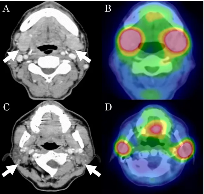
(5) Japanese Archive of cases conference of clinical nuclear medicine 02, 2020. P. 5. Fig. 1 Contrast-enhanced CT shows slight enlargement of the submandibular (A, arrows) and parotid (C, arrows) glands. A 67Ga SPECT/CT shows abnormal uptakes in each gland (B, D)..
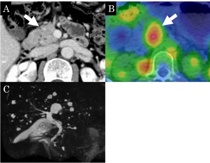
(6) Japanese Archive of cases conference of clinical nuclear medicine 02, 2020. P. 6. Fig. 2 While contrast-enhanced CT does not indicate pancreatic enlargement (A, arrow), a 67Ga SPECT/CT shows accumulation in the remnant pancreas after resection (B, arrow). Magnetic resonance cholangiopancreatography does not indicate a stricture of the main pancreatic or bile ducts (C)..
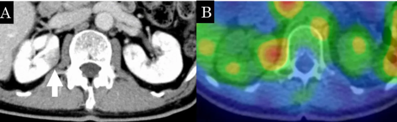
(7) Japanese Archive of cases conference of clinical nuclear medicine 02, 2020. P. 7. Fig. 3 The wedge-shaped low density area is seen on contrast-enhanced CT (A, arrow), suggesting interstitial nephritis. A 67Ga SPECT/CT shows a high accumulation in the corresponding area (B)..
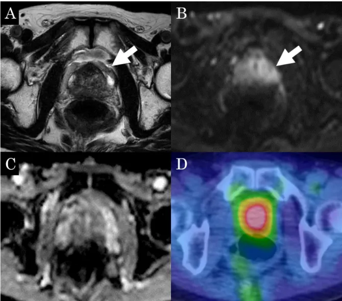
(8) Japanese Archive of cases conference of clinical nuclear medicine 02, 2020. P. 8. Fig. 4 An MRI showed low signal intensity on T2WI (A, arrow) and mild high signal intensity on the diffusion-weighted image (B, arrow), suggesting prostatitis. The corresponding area shows heterogenous signal intensity on apparent diffusion coefficient map (C). A 67Ga SPECT/CT shows high accumulation in the prostate (D).. Conclusion We report on a case of submandibular gland swelling 11 years after tumor-forming pancreatitis, which eventually led to a diagnosis of IgG4-related disease. A whole-body survey via 67Ga scan is considered very useful for IgG4-related diseases that involve organs throughout the body..
(9) Japanese Archive of cases conference of clinical nuclear medicine 02, 2020. P. 9. References 1. Kamisawa T, Zen Y, Pillai S, Stone JH. IgG4-related disease. Lancet. 2015;385:146071. doi:10.1016/s0140-6736(14)60720-0. 2. Umehara H, Okazaki K, Masaki Y, Kawano M, Yamamoto M, Saeki T, et al. Comprehensive diagnostic criteria for IgG4-related disease (IgG4-RD), 2011. Modern Rheumatology. 2012;22:21-30. doi:10.3109/s10165-011-0571-z. 3. Ishii S, Shishido F, Miyajima M, Sakuma K, Shigihara T, Kikuchi K. Whole-body gallium-67 scintigraphic findings in IgG4-related disease. Clin Nucl Med. 2011;36:542-5. doi:10.1097/RLU.0b013e318217ae16. 4. Zhang J, Chen H, Ma Y, Xiao Y, Niu N, Lin W, et al. Characterizing IgG4-related disease with (1)(8)F-FDG PET/CT: a prospective cohort study. Eur J Nucl Med Mol Imaging. 2014;41:1624-34. doi:10.1007/s00259-014-2729-3. 5. Khosroshahi A, Wallace ZS, Crowe JL, Akamizu T, Azumi A, Carruthers MN, et al. International Consensus Guidance Statement on the Management and Treatment of IgG4Related Disease. Arthritis Rheumatol. 2015;67:1688-99. doi:10.1002/art.39132..
(10)
図
関連したドキュメント
1) Tamaki N, Cuidlines for clinical use of cardiac nuclear medicine (JSC 2005). Neuronal imaging using SPECT. Eur J Nucl Med Mol Imaging. Role of MIBG myocardial scintigraphy in
The other is to find an associated lesion in patients with typical IgG4-related disease, such as autoimmune pancreatitis or IgG4-related chronic sclerosing dacryoadenitis and
Report of two cases. Post-operative herpes simplex virus encephalitis after surgical resection of acoustic neuroma: a case report. Herpes simplex virus reactivation
Tsujimoto, Yuichi; Satoh, Mototaka; Takada, Tsuyoshi; Honda, Masahito; Matsumiya, Kiyomi;. Fujioka, Hideki;
patient with apraxia of speech -A preliminary case report-, Annual Bulletin, RILP, Univ.. J.: Apraxia of speech in patients with Broca's aphasia ; A
Usefulness of positron emission tomography (PET) in a retroperitoneal primary non- seminomatous germ cell tumor: a
Correspondence should be addressed to Salah Badraoui, [email protected] Received 11 July 2009; Accepted 5 January 2010.. Academic Editor:
An example of a database state in the lextensive category of finite sets, for the EA sketch of our school data specification is provided by any database which models the
