INTRODUCTION
Although both dental pulp and dentin originate from mesenchymal dental papilla, the former re-mains unmineralized while the latter is highly min-eralized (1). Dental pulp cells are responsible for homeostasis of dental mineralized tissue. For ex-ample, pulp tissue forms reparative dentin in pro-tective response to external stimuli including caries, chemicals, and trauma (2, 3). In addition, the size of the dental pulp cavity is reduced as a result of peritubular dentin deposition with advancing age (4). On the other hand, the incidence of pulp stone formation that is also a mineralized product is not age-dependent (5). The regulatory mechanisms of
both mineralization-inducing activity and maintain-ing activity in the unmineralized state remain to be clarified.
Matrix mineralization is observed in primary culture of pulp cells. In contrast, clonal pulp cells do not easily produce mineralized nodules even though osteogenic agents such as bone morpho-genetic proteins (BMPs) are added to the culture as shown previously (6). On the other hand, mouse calvaria-derived preosteoblastic cell line MC3T3-E1 not only forms mineralized nodules steadily (7) but also secretes a variety of cytokines including BMP-2 and BMP-4 (8, 9). No other established cell line induces matrix mineralization easily. In addition, conditioned medium (CM) from
MC3T3-ORIGINAL
Soluble matrix from osteoblastic cells induces mineralization
by dental pulp cells
Akemichi Ueno
1, Kikuji Yamashita
2, Keiko Miyoshi
1, Taigo Horiguchi
1, Intan Ruspita
1,
Kaori Abe
1, and Takafumi Noma
11Department of Molecular Biology, and2Department of Oral and Maxillofacial Anatomy, Institute
of Health Biosciences, The University of Tokushima Graduate School, Tokushima, Japan
Abstract : Dental pulp cells have a capacity to differentiate into mineralization-inducing cells. To clarify the molecular mechanism, we established an in vitro mineralization-inducing system by rat clonal dental pulp cell line, RPC-C2A, and tried to purify a mineralization-inducing factor in conditioned medium (CM) from preosteoblastic MC3T3-E1 cells. The active factor was impermeable to an ultrafiltrating membrane, and sedimented by ultra-centrifugation. The sedimented factor was found as a needle-like structure about 1.3µm in average length as observed by transmission electron microscopy. The factor contained type I collagen, suggesting not a matrix vesicle, but a soluble matrix. The mineralization-inducing activity was also detected in CM from primary culture of rat calvaria (RC) cells. These results suggested that the soluble matrices from osteoblastic cells serve, at least in part, as differentiation-inducing agents. J. Med. Invest. 53 : 297-302, August, 2006 Keywords : dental pulp cells ; osteoblast ; extracellular matrix ; mineralization
Abbreviations : BMP, bone morphogenetic protein ; CBB, Coomassie brilliant blue ; CM, conditioned medium ; MW, mo-lecular weight ; RC, rat calvaria ; RT-PCR, reverse transcription-PCR ; SDS-PAGE, sodium dodecyl sulfate-polyacrylamide gel electrophoresis ; TEM, transmission electron microscopy ; TGF-β, transforming growth factor-beta ; WCE, whole cell extract
Received for publication May 17, 2006; accepted June 16, 2006. Address correspondence and reprint requests to Takafumi Noma, Department of Molecular Biology, Institute of Health Biosciences, The University of Tokushima Graduate School, Kuramoto-cho, Tokushima 770-8504, Japan and Fax: +81-88-633-7326.
The Journal of Medical Investigation Vol. 53 2006 297
E1 cells induce mineralization by clonal rat dental pulp cells, whereas CM from rat osteosarcoma cells ROS 17/2.8, normal human articular chondrocytes, or normal human dermal fibroblasts do not induce mineralization by them in vitro ((6), and unpub-lished observation by Ueno et al .). There are sev-eral diverse mechanisms of minsev-eral deposition during the formation of hard tissues. Acellular mineraliza-tion in biodegradable three-dimensional structures occurs in cell-free culture media (10) as well as at the implantation sites (11) and in the early stage of cementum formation in molars (12). On the other hand, it is generally accepted that cellular miner-alization is initiated by release of matrix vesicles from living cells and by apoptotic bodies from dying cells (13, 14). It is well known that matrix vesicles contain actin, annexins II and V, carbonic anhydrase, phosphatases (pyrophosphatase, alkaline phosphatase (ALP), 5’AMPase, Ca-ATPase), pro-teoglycan link protein, type X collagen, calbindin, lactate dehydrogenase, membrane-derived phos-pholipids, but not type I collagen (13, 14). In con-trast, the constituents of media vesicles and soluble matrix remain unknown. However, vesicle-mediated mineralization is not observed in many inverte-brates and enamel tissues (15, 16). Thus the mechanism of initiation of matrix mineral deposi-tion still remains to be determined.
In this study to investigate both mineralization-inducing and maintaining mechanisms at a mo-lecular level, we partially purified and character-ized the active substance from CM of preosteoblas-tic MC3T3-E1 cells, applying von Kossa staining as well as performing the transmission electron mi-croscopic analysis.
MATERIALS AND METHODS
RT-PCR analysisTotal RNA was isolated with TRI REAGENT (Molecular Research Center) from RDP4-1, RPC-C2A, AtT20, Balb3T3, and C3H10T1/2 cells, reverse transcribed with oligo(dT), and subjected to PCR within the log-linear range of amplification for either ALP, BMP-2, BMP-4 (30 cycles), or GAPDH (20 cycles). ALP , BMP2, BMP4, and GAPDH -specific primers were as follows : ALP forward, 5’-CTGGACCTCATCAGGATTTG-3’ ; ALP reverse, 5’- GCTGTGAAGGGCTTCTTGTC - 3’; BMP-2 forward, 5’TGAACACAGCTGGTCTCAGG - 3’; BMP-2 reverse, 5’- GCTAAGCTCAGTGGGGACAC - 3’; BMP-4 forward,
5’- CAGAGCCAACACTGTGAGG - 3’; BMP-4 reverse, 5’- TCCACTCCCTTGAGGTAACG - 3’; GAPDH forward, 5’- CATTGACCTCAACTACATGG - 3’; GAPDH reverse, 5’- CTCAGTGTAGCCCAGGATGC - 3’ .
Cell culture
Confluent RPC-C2A cells and RDP 4-1 cells were cultured for 10 days in α-MEM medium supple-mented with 0.22% NaHCO3, antibiotics, 10% FBS, 2 mMβ-glycerophsphate, 50µg/ml L-ascorbic acid, and 10-8
M dexamethasone with or without 50% CM as described previously (6). The cells were subjected to phase-contrast microscopy in a Nikon TM-D mi-croscope (Nikon).
Transmission electron micrographs of sedimented active matrices
Culture fluid from day 1 to 4 after confluency was centrifuged to remove cells and cell debris for 5 min at 800 xg and CM was collected from the su-pernatant. After the CM was centrifuged for 30 min at 10,000 xg, the supernatant was subjected to ul-tracentrifugation for 3h at 150,000 xg in a Beckman TLA 100.3 rotor. For transmission electron micros-copy, the fixed and ethanol-dehydrated pellets were embedded in Epon 812 (TAAB Laboratories) and sections were cut on an LKB Ultratome V (LKB). Ultrathin sections were double-stained with uranyl acetate in 50% ethanol and Reynolds solution and examined in a Hitachi H-800 electron microscope (Hitachi).
Western blot analysis
Samples (40µg) were separated by 7.5% SDS-polyacrylamide gel electrophoresis as described previously (17). Proteins were transferred electro-phoretically to polyvinylidene difluoride (PVDF) membranes (Immobilon-P ; Millipore). The mem-branes were blocked with TBS-T buffer (20 mM Tris-HCl, 137 mM NaCl, 0.1% Tween-20, pH 7.5) containing 5% skim milk (Difco) and labeled with rabbit anti-mouse type I collagen antibody (Calbio-chem)(1 : 500 diluted) and then with horseradish peroxidase-conjugated donkey anti-rabbit IgG (Amersham Biosciences). Immunoreactive bands were located using an ECL Western blotting de-tection kit (Amersham Biosciences) and RU X-ray film (Fujifilm). The relative intensity of the signals measured by JOYCE-LOEBL Microdensi-tometer 3CS (Joyce-Loebl).
Mineralization-inducing assay
A. Ueno, et al. Mineralization induced by soluble matrix
Confluent RPC-C2A cells were cultured for 10 days with or without CM from either RC cells or MC3T3-E1 cells. For staining of mineralized nod-ules, the cells were fixed with 10% neutral buffered formalin and stained by the von Kossa technique with 3% silver nitrate as described previously (18).
Cell growth
CM from RC cells or MC3T3-E1 cells was added to the culture of confluent RPC-C2A cells. Cell growth was measured with the metabolic indicator Alamar Blue (Biosource International)(10% of incubation volume), which was added at 24h after CM treat-ment (19). After a 4-hour incubation, part of the medium was collected and the extent of prolifera-tion was determined by measuring each plate at 570 nm and 600 nm for each plate in a Beckman DU 8 spectrophotometer. To obtain the specific absorbance for the level of the reduced state, the absorbance at 600 nm was subtracted from that at 570 nm. The cell-free blank value was then sub-tracted from each value. Data obtained from the mean of two separate experiments in triplicate are expressed as percentage of control (0% CM).
RESULTS
Expressions of osteogenic related genes
Extracellular matrix (ECM) mineralization is a physiological process in bones and teeth while a pathological process in anywhere else. Although the molecular mechanisms for ECM mineraliza-tion are not fully understood, ALPs such as tissue-nonspecific alkaline phosphatase and BMPs are possibly involved in the formation of mineralized matrix made up of hydroxyapatite and type I colla-gen (20). At first, RT-PCR analysis was performed to examine the expressions of osteogenic related genes including BMP2, BMP4, and alkaline
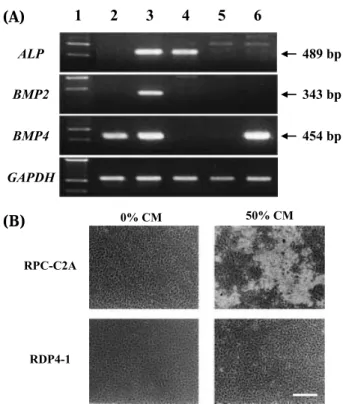
phos-phatase,in clonal cell lines without exogenous factors (Fig. 1A). BMP-2mRNA was detected in RDP4-1, another clonal rat dental pulp-derived cell line, and C3H10T1/2 cells which also differentiate to os-teoblastic cells. The only cell line that simultane-ously expressed three osteogenic mRNA (BMP-2, BMP-4, and alkaline phosphatase) was RPC-C 2 A, suggesting that this may be is the best target den-tal pulp-derived cell line for the induction of miner-alization among clonal cell lines examined.
Mineralization-inducing activity
Addition of CM from MC3T3-E1 cells has a sig-nificant effect upon bone nodule formation by RPC-C2A cells, but not by RDP4-1 cells (Fig. 1B). The active factor(s) in CM was not filtered through an ultrafiltration membrane (MW cutoff : 1,000,000), sug-gesting a large complex. Thus, CM was subjected to ultracentrifugation at 150,000 xg for 3h after cen-trifugation at 10,000 xg for 30 min. Mineralization-inducing activity was sedimented as approximately 1.3-µm-long needle-like complexes as revealed by TEM (Fig. 2). These sedimentable matrices
exhib-Fig. 1 RPC-C2A cells express osteogenic genes and produce
bone nodules after treatment with conditioned medium from MC 3T3-E1 cells. (A) RT-PCR analysis of ALP , BMP-2, BMP-4, and GAPDH mRNA. Lane 1, size marker ; lane 2, RDP-4-1 cells ; lane 3, RPC-C2A cells ; lane 4, AtT 20 cells, mouse pituitary tumor-derived cell line ; lane 5, Balb/c3T3 cells ; lane 6, C3H 10T1/2 cells. (B) Phase-contrast micrograph of rat dental pulp cells treated with conditioned medium (CM) from MC3T3-E1 cells. Bar, 200µm.
Fig. 2 Transmission electron micrographs of sedimented
active matrices. After ultracentrifugation of CM, the fixed and ethanol-dehydrated pellets were embedded in Epon 812 and sections were prepared with an LKB Ultratome V. Ultra thin sections were examined in a Hitachi H-800 electron micro-scope. Bar, 1µm.
(A)
(B)
ited mineralization-inducing activity in RPC-C2A cells. The mineralizing-inducing activity exclusively resided in the sedimented matrices not in the supernatant. In addition, we found that collage-nase treatment lost both structure and activity of sedimented matrices (data not shown).
Western blot analysis
In general, matrix vesicles do not contain type I collagen (13, 14). The CBB-stained SDS-PAGE pattern of sedimented matrices showed that the sample was not easily dissociated with a reducing agent and that the molecular weight seemed too high to run into a separation gel, suggesting that the sedimented matrices contain collagens. In order to verify whether the complex was a matrix vesicle or soluble matrix, SDS-PAGE and immunoblot analy-ses with anti-type I collagen antibody were per-formed. CBB-stained gel showed that most of the large MW sample did not move into the separation gel, suggesting that it did not dissociate easily with the SDS sample buffer (21) once after sedimented (Fig. 3A). After immunoblotting, type I collagen molecules containingα1 andα2 chains were detected at about 2 : 1 ratio in the dissociated sample in addition to the undenatured large MW collagen (lane 2 in Fig. 3B). In whole cell extract (WCE), the possible degradation products were also observed (lane 1 in Fig. 3B). These results suggest that the factor secreted is not a matrix vesicle but a soluble matrix.
von Kossa staining
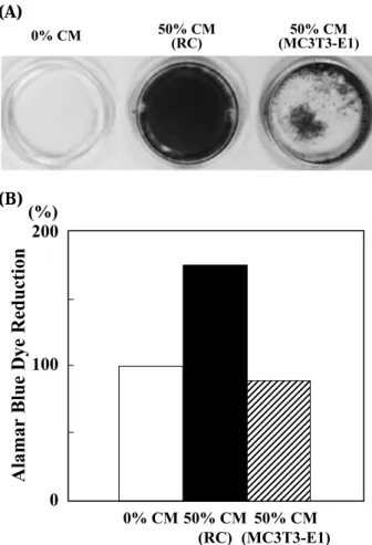
Next, in order to determine if a matrix such as this is restricted and specific to MC3T3-E1 cells, CM from primary-cultured rat calvaria (RC) cells (22) was examined for the activity of matrix miner-alization of C2A cells. The multilayered RPC-C2A cells and the mineralized nodules stained strongly by von Kossa technique (18) in their ECM were observed (Fig 4A).
Alamar Blue assay
ince the cell proliferation-promoting activity with CM from RC cells was observed by phase-contrast microscopy, Alamar Blue assay was performed to quantify the reduction ability by mitochondria (18) (Fig. 4B). CM from RC cells exhibited stimulation of the RPC-C2A cell growth by 1.8-fold, whereas CM from MC3T3-E1 cells was without effect, indi-cating that the RC CM includes a growth factor in addition to a differentiation factor.
Fig. 3 Detection of type I collagen in the sedimented matrix.
(A) Samples were separated by 7.5% SDS-polyacrylamide gel electrophoresis and the gel was stained with Coomassie bril-liant blue R250. Lanes 1 and 2, WCE of MC3T3-E1(25 and 50µg, respectively) ; lanes 3-5, 150,000xg pellet (25, 50, and 75µg, respectively) ; lane 6, broad-range molecular weight (MW) marker (Apro Science). MWs (x10-3) are shown at the right margin. (B) Western blot analysis was performed with rabbit anti-mouse type I collagen antibody and horseradish peroxidase-conjugated donkey anti-rabbit IgG. Lane 1, MC3T3-E1 WCE ; lane 2,150,000xg pellet. Star (*) indicates immunoreactive bands.
Fig. 4 Effect of CM from primary-cultured rat calvaria (RC)
cells on mineralization and proliferation of RPC-C2A cells. (A) Confluent RPC-C2A cells were cultured with or without CM from either RC cells or MC3T3-E1 cells. In order to detect min-eralized nodules, the cells were stained by the von Kossa tech-nique. (B) CM from RC cells or MC3T3-E1 cells was added to the culture of confluent RPC-C2A cells. Cell growth was evalu-ated with Alamar Blue assay.
(A)
(B)
(A) (B)
A. Ueno, et al. Mineralization induced by soluble matrix
DISCUSSION
In the present study, the mineralization-inducing factor was found to be sedimented by ultracen-trifugation and composed of at least type I colla-gen (Figs. 2 and 3B). The matrix vesicle does not contain type I collagen and shows a round shape with approximately 200 nm in diameter at maximum (13, 14). Therefore, this mineralization-inducing factor is thought to be a kind of soluble matrix that possibly contains a differentiation factor and other ECM molecules besides type I collagen. To fur-ther examine both the potency and generality of induction of matrix mineralization by CM from osteoblastic cells, we analyzed for CM from RC cells (Fig. 4A). The CM from RC cells has an ex-tremely strong effect on matrix mineralization of RPC-C2A cells with the maximal concentration at 50% in medium, demonstrating a stronger activity than that of CM from MC3T3-E1 cells. This may be because MC3T3-E1 cells lost the capacity to produce a certain growth factor and a differentia-tion factor during the course of subculture by a number of generations (7). A growth factor increases the number of precursor cells differentiating to os-teoblasts, resulting in enhanced matrix mineraliza-tion.
In conclusion, we found that not only MC3T3-E1 cells but also RC cells produce the sedimentable matrices containing at least type I collagen (Fig. 2) that have an ECM mineralization inducing activity. The both structure and activity of sedimented matrices were lost by collagenase treatment, sug-gesting the importance and an involvement of type I in mineralization inducing activity. It is well known that extracellular matrix vesicles are not always the site of initial mineral deposition (13-16). In certain cell culture systems (23) and in turkey tendon (24), matrix vesicle and collagen minerali-zation start simultaneously but distinct in site. In addition, there are no extracellular matrix vesicles in many invertebrates and in enamel (15, 16). Therefore, there must be other means of initiating mineralization besides accumulating ions in a protected environment as matrix vesicles do. For example, as one of the new paradigms for the mor-phogenesis of calcium-based biominerals, Olszta
et al proposed the mineralization of type I collagen via a polymer-induced amorphous liquid-precursor process (25, 26). We believe that the results ob-tained from the present study provide another important issues for the new basic mechanism of
inducing matrix mineralization other than well-established matrix vesicles. Further molecular char-acterization and identification of the mineralization inducing activity in the CM is required to apply for medical purpose in future, including therapy for osteoporosis.
REFERENCES
1. Linde A:The extracellular matrix of the dental pulp and dentin. J Dent Res 64 : 523-529, 1985 2. Yamamura T : Differentiation of pulpal cells
and inductive influence of various matrices with reference to pulpal wound healing. J Dent Res 64 : 530-540, 1985
3. Tziafas D : Basic mechanisms of cytodifferen-tiation and dentinogenesis during dental pulp Repair. Int J Dev Biol 39 : 281-290, 1995 4. Kvaal SI, Koppang HS, Solheim T :
Relation-ship between age and deposit of peritubular dentine. Gerodontology 11 : 93-98, 1994 5. Al-Hadi Hamasha A, Darwazeh A : Prevalence
of pulp stones in Jordanian adults. Oral Surg Oral Med Oral Pathol Oral Radiol Endod 86 : 730-732, 1998
6. Ueno A, Kitase Y, Moriyama K, Inoue H : MC3T3-E1-conditioned medium-induced min-eralization by clonal rat dental pulp cells. Matrix Biol 20 : 347-355. 2001
7. Sudo H, Kodama HA, Amagai Y, Yamamoto S, Kasai S: In vitro differentiation and calcification in a new clonal osteogenic cell line derived from newborn mouse calvaria, J Cell Biol 96 : 191-198, 1983
8. Amarnani S, Merriman HL, Linkhart TA, Baylink DJ, Mohan S : Autocrine regulators of MC3T3-E1 cell proliferation. J Bone Miner Res 8 : 157-165, 1993
9. Suzawa M, Takeuchi Y, Fukumoto S, Kato S, Ueno N, Miyazono K, Matsumoto T, Fujita T : Extracellular matrix-associated bone morpho-genetic proteins are essential for differentia-tion of murine osteoblastic cells in vitro. En-docrinology 140 : 2125-2133, 1999
10. Andre-Frei V, Chevallay B, Orly I, Boudeulle M, Huc A, Herbage D : Acellular mineral deposi-tion in collagen-based biomaterials incubated in cell culture media. Calcif Tissue Int 66 : 204-211, 2000
11. Yamashita K, Takagi T: Calcification preceding new bone formation induced by
ized bone matrix gelatin. Arch Histol Cytol 55 : 1-43, 1992
12. Yamamoto T, Domon T, Takahashi S, Arambawatta AK, Wakita M : Immunolocation of proteocans and bone-related noncollagenous gly-coproteins in developing acellular cementum of rat molars. Cell Tissue Res 317: 299-312, 2004 13. Anderson HC : Molecular biology of matrix
vesicles. Clin Orthop 314 : 266-280, 1995 14. Anderson HC : Matrix vesicles and
calcifica-tion. Curr Rheumatol Rep 5 : 222-226, 2003 15. Boskey AL : Matrix proteins and
mineraliza-tion : an overview. Connect Tissue Res 35 : 357-363, 1996.
16. Hohling HJ, Arnold S, Barckhaus RH, Plate U, Wiesmann HP : Structural relationship be-tween the primary crystal formations and the matrix macromolecules in different hard tis-sues. Discussion of a general principle. Con-nect Tissue Res 33 : 171-178, 1995
17. Islam MR, Ueno A, Nishikawa H, Nagata T, Inoue H : Osteotropic factor-stimulated syn-thesis of thrombospondin in rat dental pulp cells. FEBS Lett 393 : 193-196, 1996
18. Otsuka E, Yamaguchi A, Hirose S, Hagiwara H: Characterization of osteoblastic differentiation of stromal cell line ST2 that is induced by ascorbic acid. Am J Physiol 277 : C132-C138, 1999
19. Ahmed SA, Gogal RM, Walsh Jr JE : A new rapid and simple non-radioactive assay to monitor and determine the proliferation of lymphocytes: an alternative to [3
H] thymidine incorporation
assay. J Immunol Methods 170 : 211-224, 1994 20. Murshed M, Harmey D, Millan JL, McKee MD, Karsenty G : Unique coexpression in os-teoblasts of broadly expressed genes accounts for the spatial restriction of ECM mineraliza-tion to bone. Genes Dev 19 : 1093-1104, 2005 21. Laemmli UK : Cleavage of structural proteins
during the assembly of the head of bacterio-phage T4. Nature 227 : 680-685, 1970
22. Bellows CG, Aubin J E, Heersche JN : Physi-ological concentrations of glucocorticoids stimu-late formation of bone nodules from isostimu-lated rat calvaria cells in vitro. Endocrinology 121 : 1985-1992, 1987
23. Boskey AL, Stiner D, Leboy P, Doty S, Bindernan I : Optimal conditions for cartilage calcification in differentiating chick limb-bud mesenchy-mal cells. Bone and Mineral 14 : 11-37, 1992 24. Landis WJ : Temporal sequence of
mineraliza-tion in calcifying turkey tendon. In : WT Butler, eds. The Chemistry and Biology of Mineralized Tissues. Ebsco Media., Birmingham, 1985, pp. 360-364
25. Olszta MJ, Douglas EP, Gower LB : Scanning electron microscopic analysis of the minerali-zation of type I collagen via a polymer-induced liquid-precursor (PILP) process. Calcif Tissue Int 72 : 583-591, 2003
26. Olszta MJ, Odom DJ, Douglas EP, Gower LB : A new paradigm for biomineral formation : mineralization via an amorphous liquid-phase precursor. Connect Tissue Res 44 (Suppl 1) : 326 -334, 2003
A. Ueno, et al. Mineralization induced by soluble matrix