Yasuhiro Imamura et al.: Placenta extract modulates cell metabolism of Saos-2
Original
Effects of Placental Extract on Cell Proliferation, Type I Collagen Production,
and ALP Secretion in Human Osteosarcoma Cell Line Saos-2
Yasuhiro Imamura1), Yoshitomo Honda2), Kazuya Masuno3), Hiroe Nakamura4) and Pao-Li Wang4)
1) Department of Dental Pharmacology, Matsumoto Dental University, Shiojiri, Japan 2) Institute of Dental Research, Osaka Dental University, Osaka, Japan
3) Department of Innovation in Dental Education, Osaka Dental University, Osaka, Japan 4) Department of Bacteriology, Osaka Dental University, Osaka, Japan
(Accepted for publication, February 22, 2017)
Abstract: Porcine placenta extract (P-placenta) is widely applied in medicine and cosmetics. However, few studies have examined the effect of the extract on the cellular behavior of the osteoblastic cell line Saos-2. Here, we demonstrated that P-placenta enhances the proliferation, collagen type I production, and alkaline phosphatase (ALP) secretion of Saos-2 in vitro. Proliferation of Saos-2 was assessed by MTT and DNA synthesis assays. Type I collagen production and ALP secretion were evaluated using enzyme-linked immunosorbent assay and ALP assays. The cells were treated with/without 20, 200 and 2000 g/ml of P-placenta for 24 h. We found that 200 g/ml P-placenta significantly induced the proliferation of Saos-2 and enhanced type I collagen production and ALP secretion. The results indicate that P-placenta controls the cellular behavior of osteoblasts, resulting in the secretion of early bone-related biomarkers.
Key words: ALP, Collagen, Osteoblast, Placenta, Saos-2
Correspondence to: Dr. Pao-Li Wang, 8-1 Kuzuhahanazono, Hirakara, Osaka, 573-1121 Japan; Tel: +81-72-864-3011; Fax: +81-72-864-3101; E-mail: [email protected]
Introduction
The placenta is a temporary organ that supports fetal growth by supplying oxygen and nutrients1). The organ stores diverse
molecules such as vitamins, bioactive peptides, minerals, and growth factors2,3). The placental extract has attracted attention
worldwide owing to its potential as drugs for clinical treatments4)
and as supplements in cosmetics3,4). Previous studies showed that
placental extract has various therapeutic effects, such as wound-healing5), anti-inflammatory6), and immune-modulating potential7).
More recently, we reported that porcine placenta extract (P-placenta) controls type I collagen production, cell proliferation, and anti-inflammatory cytokine production of human gingival fibroblasts (HGFs)8). However, its pharmaceutical mechanism in
other cells, such as bone-related cells, has not been thoroughly examined.
Bone metabolism is systematically regulated by the complex iteration of bone formation and resorption. Osteoblasts mainly engage in bone formation by secreting extracellular matrix, such as type I collagen and other organic proteins. Alkaline phosphatase (ALP) secreted from osteoblasts degrades inorganic pyrophosphate to phosphate, which is associated with the mineralization of bone9).
Therefore, type I collagen and ALP are important indicators of bone metabolism10,11). However, little information is available
regarding the effect of placenta on the cellular metabolism of osteoblasts.
The aim of this study was to evaluate the effect of P-placenta on osteoblastic cells, particularly cell proliferation, type I collagen production, and ALP secretion. To evaluate these issues, we used the osteoblastic cell line Saos-2, which was first established from osteosarcoma in 198712). ALP secretion, type I collagen production,
and mineralized matrix production have been evaluated in Saos-2 cells and osteoblasts12). These cells present mature osteoblastic
characteristics13), and thus have been extensively investigated in
oncology14) and bone biology studies13). Additionally, biomaterial
function was previously evaluated using Saos-2 cells15,16).
Materials and Methods Reagents
P-placenta was purchased from Nippon Meat Packers, Inc. (Osaka, Japan). The powder-like extract was composed of natural components from the porcine placenta without any additives. Therefore, we used these reagents for the following experiments.
Cell maintenance
Saos-2 cells (HTB85) were provided by the Riken BRC Cell
Journal of Hard Tissue Biology 26[2] (2017) 157-160 © 2017 The Hard Tissue Biology Network Association Printed in Japan, All rights reserved. CODEN-JHTBFF, ISSN 1341-7649
J.Hard Tissue Biology Vol. 26(2):157 -160, 2017
Bank (Tsukuba, Japan). Cells were cultured in Dulbecco’s Modified Eagle Medium (DMEM, Nissui Pharmaceutical Co., Ltd., Tokyo, Japan) containing 10% fetal bovine serum (FBS) at 5% CO2 and 37°C.
3-(4, 5-Dimethylthial-2-yl)-2, 5-diphenyltetrazolium bromide (MTT) and DNA synthesis assay
For DNA synthesis, Saos-2 seeded at 3.1 x 10⁴/cm2 were
cultured in DMEM containing 0.5% FBS with/without P-placenta at doses of 20, 200, and 2000 g/ml for 24 h. The level of DNA synthesis in cells was determined by measuring BrdU-incorporation using the Frontier BrdU Cell Proliferation assay kit (Millipore, Billerica, MA, USA). For the MTT assay, cells were cultured in DMEM containing 10% FBS with P-placenta at the above concentrations for 24 h. The MTT assay was conducted as reported previously17).
Type I collagen production
Cells were seeded at the 3.1 x 10⁴/cm2 in DMEM with 10%
FBS, followed by treatment with DMEM containing 1% FBS with/ without P-placenta (200 g/ml) for 24 h. The levels of collagen type I in the media were measured by an enzyme-linked immunosorbent assay (ELISA), using a biotinylated anti-collagen type I antibody (0.2 mg/ml, Rockland Immunochemicals, Limerick, PA, USA). Total protein levels were quantified in the cell lysate isolated with 0.5% Triton X-100 by using a BCA protein assay kit (Thermo Fisher Scientific, Waltham, MA, USA). Collagen production was normalized to the protein content in the cell lysates.
ALP assay
Cells were seeded at a density of 3.1 x 10⁴/cm2 in DMEM
containing 10% FBS. Next, the cells were treated with DMEM containing 1% FBS with/without P-placenta (200 g/ml) for 24 h. The cells were lysed with 0.05% TritonX-100 (200 l), followed by centrifugation at 14,000 xg for 1 min, and the supernatants were collected. ALP activity in the lysates (20 l) was measured using a LabAssay ALP kit (Wako Pure Chemicals Industries, Ltd., Osaka, Japan). Protein levels in the cell lysates were also measured using the BCA protein assay kit (Thermo Fisher Scientific). ALP activity was normalized to the total protein level in the cell lysates.
Statistical analysis
Statistical analysis was carried out using StatMate software (ATMS, Tokyo, Japan). Statistical significance was evaluated by one-way analysis of variance with a Dunnett’s test or by student’s 5 4 3 2 1 0 140 120 100 80 60 40 20 0 a b R el at iv e D N A s yn th es is C el l v ia bi li ty ( % ) p-placenta no ne no ne p-placenta + ++ +++ + ++ +++
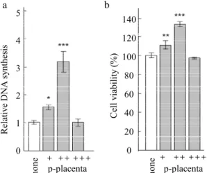
Figure 1. Effect of P-placenta on the proliferation of Saos-2 cells. (a) DNA synthesis assay. (b) MTT assay. Saos-2 seeded at a volume of 3.1 × 10⁴/cm2 were exposed to media containing 20, 200, or
2000 µg/ml P-placenta (designated as +, ++, and +++) for 24 h. All data were compared with those for cells treated with control medium without P-placenta (none). Data are shown as the mean ± standard deviation (n = 3). *P < 0.05, **P < 0.01 and ***P < 0.001, analyzed with analysis of variance with a Dunnett’s test (vs. none). 60 50 40 30 20 10 0 C ol la ge n I (p g/ g ) no ne p-pl ac en ta
Figure 2. Type I collagen production in Saos-2 cells treated with P-placenta. Saos-2 cells were seeded at a volume of 3.1 × 10⁴/cm2 and
exposed to media containing 200 µg/ ml P-placenta for 24 h. Data are shown as the mean ± standard deviation (n = 3). **P < 0.01, analyzed by Student’s t-test (vs. none). 7 6 5 4 3 2 1 0 no ne p-pl ac en ta A lp ( un it / g)
Figure 3. ALP secretion in Saos-2 cells treated with P-placenta. Saos-2 cells were seeded at 3.1 × 10⁴/cm2,
and exposed to media containing 200 µg/ml P-placenta for 24 h. Data are shown as the mean ± standard deviation (n = 3). **P < 0.01, analyzed by Student’s t-test (vs. none)
Yasuhiro Imamura et al.: Placenta extract modulates cell metabolism of Saos-2 t-test. Levels of P < 0.05, P < 0.01 and P < 0.001 were considered
to indicate statistical significance.
Results
Effect of P-placenta on Saos-2 cell proliferation
After 24 h of treatment, 200 g/ml P-placenta increased the level of DNA synthesis compared to the effect of other concentrations in Saos-2, whereas no obvious change was observed at 2000 g/ml P-placenta treatment (Fig.1a). The MTT assay showed that 200 g/ml P-placenta significantly elevated the cell viability of Saos-2 cells (Fig.1b). These results suggest that there is optimal concentration of P-placenta that facilitates Saos-2 cell proliferation.
Effect of P-placenta on type I collagen production and ALP secretion from Saos-2 cells
Based on the proliferation assay results, we further evaluated the effect of the P-placenta on type I collagen production and ALP secretion from Saos-2 cells. Addition of P-placenta to the media significantly increased the levels of type I collagen production (Fig. 2) and ALP secretion (Fig. 3) from Saos-2 cells compared to that in media without P-placenta.
Discussion
Although P-placenta may have therapeutic potential not only in medicinal drugs but also in cosmetic supplements3), its detailed
function in the cellular metabolism of osteoblasts remains unclear. In the present study, we demonstrated P-placenta enhances the cell proliferation and regulates the synthesis and secretion of bone-related proteins in osteoblastic Saos-2 cells.
Under the P-placenta stimulation, 200 g/ml condition significantly enhanced the proliferation of Saos-2 in vitro. In contrast, 2000 g/ml P-placenta showed a negligible effect on cell proliferation (Fig. 1). We previously reported that HGFs shows a similar cell proliferation tendency under the same P-placenta concentration8). Yoshikawa et al. reported that 0–100 g/ml
P-placenta elevated the proliferation of human skin fibroblasts18).
These results suggest that concentrations near 100 and 200 g/ml are sufficient for inducing the proliferation of human somatic cells originating from mesenchymal stem cells by using P-placenta in vitro.
Using 200 g/ml P-placenta, we found that the extract induced type I collagen production and ALP secretion from Saos-2 cells in vitro. The mechanism by which P-placenta elevated this secretion remains unclear. However, P-placenta is produced from porcine placenta extract without any impurities. P-placenta and human placenta extract4,19) are known to contain various minerals,
vitamins and growth factors. Including insulin-like growth factor-1, a key regulator of fetal development, are synthesized in placenta20). Type I collagen synthesis in Saos-2 cells is reportedly
upregulated by insulin-like growth factor-110). Moreover, Saos-2
cells respond to extracellular calcium21), hormones12),
microRNAs22), and growth factors10). These results support that
the multiple molecules in P-placenta synergistically, rather than solely, promote type I collagen production and ALP secretion.
A study conducted in Italy indicated that placenta treatment improved periodontal disease, a major cause of tooth loss23).
However, few studies have evaluated how placenta extract controls the condition of inflammation and bone loss in periodontal disease. More recently, we reported that P-placenta has an anti-inflammatory effect on HGFs in vitro8). The current study indicates
that P-placenta can elevate the secretion of bone-related protein (type I collagen and ALP), which are pivotal in bone formation (Figs. 2 and 3). Although there is a wide gap between in vitro and in vivo results, additional studies may reveal the mechanism underlying these effects of placenta on bone formation via protein production of osteoblastic cells, which may be useful for treating periodontal diseases. Peri-implant diseases lead to implant failure due to bone loss and loss of osseointegration24). Therefore,
preventing or treating these diseases is important in dental implant treatment. Regeneration therapy has been advocated as an approach for curing peri-implant diseases25). Given the function of P-placenta
in in vitro culture of Saos-2 cells in the present study, this extract might be effective for the treatment of not only periodontitis, but also peri-implant diseases, and in bone regenerative therapy. However, because the present study was performed under limited conditions and cell lines, additional studies of primary osteoblasts are imperative. Further detailed in vivo experiments are also required to support these results.
In conclusion, we demonstrated that P-placenta enhanced the cell proliferation, type I collagen production, and ALP secretion of Saos-2 cells in vitro. Although it remains unknown how placenta extract modulates the secretion of other bone-related proteins, our results suggest that the extract modulates the cellular functions of osteoblastic cells.
Acknowledgements
This study was supported by the Grant-in Aid for Scientific Research (15K11438) from JSPS.
Conflict of Interest The authors have declared that no COI exists.
References
1. Jash A, Kwon HK, Sahoo A, Lee CG, So JS, Kim J, Oh YK, Kim YB and Im SH. Topical application of porcine placenta extract inhibits the progression of experimental contact hypersensitivity. J Ethnopharmacol 133: 654-662, 2011 2. Cho H, Ryou J, Lee J and Lee M. The effects of placental
extract on fibroblast proliferation. J Cosmet Sci 59: 195-159
J.Hard Tissue Biology Vol. 26(2):157 -160, 2017 202, 2008
3. Intrama V, Karnchanatat A, Bunaprasert T and Vadhanasindhu P. Study of regulation approach of growth factor product from human placenta in thailand. The First International Conference on Interdisciplinary Research and Development 19: 17.1-17.4, 2011
4. Lee KK, Choi WS, Yum KS, Song SW, Ock SM, Park SB and Kim MJ. Efficacy and safety of human placental extract solution on fatigue: a double-blind, randomized, placebo-controlled study. eCAM 2012: 130875, 2012
5. Navadiya S, Patel M and Vaghani Y. Study of topical placental extract versus povidone iodine and saline dressing in various diabetic wounds. National J Med Res 2: 411-413, 2012 6. Kawakat su M, Y U, Goto S, Ono Y and Li TS.
Placental extract protects bone marrow-derived stem/ progenitor cells against radiation injury through anti-inflammatory activity. J Rad Res 54: 268-276, 2013 7. Ansari KU, Gupta N and Bapat SK. An experimental and
clinical evaluation of immuno-modulating potential of human placenta extract. Indian J Pharmacol 26: 130-132, 1994
8. Honda Y, Imamura Y, Fukui T, Masuno K and Wang P. Effect of placenta on collagen type-1and inflammatory cytokine production in human gingival fibroblasts. Oral Therap Pharmacol 34: 94-99, 2015
9. Golub E and Boesze-Battaglia K. The role of alkaline phosphatase in mineralization. Curr Opin Orthop 18: 444-448, 2007
10. Kudo Y, Iwashita M, Iguchi T and Takeda Y. The regulation of type-I collagen synthesis by insulin-like growth factor-I in human osteoblast-like SaOS-2 cells. Pflugers Arch 433: 123-128, 1996
11. Orimo H and Shimada T. Regulation of the human tissue-nonspecific alkaline phosphatase gene expression by all-trans-retinoic acid in SaOS-2 osteosarcoma cell line. Bone 36: 866-876, 2005
12. Rodan SB, Imai Y, Thiede MA, Wesolowski G, Thompson D, Bar-Shavit Z, Shull S, Mann K and Rodan GA. Characterization of a human osteosarcoma cell line (Saos-2) with osteoblastic properties. Cancer Res 47: 4961-4966, 1987
13. Pautke C, Schieker M, Tischer T, Kolk A, Neth P, Mutschler W and Milz S. Characterization of osteosarcoma cell lines MG-63, Saos-2 and U-2 OS in comparison to human osteoblasts. Anticancer Res 24: 3743-3748, 2004
14. Rasola A, Sciacovelli M, Chiara F, Pantic B, Brusilow WS and Bernardi P. Activation of mitochondrial ERK protects cancer cells from death through inhibition of the permeability transition. Proc Natl Acad Sci U S A 107: 726-731, 2010 15. Postiglione L, Di Domenico G, Ramaglia L, di Lauro AE, Di
Meglio F and Montagnani S. Different titanium surfaces modulate the bone phenotype of SaOS-2 osteoblast-like cells. Eur J Histochem 48: 213-222, 2004
16. Alcaide M, Serrano MC, Pagani R, Sanchez-Salcedo S, Nieto A, Vallet-Regi M and Portoles MT. L929 fibroblast and Saos-2 osteoblast response to hydroxyapatite-betaTCP/ agarose biomaterial. J Biomed Mater Res A 89: 539-549, 2009
17. Imamura Y, Fujigaki Y, Oomori Y, Usui S and Wang P. Cooperation of salivary protein histatin 3 with heat shock cognate protein 70 relative to the G1/S transition in human gingival fibroblasts. J Biol Chem 284: 14316-14325, 2009 18. Yoshikawa C. Effect of porcine placental extract on collagen production in human skin fibroblasts in vitro. Gynecol Obstetr 3: 1-4, 2013
19. Chakraborty P and Bhattacharyya D. Aqueous extract of human placenta as a therapeutic agent, In: Recent Advances in Research on the Human Placenta, ed by Zheng J, In Tech Europe Rijeka, 2012, pp77-92
20. Hiden U, Glitzner E, Hartmann M and Desoye G. Insulin and the IGF system in the human placenta of normal and diabetic pregnancies. J Anat 215: 60-68, 2009
21. Chang W, Tu C, Chen TH, Komuves L, Oda Y, Pratt SA, Miller S and Shoback D. Expression and signal transduction of calcium-sensing receptors in cartilage and bone. Endocrinology 140: 5883-5893, 1999
22. Qu F, Li CB, Yuan BT, Qi W, Li HL, Shen XZ, Zhao G, Wang JT and Liu YJ. MicroRNA-26a induces osteosarcoma cell growth and metastasis via the Wnt/beta-catenin pathway. Oncol Lett 11: 1592-1596, 2016
23. Calvarano G, De Polis F and Sabatini G. Treatment with placental extract in periodontal disease. Dent Cadmos 57: 85-86, 1989
24. Atieh MA, Alsabeeha NH, Faggion CM, Jr. and Duncan WJ. The frequency of peri-implant diseases: a systematic review and meta-analysis. J Periodontol 84: 1586-1598, 2013 25. Larsson L, Decker AM, Nibali L, Pilipchuk SP, Berglundh T
and Giannobile WV. Regenerative medicine for periodontal and peri-implant diseases. J Dent Res 95: 255-266, 2016