INTRODUCTION
Osseointegration is currently considered the optimal implanttobone interface.
1Natural teeth are at
tached to the surrounding alveolar bone via soft periodontal ligament tissues. With the loss of natu
ral teeth, the periodontal ligament cells are lost as well. Therefore, those cells cannot participate in wound healing around endosseous implants that are inserted for the replacement of lost teeth. At present, optimal healing around implants is consid
ered achievable via a bone anchorage with intimate bonetoimplant contacts, called osseointegration.
2Since osseointegrated implants are “ankylosed” and do not have the same mobility as natural teeth with a periodontal ligament, efforts have been made during the past few decades to compensate for this obvious difference by integrating “shock absorbing
systems” into the implant or its superstructure.
3, 4Ideally, it would be preferable to have a system for anchoring dental implants with the same functional mobility as natural teeth.
There has been a concerted effort among materi
als scientists and clinicians worldwide to improve the performance of dental implants with the aim of accelerating and maintaining their integration into hard and soft tissues and/or extending their range of applications.
5The surface characteristics of the implant material affect the rate and extent of os
seointegration.
6Vandrovcová et al.
7recently re
viewed the growing evidence demonstrating that surfacemodified materials are highly effective for promoting the adhesion, growth, and osteogenic differentiation of cells.
The structures that were used in this study are nanostructures similar to the TiO
2nanotubes cre
Effect of nano modified titanium surface on adsorption of rat periodontal ligament cells
Yukari Hara, Satoshi Komasa, Shigeki Yoshimine, Hiroshi Nishizaki and Joji Okazaki
Department of Removable Prosthodontics and Occlusion, Osaka Dental University, 8-1 Kuzuhahanazono-cho, Hirakata-shi, Osaka 573-1121, Japan
When natural teeth are lost, endosseous implants anchored via intimate bonetoimplant contacts are considered an appropriate replacement. The interactions between implants and host tissues depend on several factors. In particular, a growing body of evidence has demonstrated that the surface texture of an implant influences the response of the surrounding cells. Recent studies have shown that treating titanium with aqueous NaOH produces a nanostructured surface texture termed a titanium nanosheet (TNS). Further
more, it was recently reported that a TNS surface promoted the osteogenic differentiation of rat bone marrow cells. We investigated whether a TNS surface may improve wound healing around endosseous implants. To test this, we assessed the effects of a TNS sur
face on the adhesion and differentiation of rat periodontal ligament (RPL) cells in vitro.
The results demonstrated that RPL cells cultured on a TNS surface showed more adhe
sion and higher levels of osteogenic differentiation markers (Runx2 expression, alkaline phosphatase activity, calcium deposition, and osteocalcin secretion) than cells cultured on unmodified titanium. These findings indicate that RPL cells have the potential for in
creased bone formation on a TNS surface. Thus, TNS surface modification may warrant consideration for use in endosseous dental implants. (J Osaka Dent Univ 2018 ; 52 : 37
44)
Key words : Periodontal ligament cells ; Titanium nanostructures ; Implant
ated by titanium deposition using the process of TiO
2sputtering,
8and are called titanium nanosheets (TNS). Recently, it has been demonstrated that nanotube and TNS structures could be obtained on a titanium metal surface by treating the surface with a 10 M NaOH aqueous solution at 30° C.
9We em
ployed this method here to create TNS structures on modified titanium disks. Recent research has shown that treatment with an NaOH aqueous solu
tion produces a rough, nanoscale surface,
10and scanning electron microscopy images of our modi
fied disks demonstrated that the TNSmodified sur
face had a good surface roughness without any cracks. A previous study
11reported that TNS pro
duced via chemical processing promoted the osteo
genic differentiation of rat bone marrow cells. The surface properties and structures of materials influ
ence the adsorption of proteins. Thus, the in
creased adsorption of proteins on a TNSmodified surface might influence cell behavior.
Schroeder et al. demonstrated the successful placement of titanium implants using a nonsub
merged technique and showed for the first time un
dermineralized histological sections of titanium with soft and hard tissues.
12The reaction of soft tissues to the implant surface has been further examined by Schroeder et al.,
12Buser et al.,
13and Listgarten et al.
14These studies indicated that the surface tex
ture of an implant significantly affects the type of connective tissue that attaches to it. The purpose of this study was to analyze the reaction of soft tissue cells to an alkalimodified titanium surface.
MATERIALS AND METHODS Specimen production
In the test group, titanium disks that were treated to produce nanostructures on their surfaces were used as the experimental material. Titanium disks (15 mm diameter) were punched from 1mm thick grade 2 unalloyed titanium sheets (Daido Steel, Osaka, Japan). These disks were immersed in 10 M NaOH (aq) and placed in an oil bath maintained at 30° C for 24 h. Unprocessed titanium disks were used as controls. The NaOH solution in each flask was replaced with distilled water (200 mL), and the
NaOH treatment and wash procedure was repeated until the wash solution reached a conductivity of 5 μS/cm. Specimens were then dried at room tem
perature. The surface topology and roughness of those groups were evaluated using a scanning electron microscope (S4800 ; Shimadzu, Kyoto, Japan) and scanning probe microscopy (SPM
9600 ; Shimadzu) over a surface area of 2×2 μm.
The composition of the coating was analyzed by X
ray photoelectron spectroscopy (XPS ; ESCA 5600, UlvacPhi, Kanagawa, Japan), using surface etch
ing with ionized argon. XPS analysis was also used in the surface analyses, with an Al K line (15 kV, 300 W) used as an Xray source. Argon ion sput
tering was performed during XPS to estimate the thickness and structure of the surface layers.
Cell culture
Rat periodontal ligament (RPL) cells were pur
chased from Lonza (Walkersville, MD, USA). The medium (BulletKit
TM; Lonza) was prepared at an appropriate concentration (1 mL/cm
2) and the cells were carefully thawed from cryovials. According to the recommended seeding density (3500 cells/cm
2), the cells were placed into prepared tissue culture flasks and incubated in a 5% CO
2humidified incu
bator at 37° C. The growth medium was changed the day after seeding and every other day thereaf
ter. When the cells reached 7080% confluency throughout the flask, they were subcultured using 4
(2hydroxyethyl)1piperazineethanesulfonic acidbu
ffered saline solution, trypsin/ethylenediaminete
traacetic acid, and trypsin neutralizing solution.
Then, the harvested cells were pelleted by centrifu
gation at 220×g for 5 min. This study protocol was performed following the Guidelines for Animal Ex
perimentation at Osaka Dental University (Approval No.1608001).
Cell adhesion
Cell adhesion was measured using the CellTiter
Blue
Ⓡcell viability assay (Promega, Madison, WI, USA) according to the manufacturer’s protocol.
RPL cells were seeded on the samples at a density
of 4×10
4cells/cm
2and allowed to attach for 1, 3, 6
or 24 h. At each prescribed time point, nonadherent cells were removed by rinsing with phosphate
buffered saline (PBS). CellTiterBlue
Ⓡreagent (50 μL) and PBS (250 μL) were then added to each well. After incubation at 37° C for 1 h, the solution was removed from the 24well tissue culture plates (Falcon
Ⓡ, Corning, Corning, NY, USA) and 100 μL was added to a new 96well tissue culture plate (Falcon
Ⓡ; Corning). The optical densities of the re
maining solution at 560 and 590 nm were meas
ured. The difference between the two optical densi
ties was defined as the proliferation value.
Runx2 mRNA expression
Total RNA was extracted from cells and 1 μg was used to synthesize cDNA using a highcapacity cDNA archive kit (Applied Biosystems, Foster City, CA, USA) after 3 days. Runx2 mRNA expression was investigated by quantitative reverse transcrip
tion (RT)polymerase chain reaction (PCR) on a StepOnePlus
TMrealtime RTPCR system (Applied Biosystems). TaqMan
Ⓡfast universal PCR master mix (10 μL), 1 μL of the primer probe set (20×
TaqMan
Ⓡgene expression assays), sample cDNA (2 μL), and diethylpyrocarbonatetreated water (7 μL ; Nippongene, Toyama, Japan) were added to each well of a fast 96well reaction plate (0.1mL well volume ; Applied Biosystems). The plate was subjected to 40 reaction cycles of 95° C for 1 s and 60° C for 20 s. The expression level of each target gene was calculated with the
ΔΔCt method and nor
malized to that of the negative control group.
Alkaline phosphatase (ALP) activity
RPL cells were washed with PBS and lysed with 200 μL of 0.2% Triton
TMX100 (SigmaAldrich, St.
Louis, MO, USA). The lysate was transferred to a microcentrifuge tube containing a 5mm hardened steel ball after 7 and 14 days of culture. Tubes were agitated on a shaker (Mixer Mill Type MM 301 ; Retsch GmbH, Haan, Germany) at 29 Hz for 20 sec to homogenize the sample. ALP activity was measured using an ALP luminometric enzyme
linked immunosorbent assay (ELISA) kit (Sigma
Aldrich) according to the manufacturer’s protocol.
The reaction was terminated by adding 3 N NaOH to achieve a final concentration of 0.5 N NaOH and p nitrophenol production was determined by meas
uring the absorbance at 405 nm on a 96well mi
croplate reader (SpectraMax
ⓇM5 ; Molecular De
vices, Sunnyvale, CA, USA). DNA content was measured with the PicoGreen
ⓇdsDNA assay kit (Invitrogen/Life Technologies) according to the manufacturer’s protocol. The amount of ALP was normalized to the amount of DNA in the cell lysate.
Osteocalcin production
After 21 or 28 days of culture, a commercial sand
wich ELISA kit (Rat Osteocalcin ELISA kit ; DS Pharma Biomedical, Osaka, Japan) was used to determine osteocalcin levels directly in the cell cul
ture supernatant according to the manufacturer’s in
structions.
Calcium deposition
Calcium deposition was assessed using a calcium Etest kit (Wako Pure Chemical Industries, Osaka, Japan). At each tested time point (21 or 28 days of culture), 1 mL calcium ETest reagent and 2 mL buffer were added to 50 μL of the collected me
dium. The absorbance of the reaction products was measured at 610 nm using a 96well microplate reader (Falcon
Ⓡ; Corning). The concentration of calcium ions was calculated according to the manu
facturer’s instructions.
Statistical analysis
All experiments were performed in triplicate. Data are presented as mean and standard deviation. In all analyses, statistical significance was determined by Student’s ttest at p<0.05.
RESULTS
Characterization of materials
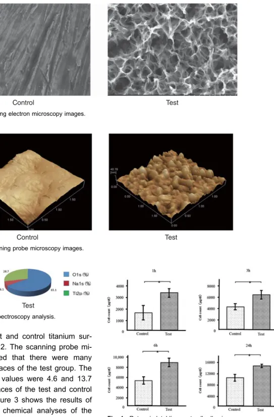
Scanning electron microscopy images are shown in Fig. 1. After modification in NaOH at 30° C, the tita
nium surface of the test disks showed a nanometer
scale network structure. Scanning probe micros
copy was used to estimate the vertical height of the
surface roughness of the samples. The surface
Control Test
Control Test
Control Test
morphologies of the test and control titanium sur- faces are shown in Fig. 2. The scanning probe mi- croscopy images showed that there were many nanonodules on the surfaces of the test group. The surface roughness (Ra) values were 4.6 and 13.7 nm for the titanium surfaces of the test and control groups, respectively. Figure 3 shows the results of wide-scan XPS surface chemical analyses of the test and control titanium surfaces. The presence of Ti and O was confirmed on both titanium surfaces.
In addition, the presence of Na was confirmed on the surface of the test group. The concentrations of Ti and O on the surface of the test group were greater than those on the surface of the controls.
Cell adhesion
Cell adhesion on the disks after 1, 3, 6 and 24 h of incubation was assessed (Fig. 4). There were sig- nificant differences in cell adhesion and proliferation
Fig. 1 Scanning electron microscopy images.
Fig. 2 Scanning probe microscopy images.
Fig. 3 X-ray photoelectron spectroscopy analysis.
Fig. 4 Rat periodntal ligament cell adhesion.
between the alkali-treated and control samples for all time periods.
Gene expression
The mRNA expression levels of osteogenesis- related genes including runt-related transcription factor (Runx2) in RPL cells grown on the different surfaces for 3 days were assessed by qRT-PCR.
The expression of Runx2 was upregulated in the cells grown on the test disks as compared to those grown on the control disks (Fig. 5).
ALP activity
ALP activity was observed in RPL cells grown on the different substrates for 7 days, and was found to increase over time. There were significant differ- ences in ALP activity between the alkali-treated and untreated titanium surfaces at 7 and 14 days (Fig.
6), with greater activity levels in the former group.
Osteocalcin secretion
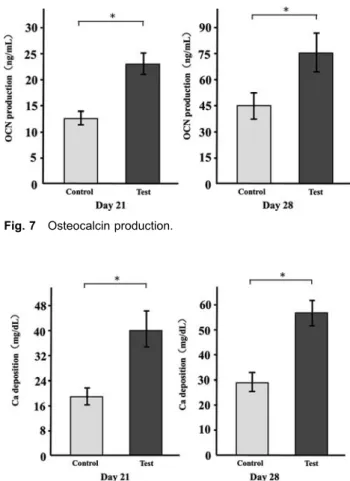
We measured osteocalcin levels in the culture su- pernatant of RPL cells grown on the different sub- strates for 21 and 28 days. The supernatants of cells grown on the test disks showed higher levels of secreted osteocalcin at both time points as com- pared with the control cultures (Fig. 7).
Calcium deposition
Calcium deposition in cells cultured on the different surfaces for 21 and 28 days was assessed as a measure of osteogenic differentiation. Mineraliza- tion was greater in the cells grown on the test disks than in those grown on the control disks (Fig. 8).
DISCUSSION
Dental implants, which are artificial tooth roots that support dental prostheses, are a popular approach for dental restoration.
15The immediate placement of dental implants into fresh extraction sockets is an
Fig. 5 Expression of Runx2 mRNA (p<0.05).
Fig. 6 Alkaline phosphatase activity.
Fig. 7 Osteocalcin production.
Fig. 8 Calcium deposition.
attractive alternative to delayed implant placement after alveolar healing because of advantages that include a shorter rehabilitation time, fewer surgical episodes, an improved preservation of bone tis- sues, and improvement in the peri-implant gingival tissue aesthetics.
16Immediate implantation, which involves the placement of the implant in the peri- odontium before the healing of the tooth extraction site, is followed by the formation of connective tis- sue by periodontal ligament cells in the fresh ex- traction socket.
Periodontal ligament cells are involved in the complex process of osseointegration, i.e., the es- tablishment of a direct bone-to-implant anchorage without intervening connective tissue, which is es- sential for the success of immediate implantation.
17This study investigated whether RPL cells would re- spond differently to titanium implants that had un- dergone a chemical surface modification compared with those that had not. We found that the initial adhesion of cells, the expression of the Runx2 tran- scription factor, and the concentrations of RPL cell differentiation markers such as ALP and osteocal- cin were elevated in cells grown on a TNS-modified titanium disk as compared with those grown on an unmodified, polished titanium disk. We also found that calcium deposition in the extracellular matrix of the RPL cells was increased in the presence of a TNS-modified disk as compared with the cultures grown on unmodified control disks. Our results sug- gest that a TNS-structured titanium surface pro- motes RPL cell adhesion, differentiation, and acti- vation, which augments calcium deposition.
The surface properties of dental implants have long been recognized as a critical factor for achiev- ing clinical success.
18-21The topographical proper- ties of nanostructures on titanium alloy surfaces play important roles in modulating cell responses at the implant-tissue interface, which can have a large effect on tissue integration with the implant.
22Re- cently, we showed that TiO
2nanotubes and TNSs could be formed on titanium metal surfaces by treatment with 10 M NaOH aqueous solution at 30° C.
11We used this method here to prepare TNS- modified disks. Komasa et al.
11suggested that TNS
-structured titanium surfaces modulate the osteo- genic differentiation of bone marrow cells and en- hance mineralization.
Ra is a height parameter that is commonly used to describe the surface roughness of implants. The Ra of the TNS-modified titanium surface was 17 nm, which was smaller than that of the untreated ti- tanium surface. Xing, Fujino, and colleagues showed that a surface roughness between 13-16 nm was optimal for RBM cell culture.
23, 24The nanonetwork structure formed on the titanium disks here is similar to the hierarchical structure reported by Lingzhou and coworkers.
25In their work, hierar- chical nanotextured titanium surface topographies with titania nanostructures that mimicked the hierar- chical structure of bone tissues were produced by etching followed by anodization.
Natural tissues are hierarchical structures of nanoscale building blocks assembled in an organ- ized manner. Hierarchical structures composed of nanocomponents may provide a more suitable sur- face topography for bone marrow cell functions than simpler structures because they can better mimic the structure of natural tissues. In this study, our research revealed that NaOH treatment led to the formation of a Ti-O-Na titanate layer on the tita- nium surface.
11Thus, we expect that NaOH treat- ment caused a thick oxide film to form on the TiO
2layer on the titanium surface. The deconvolution procedure suggested that this may have been due to surface contamination resulting from the binding of O to C.
25Our findings demonstrated that an alkali-modified
titanium surface induced more adhesion of RPL
cells than an untreated titanium surface. Cell adhe-
sion influences several cellular functions including
proliferation, migration, and extracellular matrix pro-
duction. The enhanced adhesion may be related to
the increased adsorption from the culture medium
of serum proteins such as fibronectin and vi-
tronectin, which play important roles in cell adhe-
sion. In a recent study, a TNS surface promoted
the adsorption of albumin and fibronectin.
11This ob-
servation suggests that the TNS structure promotes
cell adhesion and spreading on the titanium sur-
face. The periodontal ligament cells that support the teeth within the alveolar socket are composed primarily of osteoblasts, osteoclasts, cementoblasts, and fibroblasts. These results show that TNS struc- tures have the potential to improve periodontal re- generation in dental clinics.
Runx2 mRNA expression, ALP activity, osteocal- cin production, and calcium deposition were all ele- vated by the presence of TNSs on the implant sur- face. Runx2 mediates several early osteogenic gene responses for cellular adhesion.
26Several lines of evidence have shown that certain surface modifications lead to a high expression of Runx2 mRNA.
27, 28The data from this study suggests that the elevated expression of Runx2 mRNA in cells grown on the nanostructured titanium surfaces compared with that on untreated surfaces is a causative factor in the differentiation of RBM cells into osteogenic cells. Importantly, the functional phenotypes expressed in the middle and late stages of culture, such as ALP activity and miner- alization, were considerably increased.
A substantial body of research has confirmed the ALP-activating effect of surface-modified materi-
als.
29-31The surface modification of implants has
been found to affect ALP activation,
32-33and our re- sults support this conclusion. The increase of os- teocalcin production in the presence of a nanos- tructured surface that we observed is also in agree- ment with previous findings.
34Periodontal ligament cells are useful cells for the reconstruction of peri- odontal tissues because they contain osteogenic and fibrogenic progenitor cells, which differentiate into fibroblasts, cementoblasts, or osteoblasts de- pending on the surrounding conditions.
Another study showed that a rough titanium sur- face improved periodontal ligament cell and osteo- genic differentiation. To avoid the interference of connective tissues, the implant surface should be engineered to promote osteogenic differentiation and a faster healing process. In our previous study, the investigation of several different implant surface nanostructures demonstrated that modifying the im- plant surface at the nanometer scale can enhance the adhesion of endothelial cells and the expres-
sion of genes encoding angiogenic factors and ad- hesion molecules.
35Therefore, applying the TNS structural modification to a titanium surface could be useful for dental implants because it can pro- mote osteogenic differentiation and a faster healing process.
Titanium and titanium alloy implants have be- come an essential treatment modality in reconstruc- tive surgery in orthopedics and dentistry. However, patient morbidity and treatment complications need to be minimized, and outcome predictability and treatment indications should be maximized. There- fore, considerable effort has been expended on de- veloping new technologies to modify the surface of titanium to assist the biointegration of titanium im- plants with bone. The surface modification method used here is effective and simple, involving incuba- tion in NaOH at room temperature without a tem- plate. The resulting TNS-modified titanium surface induces the osteogenic differentiation of RBM cells.
CONCLUSIONS
Our study demonstrated that periodontal ligament cells cultured on a nano-modified titanium surface showed higher adhesion and were osteogenic dif- ferentiation than those cultured on a non-modified titanium surface. These findings indicate that RPL cells have the potential for increased bone forma- tion on a TNS surface. The TNS surface modifica- tion may warrant consideration when manufacturing the materials for use in dental implants intended for immediate implantation.
We would like to thank Tohru Sekino of Osaka University for preparing the nanosheets and for helpful suggestions.
We are also grateful to the members of the Department of Removable Prosthodontics and Occlusion. We also thank Toshio Tamaki and Hirokazu Hojyo. This work was sup- ported by a grant from the Japan Society for the Promotion of Science (16K20524) and an Oral Implant Research Grant (17-01) from Osaka Dental University.
REFERENCES
1. Moradian-Oldak J, Wen HB, Schneider GB, Stanford CM.
Tissue engineering strategies for the future generation of dental implants.Periodontol 20002006; 41: 157-176.
2. Branemark PI, Zarb GA, Albrektsson T. Tissue integrated prostheses. Osseointegration in clinical dentistry. Chicago : Quintessence, 1985.
3. Kirsch A. The two-phase implantation method using IMZ in- tramobile cylinder implants. J Oral Implantol 1983; 11: 197-210.
4. Skalak R. Biomechanical considerations in osseointegrated prostheses.J Prosthet Dent1983; 49: 843-848.
5. Wadamoto M, Akagawa Y, Sato Y, Kubo T. The three- dimensional bone interface of an osseointegrated implant. I : A morphometric evaluation in initial healing.J Prosthet Dent 1996; 76: 170-5.
6. Annunziata M, Oliva A, Buosciolo A, Giordano M, Guida A, Guida L. Bone marrow mesenchymal stem cell response to nano-structured oxidized and turned titanium surfaces.Clin Oral Implants Res2010; 23(6): 733-740.
7. Vandrovcová M, Bačáková L. Adhesion, growth, and differ- entiation of osteoblasts on surface-modified materials devel- oped for bone implants.Physiol Res2011; 60: 403-17.
8. Kasuga T, Hiramatsu M, Hoson A, Sekino T, Niihara K. Ti- tania nanotubes prepared by chemical processing.Adv Ma- ter1999; 11: 1307-11.
9. Miwa T, Nishida H, Egusa H, Park DJ, Sekino T, Tanaka S.
Direct synthesis and biocompatibility of titania nanotube lay- ers on metal substrates for implant biomaterial. The 21st JAPS-KOSEF Core University Program Seminar between Japan and Korea, 2008 : 36.
10. Perla V, Webster TJ. Better osteoblast adhesion on nano- particulate selenium−A promising orthopedic implant mate- rial.J Biomed Mater Res A2005; 75: 356-364.
11. Komasa S, Taguchi Y, Nishida H, Tanaka M, Kawazoe T.
Bioactivity of nanostructure on titanium surface modified by chemical processing at room temperature. J Prosthodont Res2012; 56: 170-177.
12. Schroeder A, Zypen E, Stich H, Sutter F. The reaction of bone, connective tissue, and epithelium to endosteal im- plants with titanium-sprayed surfaces. J Maxillofac Surg 1981; 9: 15-25.
13. Buser D, Weber HP, Donath K, Fiorellini JP, Paquette DW, William RC. Soft tissue reactions to non-submerged un- loaded titanium implants in beagle dogs. J Periodontol 1992; 63: 226-236.
14. Listgarten MA, Buser D, Steimann SG, Donath K, Lang NP, Weber HP. Light and transmission electron microscopy of the intact interfaces between nonsubmerged titanium-coated epoxy resin implants and bone or gingiva. J Dent Res 1992; 71: 364-371.
15. Van Steenberghe D. A retrospective multicenter evaluation of the survival rate of osseointegrated fixtures supporting fixed partial prostheses in the treatment of partial edentu- lism.J Prosthet Dent1989; 61: 217-223.
16. Boix D, Gauthier O, Guicheux J, Pilet P, Weiss P, Grimandi G, Daculsi G. Alveolar bone regeneration for immediate im- plant placement using an injectable bone substitute : an ex- perimental study in dogs. J Periodontol 2004; 75: 663- 671.
17. Pivodova V, Frankova J, Ulrichova J. Osteoblast and gingi- val fibroblast markers in dental implant studies.Biomed Pap Med Fac Univ Palacky Olomouc Czech Repub2011; 155: 109-116.
18. Mendonça G, Mendonca D, Aragao FJ, Cooper LF. Advanc- ing dental implant surface technology−from micron- to nano- topography.Biomaterials2008; 29: 3822-35.
19. Bell B, Schuler M, Tosatti S, Textor M, Schwartz Z, Boyan B. Osteoblast response to titanium surfaces functionalized with extracellular matrix peptide biomimetics.Clin Oral Im-
plants Res2011; 22: 865-72.
20. Albrektsson T, Brånemark P-I, Hansson H-A, Lindström J.
Osseointegrated titanium implants : requirements for ensur- ing a long-lasting, direct bone-to-implant anchorage in man.
Acta Orthopaedica1981; 52: 155-70.
21. Baier R, Meyer A, Natiella J, Natiella R, Carter J. Surface properties determine bioadhesive outcomes : methods and results.J Biomed Mater Res1984; 18: 337-55.
22. Boyan B, Bonewald L, Paschalis E, Lohmann C, Rosser J, Cochran DL, Dean DD, Schwartz Z, Boskey AL. Osteoblast- mediated mineral deposition in culture is dependent on sur- face microtopography.Calcif Tissue Int2002; 71: 519-29.
23. Xing H, Komasa S, Taguchi Y, Sekino T, Okazaki J. Osteo- genic activity of titanium surfaces with nanonetwork struc- tures.Int J Nanomed 2014; 9: 1741-55.
24. Fujino T, Taguchi Y, Komasa S, Sekino T, Tanaka M. Cell differentiation on nanoscale features of a titanium surface : effects of deposition time in NaOH solution. J Hard Tissue Biol 2014; 23: 63-70.
25. Zhao L, Mei S, Chu PK, Zhang Y, Wu Z. The influence of hierarchical hybrid micro/nano-textured titanium surface with titania nanotubes on osteoblast functions. Biomaterials 2010; 31: 5072-82.
26. Klein MO, Bijelic A, Ziebart T, Koch F, Kämmerer PW, Wie- land M, Konerding MA, Al-Nawas B. Submicron scale- structured hydrophilic titanium surfaces promote early osteo- genic gene response for cell adhesion and cell differentia- tion.Clin Implant Dent Relat Res2013; 15: 166-75.
27. Masaki C, Schneider GB, Zaharias R, Seabold D, Stanford C. Effects of implant surface microtopography on osteoblast gene expression.Clin Oral Implants Res2005; 16: 650-6.
28. Schneider G, Perinpanayagam H, Clegg M, Zaharias R, Seabold D, Keller J, Stanford, C. Implant surface roughness affects osteoblast gene expression.J Dent Res 2003; 82: 372-6.
29. Att W, Kubo K, Yamada M, Maeda H, Ogawa T. Biome- chanical properties of jaw periosteum-derived mineralized culture on different titanium topography.Int J Oral Maxillofac Implants2009; 24: 831-41.
30. Brammer KS, Choi C, Frandsen CJ, Oh S, Johnston G, Jin S. Comparative cell behavior on carbon-coated TiO2 nano- tube surfaces for osteoblasts vs. osteo-progenitor cells.
Acta Biomaterialia2011; 7: 2697-703.
31. Göransson A, Arvidsson A, Currie F, Franke-Stenport V, Kjellin P, Mustafa K, Sul YT, Wennerberg A. An in vitro comparison of possibly bioactive titanium implant surfaces.
J Biomed Mater Res A2009; 88: 1037-47.
32. Iwasa F. TiO2micro-nano-hybrid surface to alleviate biologi- cal aging of UV-photofunctionalized titanium.Int J Nanomed 2011; 6: 1327-41.
33. Yu WQ, Jiang XQ, Zhang FQ, Xu L. The effect of anatase TiO2nanotube layers on MC3T3-E1 preosteoblast adhesion, proliferation, and differentiation. J Biomed Mater Res A.
2010; 94: 1012-22.
34. Dalby MJ, McCloy D, Robertson M, Agheli H, Sutherland D, Affrossman S, Oreffo RO. Osteoprogenitor response to semi -ordered and random nanotopographies.Biomaterials2006; 27: 2980-7.
35. Nakano Y, Komasa S, Taguchi Y, Sekino T, Okazaki J. In- itial attachment, behavior, and gene expression of rat endo- thelial cells in response to titanium surface treated by NaOH solution.J Oral Tis Eng2014; 11(3): 189-200.