Kyushu University Institutional Repository
Genetic assessment of recurrent pancreatic high-risk lesions in the remnant pancreas:
Metachronous multifocal lesion or local recurrence?
後藤, 佳登
http://hdl.handle.net/2324/2236124
出版情報:九州大学, 2018, 博士(医学), 課程博士 バージョン:
権利関係:
ContentslistsavailableatScienceDirect
Surgery
journalhomepage:www.elsevier.com/locate/surg
Genetic assessment of recurrent pancreatic high-risk lesions in the remnant pancreas: Metachronous multifocal lesion or local
recurrence? ✩
Yoshitaka Gotoh, MD
a, Takao Ohtsuka, MD, PhD
b,∗, So Nakamura, MD
a, Koji Shindo, MD, PhD
a, Kenoki Ohuchida, MD
a, Yoshihiro Miyasaka, MD, PhD
a, Yasuhisa Mori, MD, PhD
a, Naoki Mochidome, MD
a,b, Yoshinao Oda, MD, PhD
b, Masafumi Nakamura, MD, PhD, FACS
aaDepartment of Surgery and Oncology, Graduate School of Medical Sciences, Kyushu University, Fukuoka, Japan
bDepartment of Anatomic Pathology, Graduate School of Medical Sciences, Kyushu University, Fukuoka, Japan
a rt i c l e i n f o
Article history:
Accepted 16 October 2018 Available online xxx
a b s t r a c t
Background: It isdifficulttodeterminewhetherasecondhigh-risklesion,includingpancreaticductal adenocarcinomaorhigh-gradepancreaticintraepithelialneoplasm,isametachronousmultifocallesion orrepresentslocalrecurrenceafterresectionofthefirsthigh-risklesion.Thisstudy attemptstoclarify thecharacteristicsofsecondhigh-risklesionsintheremnantpancreasusinggeneticanalyses.
Methods: Clinicopathologicdatawerecollectedfrom12patientswhounderwentpancreatectomy fora secondhigh-risklesioninthe remnantpancreas.We performedmutationaland immunohistochemical analysesof4majorgenes—KRAS,TP53,CDKN2A,andSMAD4—associatedwithpancreaticductaladenocar- cinomaprogression,aswellastargetednext-generationsequencing.
Results: Mutationsinthefourgenesinthesecondhigh-risklesionwereconsistentwiththefirstlesion infourpatientsbutwereinconsistentintheremainingeightpatients,andthusweconsideredthatthe lattereightpatientslikelyhadmetachronousmultifocalhigh-risklesionsandtheotherfourpatientshad localrecurrence.Theestimatedcumulativerecurrencerateafterresectionofthesecondhigh-risklesion wasgreater inthelocalrecurrence groupcomparedwiththemetachronousmultifocal group,and the estimated cumulativedisease-specificsurvival rate wasgreater inthe metachronousmultifocal group.
Targetednext-generationsequencingdemonstratedthatthesecondlesionsinthemetachronousmultifo- calhigh-risklesiongroupshoweddifferencesinfoundermutationscomparedwiththefirstlesion.Inthe localrecurrencegroup,thefoundermutationsinthesecondlesionwerecommonwiththoseinthefirst lesion.
Conclusion: Geneticassessmentmighthelpdiscriminatemetachronousmultifocalhigh-risklesionsfrom localrecurrence.
© 2018 Elsevier Inc. All rights reserved.
Introduction
Pancreatic ductal adenocarcinoma (PDAC) is a most lethal solid neoplasm because of its aggressive behavior and the diffi- culty in early detection. Despite the improvements in operative techniques andperioperative management duringrecent decades and the slightly increased survival time from newly developed
✩Supported by a Grant-in-Aid from the Japan Society for the Promotion of Sci- ences for Scientific Research (B) (Grant Number 16H05417 ).
∗ Corresponding author: Department of Surgery and Oncology, Graduate School of Medical Sciences, Kyushu University, 3-1-1 Maidashi, Higashi-ku, Fukuoka 812- 8582, Japan.
E-mail address: [email protected] (T. Ohtsuka).
chemotherapeutic agents, the rateof cancer-related death in pa- tientswithPDACremainshigh.1
Geneticassessmentcanhelp evaluatetheprogression ofPDAC andtherebyprovideimportantinformationformoleculartargeted diagnosis and treatment. “Clonal evolution” is a theory of can- cer progression in which founder mutations are passed on to any sites of the next-generation lesions, and progressor muta- tions vary in the primary lesion or metastatic site of the next generation. This concept of clonal evolution has been used to describethemechanismofmalignantprogressionofPDAC.2,3 Mu- tationsinfourgenes—KRAS,TP53,CDKN2A,andSMAD4—occurdur- ingtheprogression ofPDAC.4,5 Hosodaetal6 usedtargetednext- generationsequencing (NGS)to show that KRASmutationsact as foundermutationswhichinitiatethedevelopmentofpremalignant https://doi.org/10.1016/j.surg.2018.10.025
0039-6060/© 2018 Elsevier Inc. All rights reserved.
2 Y. Gotoh, T. Ohtsuka and S. Nakamura et al. / Surgery xxx (xxxx) xxx
JID:YMSY [m5G;November26,2018;17:26]
pancreaticintraepithelialneoplasia(PanIN),andmutationsinTP53 andSMAD4arelater eventsthatmightacceleratetheinvasivebe- haviorofPDAC.
Recentadvances indiagnosticsforearlydetectionofPDACand multidisciplinarytreatmentforadvancedPDAChaveledtoaslight increaseinthenumberoflong-termsurvivorsafterresection.1 In thesepatients,asecondPDACorhigh-gradePanINintheremnant pancreasafter partial pancreatectomyfor the firstlesion is often observed.7–11 Usingclinicopathologicfindingsalone,however,itis usuallydifficulttodeterminewhetherthissecondlesionisanew metachronous,multifocal lesion ora local recurrence of thefirst lesion.Itmayprovetobeimportanttodistinguishbetweenthese two entities,both for clinical management andto further under- standthedevelopmentandprogressionofPDAC,butmostreports havecombinedthesetwoentitiesintoasinglecategoryof“recur- rence.”7–9Therefore,theaimofthisstudywastodistinguishthese twoentitiesthroughmolecularassessments,includingNGS.
PatientsandMethods
This study was approved by the Ethics Committee of Kyushu University(No.27-126)andconductedaccordingtoEthicalGuide- linesforHumanGenome/GeneResearch enacted bythe Japanese GovernmentandtheHelsinkiDeclaration.
Patientsandclinicopathologicdata
The medicalrecordsof411consecutivepatientswho were di- agnosedhistologically witha high-risk lesion (HRL) involving ei- therPDAC ora high-grade PanINafter partial pancreatectomy at theDepartmentofSurgeryandOncology,Kyushu UniversityHos- pital,Fukuoka, Japan,between2000and2014were reviewedret- rospectively.High-gradePanINwasincludedinthisstudybecause this lesion is considered to be equivalent to “carcinoma in situ” accordingto the recommendationfrom the Baltimore Consensus Meeting12 and because many or most pancreatologists consider itasindication forresection.6,10,11 Patients who hadPDACpossi- blyarising fromintraductal papillary mucinous neoplasm(IPMN) andthosewhounderwenttotalpancreatectomywerenotincluded in this study. We collected data on age, sex, history of smok- ing, history of alcohol consumption, comorbid diseases, preoper- ativevaluesofserumcarcinoembryonicantigen andcarbohydrate antigen 19-9, tumor location, operation performed, postoperative surveillance,andpathologicfindings,includingtumor size,patho- logic type, T factor, N factor, stage, lymphatic invasion, vascu- larinvasion,neural invasion,andresidual tumor (R). Allresected specimens were rereviewed and revised, if necessary, by two pathologists(Y.O. andN.M.) experienced inthe histopathology of pancreatic neoplasms. Tumor location, operation performed, and pathologicfindingsweredeterminedaccordingtotheClassification ofpancreaticcarcinoma.13Chest–abdominalcomputedtomography (CT)andserumcarcinoembryonic antigen andcarbohydrateanti- gen19-9levels wereexaminedperiodicallyduringthepostopera- tive surveillance periodaccordingto the NationalComprehensive CancerNetworkguidelinesforPDAC.14
KRASmutationalanalyses
Formalin-fixed, paraffin-embedded (FFPE) tissue samples were collected for each patient. Appropriate tissue blocks were se- lected, multipleserial sections(10-μm thick)were prepared, and laser microdissection was performed manually. using the Leica LMD6500 system (Leica Microsystems, Wetzlar, Germany). Ge- nomic DNA was extracted using the QIAamp DNA FFPE Tis- sue Kit (Qiagen, Hilden, Germany), according to the manufac- turer’s instructions. The status of the KRAS mutation at codons
12 and 13 was assessed by a direct sequencing method as de- scribedelsewhere.15–17Briefly,theKRASouterprimersforgenomic deoxyribonucleic acid (DNA) were 5-AGGCCTGCTGAAAATGACT-3 (forward)and5-TTGTTGGATCATATTCGTCCAC-3(reverse),andthe sequencingprimerwas5-CCTGCTGAAAATGACTGAA-3.Afterpoly- merase chain reaction (PCR), the amplified products were puri- fiedusingthe QIAquickPCRPurificationKit(Qiagen),andthese- quence ofKRAS at codons 12 and 13 (point mutation sites) was determined by the dideoxy chain-termination method, using the BigDyeTerminator CycleSequencingKit(Applied Biosystems,Fos- terCity,CA,USA),accordingtothemanufacturer’sinstructions.The products were analyzedusinga 3130xl GeneticAnalyzer (Applied Biosystems).
In four patients, KRAS mutational analyses of pancreatic juice sampleswerealsoperformedaccordingtoourearlierreport,16and theresultswerecomparedwiththeresectedspecimenofthesec- ondlesions.
ImmunohistochemistryforTP53,CDKN2A,andSMAD4
FFPEtissuesamplesfromeachpatientwereimmunostainedfor TP53,CDKN2A,andSMAD4.Aminimumofthreeslidesfromeach patientwerestainedtoevaluateheterogeneitywithinthefirstand second HRL. An antihuman,TP53, rabbit polyclonalantibody(FL- 393, 1:100, Santa Cruz Biotechnology, CA, USA), an antihuman, CDKN2A, mouse monoclonal antibody (clone E6H4, 1:100, Roche MTM laboratories AG, Heidelberg, Germany), and an antihuman, SMAD4,mousemonoclonalantibody(cloneB-8,1:100,SantaCruz Biotechnology) were used. TP53 staining was considered abnor- mal inlesions withdiffuse (≥60% neoplastic cells) nuclearstain- ing (overexpression) or those with no nuclear staining (lack of expression).6 Scatteredacinar andductal cellswith nuclear TP53 expression were typically present in the adjacent normal tissue and used as an internal control. Islet cells served as an internal control for positive CDKN2A expression, andCDKN2A staining in eitherinthecytoplasmorinthenucleiwasscoredaspositive,in- dicating an intactCDKN2A gene, andlack of stainingwas scored asnegative, indicatingthat a deletionor inactivatingmutationof CDKN2A had occurred.5 Normal acinar, ductal, islet, and stromal cells served as internal controls for positive SMAD4 expression.
SMAD4expression,eitherinthenucleusorcytoplasm,wasscored aspositive,indicating thepresenceof anintactSMAD4gene,and absenceofstainingwasscoredasnegative,indicatingthatadele- tion orinactivating mutation of SMAD4hadoccurred.5,6 Samples processedwithnonimmuneseruminsteadoftheprimaryantibod- ieswereusedasanegativecontrol.
Estimationofprogressionofsecondlesions
To determine whether second HRLs might result from a metachronousmultifocallesionorlocalrecurrence ofthefirstle- sion, themargin status atthe time of the first operation,patho- logic findings, KRAS mutational status, and TP53, CDKN2A, and SMAD4 immunostaining of FFPE samples from first and second lesionswerecompared.
Clinicalcharacteristics,includingpostoperativeprognosis,were alsocomparedamongpatientswithsuspectedmetachronousmul- tifocal lesion or local recurrence of the first lesion in the rem- nantpancreas.ToassessprognosisafterresectionofasecondHRL, theestimateddisease-freesurvivalrateforpatientswascompared amongpatientshavingresection oflocalrecurrence,patientshav- ingresection ofmetachronousmultifocalHRL,patientswithdiag- nosisofunresectablePDACintheremnantpancreas,includingad- vancedlocalinvasionandconcomitant distantmetastasis(n=10), andpatientswithadiagnosisofextrapancreaticrecurrence,includ- ingdistantmetastasesandperitonealmetastases(n=304,Fig.1).
Fig. 1. Study flow of lesion identification in 411 patients who underwent pancrea- tectomy for high-risk lesions. A total of 411 patients with high-risk lesions (HRLs) were included in this study. The 85 patients with any sign of recurrence were ex- cluded, and the remaining 326 patients were further analyzed. After the first partial pancreatectomy, 22 patients (5%) were diagnosed as having HRLs in the remnant pancreas, 12 of whom underwent a second pancreatectomy. Resected specimens of both initial and second HLRs were obtained to determine whether the second HLR was a metachronous multicentric lesion or local recurrence.
TargetedNGS
Frozentissue sampleswere collectedbytissue tabletmethods accordingtoourearlierreport,18and10-μmsectionswere cuton acryostatandstoredat–20°Cuntiluseorfixed in5%aceticacid andstainedwithtoluidineblue.Cancercellswerecollectedimme- diately by laser microdissection in200-μltubes. DNAwassubse- quently extracted using the DNeasyBlood & Tissue Kit(Qiagen), according to themanufacturer’s instructions, andresuspended in 30μL of nuclease-free water.DNA quantity wasassessed using a Qubitphotometer(LifeTechnologies,Carlsbad,CA,USA),andDNA quality was determined using the Qubit dsDNA High Sensitivity Assay Kit (Life Technologies) and a NanoDrop spectrophotometer (Thermo Fisher Scientific, Waltham, MA, USA), according to the manufacturer’sinstructions.
LibrarieswerepreparedusingtheIonAmpliSeqLibraryKit2.0- 96LV (Life Technologies) and Cancer Hotspot Panel v2 (CHPv2).
The minimum DNA concentration requiredto obtain 10ng ofto- tal DNAinputwas1.6ng/μL.In samplescontaininga lowamount of DNA (DNA<1.6ng/μL), the DNA was not diluted further, and the quantity of nuclease-free water was decreased accordingly.
The numberofmultiplex PCRswasincreasedfromthesuggested 20 cycles to 29 cycles. The CHPv2 covers approximately 2,800 Catalogue of Somatic Mutations in Cancer mutations from 50 oncogenes and tumor suppressor genes (ABL1, EGFR, GNAS, KRAS, PTPN11, AKT1, ERBB2, GNAQ, MET, RB1, ALK, ERBB4, HNF1A, MLH1, RET, APC, EZH2, HRAS, MPL, SMAD4, ATM, FBXW7, IDH1, NOTCH1, SMARCB1,BRAF,FGFR1,JAK2,NPM1,SMO,CDH1, FGFR2,JAK3,NRAS, SRC,CDKN2A,FGFR3,IDH2,PDGFRA,STK11,CSF1R,FLT3,KDR,PIK3CA, TP53, CTNNB1, GNA11, KIT, PTEN, and VHL). The CHPv2 amplicon reference range wasreported as111–187 bp, with an average of 154 bp. Each library wasbarcoded with the Ion Xpress Barcode Adapters 1-16Kit (Thermo FisherScientific), quantified usingthe Qubit dsDNA High Sensitivity Assay Kit (Life Technologies) and NanoDropLife(ThermoFisherScientific),anddiluted innuclease- freewatertoobtainafinalconcentrationof100μM.
Ion Torrent Suite Software v 5.0.4 (Thermo Fisher Scien- tific) was used to sequence genes involved in pancreatic can- cer (KRAS, TP53, CDKN2A, and SMAD4). After alignment to the hg19 human reference genome, the Variant Caller plug- in (version 5.0.28-1) (Thermo Fisher Scientific) was applied us- ing the Hotspot Cancer Panel file as reference. The Variant Caller plug-in reports by default a given variant when it is detectedin≥1%ofthetotalampliconreadswiththerequirement ofat least the presence of one forward andone reverse read to avoidstrandbias.TheIonReportersuite(ThermoFisherScientific) wasusedto filteroutknown polymorphicvariants. BinaryAlign- ment Map (BAM) files were inspected visually using the Golden HelixGenomeBrowser(version2.0.7;GoldenHelix,Bozeman,MT, USA), and only the mutations reported in the Catalogue of So- matic Mutations in Cancer database (http://cancer.san-ger.ac.uk/) weretakenintoaccount. Postsequencingmetrics(readlength,the percentageofon-target reads, qPCR,andthe averagebasedepth) wererecordedforeachsample.
Samplingoffrozentissueanddefinitionoflocalrecurrenceor multifocallesion
Twocategoriesofmutationswereidentified.Thefirstcategory consistedoffoundermutationsthatwereinallsubclonesfromthe parental clone. The second category consisted of progressormu- tationsthatcreateheterogeneity.Inpancreaticcancer,thefounder mutationsaremutationsindrivergenes,shownelsewheretodrive pancreatictumorigenesis.2,19 Inthisstudy,DNAwasisolatedfrom frozentissuesamplestakenfromthreeregionsofaprimarylesion and analyzed by targeted NGS. The common mutations in these threeregionswerecomparedwithmutationsfromoneregionofa second lesionthat wasanalyzed by targetedNGS.The second le- sionwaslikelytobe arecurrenceofthefirstlesioniftherewere commondrivermutations inbothprimary andsecondary lesions, and multifocal development was probable if common mutations werenotobservedinthesecondlesion.
Statistics
Statistical analysis was performed using JMP statistical soft- ware(version13.0.2;SASInstitute,Cary,NC,USA).TheFisherexact probabilitytestorthe
χ
2 testwasusedtoevaluatedifferencesin clinicalfactorsbetweenthetwogroups.TheMann-WhitneyUtest wasused forcontinuousdata, which were expressedas theme- dian withrange. The estimateddisease-specific survival rateand estimated overall survival rate were analyzed using the Kaplan- Meier method with the Wilcoxon test. No formal statistics were appliedtothecomparisonofthefourpatientswithpresumedlocal recurrencesandtheeightpatientsbelievedtohavemetachronous lesionsbecauseofthesmallnvaluesineachgroup.Results
Clinicopathologiccharacteristicsofthestudypopulation
Figure1presentsthestudyflowforanalysisofthe411patients inthisstudywhounderwentpartialpancreatectomyforHRLs.The 85 patients with anysign of recurrence were excluded, and the remaining326patientswerefurtheranalyzed.Afterthefirstpan- createctomy,22patients(5%) were diagnosedashaving asecond HRLintheremnantpancreas,and12ofthesepatientsunderwent asecondpancreatectomy.Resectedspecimensofboththefirstand secondHRLswereobtainedfromthese12patients.
Table 1 presents the clinicopathologic characteristics of the 12 patientswho were diagnosed histologically withan HRL after partial pancreatectomy and underwenta second pancreatectomy.
4 Y. Gotoh, T. Ohtsuka and S. Nakamura et al. / Surgery xxx (xxxx) xxx
JID:YMSY [m5G;November26,2018;17:26]
Table1 Clinicopathologicandmolecularcharacteristicsofthe12patientswhounderwentasecondpancreatectomyoftheremnantpancreas. NoAge/SexComorbidityFirstlesionMonths afterfirst operation
SecondlesionEstimated patternof progressionLocation∗DiagnosisMargin∗KRAS mut†TP53 IHCCDKN2A IHCSMAD4 IHCDistancefrom PCM(mm)∗DiagnosisKRAS mutTP53 IHCCDKN2A IHCSMAD4 IHC (T/N/P)∗T/N/P 163/FDMPhT3/N1/modR0G12AAbn+–7215T3/N1/wellG12DAbn++Multifocal 273/MIPMNPbtT3/N0/modR0WTN++4220T3/N0/modG12DN++Multifocal 367/FDMPhT3/N1/wellR0G12DAbn+–480T3/N1/modG12VAbn++Multifocal 465/MIPMN,DMPbtT1/N0/modR0G12DN+–3120T3/N0/modG12AAbn++Multifocal 571/MChronic pancreatitisPbtTis/N0/wellR0WTAbn+–2410Tis/N0/wellG12VN++Multifocal PbtTis/N0/wellR0G12DN++30Tis/N0/wellG12DAbn+–Multifocal 658/FIPMNPbtT1/N0/modR0G12VAbn+–5315Tis/N0/wellG12DAbn++Multifocal 758/M–PhT3/N1/porR0G12VN––378T1/N0/porG12DAbn++Multifocal 867/F–PbtT2/N0/porPCM+G12DAbn++4415T3/N0/porWTN+–Multifocal 966/FIPMNPbtT3/N1/modR0G12DAbn–+350T3/N1/porG12DAbn–+Recurrence 1079/F–PhT3/N0/modDPM +G12AAbn++245T3/N1/modG12AAbn++Recurrence 1176/M–PbtT3/N0/modR0G12DAbn++180T3/N0/modG12DAbn++Recurrence 1260/M–PhT3/N0/modR0G12VAbn++90T3/N1/modG12VAbn++Recurrence ∗DeterminedaccordingtotheJapanPancreasSociety. 13 †KRASmut:KRASmutationwiththehighestconcentration.IHC , immunohistochemistry;DM ,diabetesmellitus;Ph ,pancreatichead;Pbt ,pancreaticbodytotail;T ,tumorfactor;N ,nodalfactor;P , pathologic type.Well,mod ,andporindicatewell,moderately,andpoorlydifferentiatedadenocarcinoma,respectively.R0 , negativemargin;WT ,wild-type;PCM , pancreaticcutendmargin;DPM , dissectedperipancreatic tissuemargin;Abn , abnormalexpression;N , normalexpression.
One patient (number 5) had 2 synchronous and metachronous multifocal HRLs. One of the high-grade PanIN lesions was diag- nosedatthetimeoffirstoperationbybrushcytology(classV)for a strictureinthemain pancreatic duct,andthe other high-grade PanINwas found by chance during pathologic assessmentof the resected specimenofthe first pancreatectomy. We oftenperform follow-upendoscopicretrogradepancreatography(ERP)/pancreatic juicecytologyinpatientswithhighriskforthedevelopmentofa secondHRLintheremnantpancreas,suchaspatientswithmulti- ple,high-gradePanINlesionsorwithHRLconcomitantwithIPMN, becauseERP/pancreaticjuicecytologyistheonlywaytodiagnose earlystage HRLthat cannotbe detectedby other imagingmodal- ities.10,20 Thus,a second pancreatectomy wasperformedin2 pa- tients (numbers5 and 6) because follow-upERP/pancreatic juice cytology showed class V even without any sign of the HRL by three-dimensionalradiologicfindings.
KRAS mutation status assessed in FFPE samples was different betweenthefirstandsecondlesionsin8patients(numbers1–8) butwasthesameintheremaining4patients(numbers9–12).No- tably,allKRASmutationsdetected inthisstudywereatcodon12 andnotatcodon13.Inaddition,patternsofimmunohistochemical stainingforTP53, CDKN2A,andSMAD4were not alwaysincom- pleteaccord between the first andsecond HRLs inpatient num- bers1−8 butwere consistent inpatient numbers9−12. Together theseresultsshowthatthemutationstatusorexpressionofthese 4majorgenesdifferedbetweenthefirstandsecondlesionsinpa- tientnumbers1–8,andtherewerenosuch differencesinpatient numbers9−12.
DistinctionofsecondHRLsandcomparisonoftheirclinicopathologic characteristics
Based on the mutational and immunohistochemical analyses andclinicopathologic findings discussedearlierin thisreport, we considered the progression pattern of the second HRLs in pa- tientnumbers1–8tobemetachronousmultifocallesions,andthe HRLs in patient numbers 9−12 were determined as local recur- rence (Table 1). The time to diagnosis of the second HRL after resection of first lesiontended to be less inthe local recurrence group (median 21 months, range 9–35 months) compared with themetachronousmultifocalgroup(median43months,range24–
72 months) The distance from the pancreatic cut margin to the second HRL inthe remnantpancreas tended tobe greater inthe metachronousmultifocalgroup(median 15mm,range0–30mm) compared withthe local recurrence group (median 0mm, range 0–5mm).
No differences in clinicopathologic characteristics of the first HRLs were found between the metachronous multifocal and lo- cal recurrence groups (Table 2). In contrast, after resection of thesecond HRL,the estimatedcumulativerecurrence ratetended to be greater in the local recurrence group compared with the metachronous multifocal group (Fig. 2, A). In addition, the esti- mated cumulative disease-specific survival rate after resection of thesecondHRLtendedtobegreaterinthemetachronousmultifo- calgroupcomparedwiththelocalrecurrencegroup(Fig.2,B).The disease-specific survivalrateofthepatientswithlocalrecurrence aftersecondpancreatectomywasnotdifferentfromthe10patients withunresectablesecondPDACintheremnantpancreas(P=0.71) orthe304patientswithextrapancreaticrecurrence(P=0.15).
AssessmentofKRASmutationsinpancreaticjuice
SomepatientsunderwentsurveillanceERPandsubsequentcol- lection of pancreatic juice before the second pancreatectomy. To clarifywhetherthestatusoftheKRASmutationinthesecondle- sionscouldbepredictedandtodeterminewhetherthesecondle-
Table 2
Characteristics of patients in the possible metachronous multifocal and local recurrence groups.
Metachronous multifocal ( n = 8) Local recurrence ( n = 4)
Sex (M / F) 4 / 4 2 / 2
Age in years (range) 65 (58–73) 70 (68–79)
History of smoking, positive 4 (50%) 1 (25%)
History of alcohol consumption, positive 2 (25%) 3 (75%)
Comorbidity
IPMN 3 (37.5%) 1 (25%)
Chronic pancreatitis 1 (12.5%) 0 (0%)
Diabetes mellitus 3 (37.5%) 0 (0%)
Tumor marker
CEA, ng/mL (range) 3.0 (0.5–19.1) 7.4 (2.8–15.3)
CA19-9, U/L (range) 30 (0.6–62) 855 (0.9–3352)
Operation
PD / DP 2 / 6 2 / 2
Pathologic findings ∗
Tumor size (mm) 17 (13–35) 25 (13–45)
T factor, ≥T3 4 (50%) 4 (100%)
N factor, ≥N1 3 (38%) 1 (25%)
Stage, ≥IIB 4 (50%) 1 (25%)
Lymphatic invasion + 4 (50%) 2 (50%)
Vascular invasion + 2 (25%) 2 (50%)
Neural invasion + 4 (50%) 4 (100%)
Residual tumor, R1 1 (13%) 1 (25%)
∗ Pathologic findings were determined according to the Japan Pancreas Society. 13IPMN , intraductal papillary mucinous neo- plasm; CEA , carcinoembryonic antigen; CA19-9 , carbohydrate antigen 19-9; PD , pancreatoduodenectomy; DP , distal pancreatec- tomy.
Fig. 2. Clinical course after resection of first or second lesion. ( A ) Estimated cumulative recurrence rate after resection of the second high-risk lesion (HRL) tended to be greater in the local recurrence group compared with the metachronous multifocal group. ( B ) Estimated cumulative disease-specific survival rate after resection of the second HLR tended to be greater in the metachronous multifocal group compared with the local recurrence group ( ∗P < .01). Disease-specific survival rate of the patients with local recurrence after second pancreatectomy was not different from patients with unresectable second PDAC in the remnant pancreas ( n = 10; ∗∗P = .71) or patients with extrapancreatic recurrence ( n = 304; ∗∗∗P = .15).
Table 3
KRAS mutation status in the resected second high-risk lesion and pancreatic juice before the second operation.
Patient number Second lesion Pancreatic juice
G12A G12D G12V G12A G12D G12V
4 (possible multifocal) 42% 4% 0% 46% 8% 0%
5 (possible multifocal) 0% 40% 6% 0% 54% 10%
11 (possible recurrence) 10% 18% 0% 14% 22% 0%
12 (possible recurrence) 6% 0% 26% 8% 0% 66%
Patient with chronic pancreatitis 0% 0% 0% 0% 0% 0%
sion mightbe recurrence ora metachronous multicentericlesion beforethesecond pancreatectomy,we comparedKRASmutational analysesofpancreaticjuicesampleswiththeresectedspecimenof thesecondlesions.MutationsinKRASatcodon12and13werede- terminedinpancreaticjuiceobtainedfrom4patients(numbers4, 5,11, and12)beforetheir second operation.Asimilar mutational statusatcodon12wasobservedinthesecondHRLsandpancreatic juice samplesforall4patients(Table3).Patientswhounderwent
pancreatectomyformass-forming pancreatitisserved asacontrol andhadwild-typeKRASinbothresectedspecimensandpancreatic juice.
TargetedNGS
TargetedNGSwasperformedinfrozensamplesfromthreepa- tients (numbers 8, 9, and 12). In patient number 8, the status
6 Y. Gotoh, T. Ohtsuka and S. Nakamura et al. / Surgery xxx (xxxx) xxx
JID:YMSY [m5G;November26,2018;17:26]
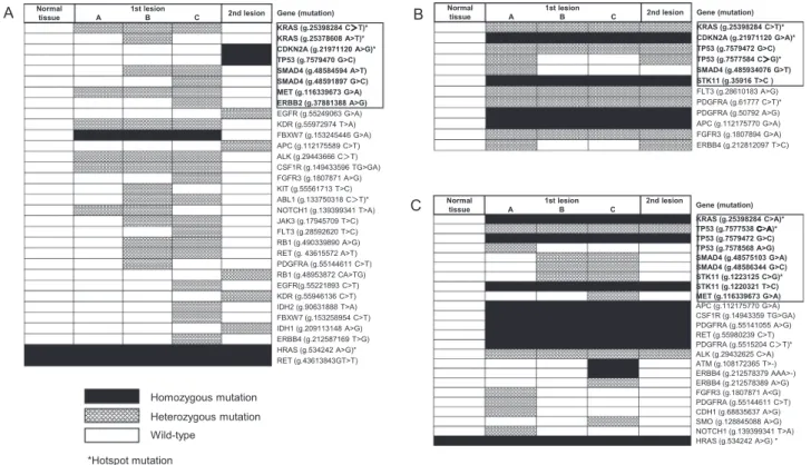
Fig. 3. Next-generation sequencing results. Next-generation sequencing was performed in three patients in frozen samples from three sampling regions (columns A, B, and C) from the first lesion, the second lesion, and normal tissue. ( A ) In patient number 8, the main founder mutations in KRAS, CDKN2A, TP53 , and SMAD4 were different between the first and second lesions, indicating that these two lesions were metachronous independent lesions. Mutations in bold and within the box indicate founder mutations of pancreatic ductal adenocarcinoma. ∗Hotspot mutations include KRAS at codon 12 and 61, CDKN2A at codon 80, TP53 at codon 233 and 248, and STK11 at codon 354. ( B ) In patient number 9, the KRAS, CDKN2A, TP53 , and STK11 founder mutations were detected in the primary and second lesion, indicating that the second lesion was likely to be a recurrence of the first lesion. ( C ) In patient number 12, the founder mutations in KRAS, TP53 , and STK11 were detected in both primary and secondary lesions, indicating that the second lesion was likely to be a recurrence of the first lesion.
ofthe four main founder mutations inKRAS, CDKN2A,TP53, and SMAD4wasdifferentinthefirstandsecondlesions,indicatingthat the two lesions were likely to be independent metachronous le- sions.Inpatient number8,twoKRASmutationswere observedin thefirstHRL,withamutationatcodon12(g.25398284C>T)de- tectedin all three samplingregions and a mutationat codon61 (g.25378608A >T) detected inonlyone samplingregion(Fig.3, A). Inpatient number9,founder mutations inKRASatcodon 12, CDKN2A,TP53,andSTK11werethesameinboththefirstandsec- ondlesion,indicatingthatthesecondlesionwaslikelytobeare- currence ofthe first lesion(Fig. 3,B). In patient number 12, the founder mutations in KRAS at codon 12, TP53, and STK11 in the firstlesionwere the sameasthose inthe second lesion,indicat- ingthatthesecondlesionwasalsolikelytobearecurrenceofthe firstlesion(Fig.3,C).Other mutationpatternsinpatient numbers 9and12werealsosimilarbetweenthefirstandsecondHRLs.
Discussion
This study demonstrates that the clinical course of metachronous multifocal HRLs appears to be different from that of local recurrence in the remnant pancreas after resec- tion of a first HRL and that genetic assessment using NGS may possibly discriminate between these two entities. Assessment of KRAS mutation status in pancreatic juice may be helpful for preoperative differentiation between metachronous multifocal HRLsandlocalrecurrence intheremnant pancreas,whichwould informappropriatemanagementofthesecondHRLintheremnant pancreas.
Genomic assessmentusing NGSprovides a detailedprocess of progressionofPanINtoPDAC,indicatingtheprocessofmultistage
carcinogenesis via KRAS→CDKN2A→TP53→SMAD4mutations in a stepwisemanner.2–6,21,22Mutationsinthesefourgenesaretermed
“foundermutations” for PDACandare consistent duringthepro- gressionofPanINtoPDAC.Thesemutationscanleadtomalignant characteristics of PanIN that drive progression to PDAC. Multiple IPMNshavebeenreportedtoshowtwopotentialprogressionpat- terns,namelymonoclonalskipprogressionviaintraductaldissemi- nation23and multicentricoccurrence via field defecttheory.24This studyisthefirstmolecularassessmentoftwopossibleprogression patternsinsecondHRLsintheremnantpancreas.
Despite the limited size of this study population, our results suggestthat mutationalandimmunohistochemicalassessmentsof thefourmajorgenesmaypossiblydiscriminatesynchronousmul- tifocalHRLfromlocalrecurrenceintheremnantpancreas.Among thePDACmutationhotspots inKRAS,onlymutations atcodon12 were detectedintheFFPEsamplesoftheexamined patients,and these results that were confirmed by NGS. In patient number 8, KRASmutationatcodon61 wasalsodetected inoneofthethree samplingregions. This discovery wasin addition tomutations at codon12inallthreesamplingregionsofthefirstHRL,indicating thepossibilitythat,withtheexceptionofcodon12mutation,KRAS pointmutationsmightoccuraslateeventsduringtheprogression ofPDAC.
AJapanese, multicenterstudy ofstage 0/IHRL11 showed that, although the estimated overall survival rates after resection of stage 0high-grade PanIN, stage I(TS1a) andstage I(TS1b)PDAC were 94.7%, 93.8%, and 78.9%, respectively, 31 of 200 patients (15.5%)withstage0/IPDACexperiencedasecondHRLintherem- nantpancreas duringthe postoperativesurveillance period.There aremanytypesofPanINother thanHRL,andthereforeitislikely that HRL may potentially develop multifocally in the same pan-
creas.Wehavenotbeenabletoconfirmthisphenomenontodate becausemostPDACpatientsdiebeforethedevelopmentofasec- ondHRL.Furtherexaminationisnecessarytoclarifywhethersuch lesions are really multicentric independent,and careful attention should be paid to possible occurrence of a distinct second HRL inthe remnantpancreas inlong-termsurvivorsafter resectionof HRL,evenifthereisnofindingofextrapancreaticrecurrence.
A totalof 7of the12 patients (58%)in ourstudy hadone or more pancreas-related comorbid diseases, such as IPMN, chronic pancreatitis,ordiabetesmellitus.Thesediseasesarepredictivefac- torsfortheearlydetectionofHLRs,10,11andmanyearlystageHLRs described in the literature were found duringthe assessmentor surveillance of these comorbid diseases.10,11 In addition, Date et al25 demonstrated recently that patients who underwent partial pancreatectomy for HRL concomitant with IPMN had a high risk ofdevelopingasecondconcomitantHLRintheremnantpancreas.
Detailedmolecular andgeneticassessmentsofHLR mightleadto furtherunderstandingofthemechanismsofdevelopmentofPanIN and PDAC in patients with such comorbidities andto the selec- tion ofpatientsforwhom preventive totalpancreatectomywould bebeneficial.
The present studyhas shown that assessment of KRASmuta- tions in pancreatic juice of patients with second HRLs and sub- sequent comparison with the KRAS mutations in the first HLR may contribute to the distinction of first and second HLRs pre- operatively. We prefer to use pancreatic juice rather than en- doscopic ultrasonographic-guided fine-needle aspiration samples for potentially resectable HLR26 because the latter has a risk for needle-tractimplantationordissemination.Incontrast,endoscopic ultrasonographic-guided fine-needle aspiration can also be used forKRASmutational assessment.Despitethe smallstudypopula- tion, ourcurrentstudydemonstrates thatthe prognosis ofrecur- rentHLRintheremnantpancreasispoor,andtherefore,theman- agementstrategyforrecurrentHLRintheremnantpancreasmight bedifferentfromthatofmetachronousmultifocalHLR(ie,upfront resection wouldbe chosen formetachronousmultifocal HLR, and neoadjuvant chemoradiotherapy and subsequent resection would bechosenforrecurrentlesions).
Although it remains unclear whether PanIN lesions will progress toinvasive PDAC, we havetakentheposition that high- grade PanINiscompatible forcarcinoma insitu, asstated inthe recommendation by theBaltimore Consensus Meeting.12 In addi- tion,weoftencannotdeterminewhetherthesmalllesionofinter- estishigh-gradePanINorinvasivePDACwhenonlyERP/pancreatic juice cytology provides evidence for the presence of carcinoma cells (class V), and the final diagnosis of high-grade PanIN (car- cinomainsitu)canonlybeobtainedafterresection,aspresented inourcurrentstudy.Prognosisafterresectionofhigh-gradePanIN isfavorable, andthereforewe considerthat resectionatthe time ofhigh-gradePanINwouldbeideal.Furtherinvestigationisneces- sarytoclarifywhetherastepwiseprogressionprocessfromPanIN toinvasivePDACexists,asinIPMN.
Thisstudyhasseverallimitations.First,asdescribedearlier,the study population was small because patients undergoing a sec- ondpancreatectomyforsecondHLRintheremnantpancreasrep- resent a small population.Therefore, the present studydoes not provide valid statistical evidenceto help determine the manage- ment ofthis rarecondition. Second, this isa retrospectivestudy, andfrozensectionsfromfirstandsecondHLRsforNGSwereonly available forthree patients, and there were no stage 0 orI HLR samples. Inaddition, multiple samplingscould not be performed foranyof thesecond HLRs. It isdifficult toobtain largesamples fromsmallHLRs, especiallyfromhigh-gradePanIN(carcinomasin situ), because noninvasive lesions can only be identified micro- scopically.WehavealsobeenattemptingtopurifyDNAsuitablefor NGSfromFFPEorpancreaticjuicesamples;however,wehavenot
yet been able to obtain stable, high-quality DNA samples.Third, although immunohistochemistry for TP53, CDNK2A, and SMAD4 wasperformedtoevaluatethemutationstatusfortheclinicaldif- ferentiation of the second HRL from the first lesion, the protein expressionleveldoesnotalways reflecttheprecise mutationsta- tus.Inaddition,assessmentofKRASstatusinonlypancreaticjuice mayalso beinsufficientfordifferentiationbecauseKRASisa mu- tationalhotspot,andthereisapossibilitythatthesamemutation mayoccurbychanceinthesecondlesion;however,comparisonof thecombination ofthe mutationalandimmunohistochemical as- sessmentsbetween first andsecond lesions might give some in- formationtohelpdiscriminatethesecond lesionfromthefirstle- sion.Aprospective studydesign,includingan adequate sampling methodandfurthereffortstoimprovethequalityofDNAsamples fromFFPEorpancreaticjuiceforNGS,isneededtoovercomethese limitations.
Inconclusion, carefulattentionshouldbe paid tothe possible developmentofmetachronousmultifocalsecondHRLsintherem- nantpancreasinadditiontotherecurrenceofthefirstPDACafter partialpancreatectomy.Geneticassessmentmaypossiblydiscrimi- natemultifocalHLRfromlocalrecurrence.
Acknowledgments
We thank Jeremy Allen, PhD, from Edanz Group (www.
edanzediting.com/ac) for editing a draft of this manuscript. This studywassupported byaGrant-in-AidfromtheJapanSocietyfor thePromotionofSciencesforScientificResearch(B)(Grant Num- ber16H05417).
Becauseof the sensitive natureof the questionsasked inthis study, survey respondents were assured raw data would remain confidentialandwouldnotbeshared.Dataarenotavailable.
References
1. Egawa S , Toma H , Ohigashi H , Okusaka T , Nakao A , Hatori T , et al. Japan Pan- creatic Cancer Registry; 30th year anniversary: Japan Pancreas Society. Pancreas . 2012;41:985–992 .
2. Yachida S , Jones S , Bozic I , Antal T , Leary R , Fu B , et al. Distant metas- tasis occurs late during the genetic evolution of pancreatic cancer. Nature . 2010;467:1114–1117 .
3. Jones S , Hruban RH , Kamiyama M , Borges M , Zhang X , Parsons DW , et al. Ex- omic sequencing identifies PALB2 as a pancreatic cancer susceptibility gene. Sci- ence . 2009;324:217 .
4. Yachida S , Iacobuzio-Donahue CA . Evolution and dynamics of pancreatic cancer progression. Oncogene . 2013;32:5253–5260 .
5. Oshima M , Okano K , Muraki S , Haba R , Maeba T , Suzuki Y , et al. Immuno- histochemically detected expression of 3 major genes (CDKN2A/p16, TP53, and SMAD4/DPC4) strongly predicts survival in patients with resectable pancreatic cancer. Ann Surg . 2013;258:336–346 .
6. Hosoda W , Chianchiano P , Griffin JF , Mittman ME , Brosens LA , Noë M , et al. Genetic analyses of isolated high-grade pancreatic intraepithelial neo- plasia (HG-PanIN) reveal paucity of alterations in TP53 and SMAD4. J Pathol . 2017;242:16–23 .
7. Thomas RM , Truty MJ , Nogueras-Gonzalez GM , Fleming JB , Vauthey JN , Pis- ters PW , et al. Selective reoperation for locally recurrent or metastatic pancre- atic ductal adenocarcinoma following primary pancreatic resection. J Gastroin- test Surg . 2012;16:1696–1704 .
8. Strobel O , Hartwig W , Hackert T , Hinz U , Berens V , Grenacher L , et al. Re-re- section for isolated local recurrence of pancreatic cancer is feasible, safe, and associated with encouraging survival. Ann Surg Oncol . 2013;20:964–972 . 9. Miyazaki M , Yoshitomi H , Shimizu H , Ohtsuka M , Yoshidome H , Furukawa K ,
et al. Repeat pancreatectomy for pancreatic ductal cancer recurrence in the remnant pancreas after initial pancreatectomy: Is it worthwhile? Surgery . 2014;155:58–66 .
10. Kimura H , Ohtsuka T , Matsunaga T , Watanabe Y , Tamura K , Ideno N , et al. Pre- dictors and diagnostic strategies for early-stage pancreatic ductal adenocarci- noma. A retrospective review. Pancreas . 2015;44:1148–1154 .
11. Kanno A , Masamune A , Hanada K , Maguchi H , Shimizu Y , Ueki T , et al. Multi- center study of early pancreatic cancer in Japan. Pancreatology . 2018;18:61–67 . 12. Basturk O , Hong SM , Wood LD , Adsay NV , Albores-Saavedra J , Biankin AV ,
et al. A revised classification system and recommendations from the Baltimore Consensus Meeting for neoplastic precursor lesions in the pancreas. Am J Surg Pathol . 2015;39:1730–1741 .
13. Japan Pancreas Society . Classification of pancreatic cancer . 4th English ed. Tokyo:
Kanehara & Co; 2017 .
8 Y. Gotoh, T. Ohtsuka and S. Nakamura et al. / Surgery xxx (xxxx) xxx
JID:YMSY [m5G;November26,2018;17:26]
14. NCCN clinical practice guidelines in oncology; Pancreatic adenocarcinoma, v 3; 2017, Web site. https://www2.tri-kobe.org/nccn/guideline/pancreas/english/
pancreatic.pdf . Accessed November 21, 2018.
15. Tamura K , Ohtsuka T , Ideno N , Kimura H , Watanabe Y , Ideno N , et al. As- sessment of clonality of multisegmental main duct intraductal papillary mu- cinous neoplasms of the pancreas based on GNAS mutation analysis. Surgery . 2015;157:277–284 .
16. Tamura K , Ohtsuka T , Date K , Fujimoto T , Matsunaga T , Kimura H , et al. Dis- tinction of invasive carcinoma derived from intraductal papillary mucinous neo- plasms from concomitant ductal adenocarcinoma of the pancreas using molec- ular biomarkers. Pancreas . 2016;45:826–835 .
17. Ideno N , Ohtsuka T , Matsunaga T , Kimura H , Watanabe Y , Tamura K , et al. Clin- ical significance of GNAS mutation in intraductal papillary mucinous neoplasm of the pancreas with concomitant pancreatic ductal adenocarcinoma. Pancreas . 2015;44:311–320 .
18. Torata N , Ohuchida K , Akagawa S , Cui L , Kozono S , Mizumoto K , et al. Tissue tablet method: An efficient tissue banking procedure applicable to both molec- ular analysis and frozen tissue microarray. Hum Pathol . 2014;45:143–152 . 19. Waddell N , Pajic M , Patch AM , Chang DK , Kassahn KS , Bailey P , et al. Whole
genomes redefine the mutational landscape of pancreatic cancer. Nature . 2015;518:495–513 .
20. Ohtsuka T , Ideno N , Aso T , Nagayoshi Y , Kono H , Mori Y , et al. Role of endo- scopic retrograde pancreatography for early detection of pancreatic ductal ade- nocarcinoma concomitant with intraductal papillary mucinous neoplasm of the pancreas. J Hepatobiliary Pancreat Sci . 2013;20:356–361 .
21. Hansel DE , Kern S , Hruban RH . Molecular pathogenesis of pancreatic cancer.
Annu Rev Genomics Hum Genet . 2003;4:237–256 .
22. Kanda M , Matthaei H , Wu J , Hong SM , Yu J , Borges M , et al. Presence of so- matic mutations in most early-stage pancreatic intraepithelial neoplasia. Gas- troenterology . 2012;142:730–733 .
23. Date K , Ohtsuka T , Fujimoto T , Tamura K , Kimura H , Matsunaga T , et al. Molec- ular evidence for monoclonal skip progression in main duct intraduc- tal papillary mucinous neoplasms of the pancreas. Ann Surg . 2017;265:
969–977 .
24. Pea A , Yu J , Rezaee N , Luchini C , He J , Dal Molin M , et al. Targeted DNA sequenc- ing reveals patterns of local progression in the pancreatic remnant following resection of intraductal papillary mucinous neoplasm (IPMN) of the pancreas.
Ann Surg . 2017;266:133–141 .
25. Date K , Ohtsuka T , Nakamura S , Mochidome N , Mori Y , Miyasaka Y , et al. Surveillance of patients with intraductal papillary mucinous neoplasm with and without pancreatectomy with special reference to the incidence of concomitant pancreatic ductal adenocarcinoma. Surgery . 2018;163:291–299 . 26. Ohtsuka T , Tamura K , Ideno N , Aso T , Nagayoshi Y , Kono H , et al. The
role of ERCP in the era of EUS-FNA for preoperative cytological confirma- tion of resectable pancreatic ductal adenocarcinoma. Surg Today . 2014;44:
1887–1892 .