A
nterior cruciate ligament (ACL) injuries are a common type of sports-related knee injury. When an ACL injury occurs, it is accompanied by meniscus damage in 10-20% of cases, and cartilage damage in 16% of cases. If left untreated, the rate of meniscus injury increases to 63%, and that of cartilage injury increases to 29% at 6 months after injury. As 60% of cases progress to knee osteoarthritis at 10 years after injury, early ACL reconstruction is necessary [1].The ACL can be divided anatomically into the anteromedial (AM) bundle and the posterolateral (PL) bundle. While conventional single-bundle
reconstruc-tion has mainly involved construcreconstruc-tion of the AM bun-dle, the PL bundle is known to play an important role in anterior damping properties during extension and in rotational stability [2, 3]. Thus, anatomic double-bun-dle ACL reconstruction is increasingly being performed with the goal of more anatomical ACL reconstruction [4-6].
When tibial tunnel coalition occurs, even if the AM tunnel has been created in an ideal position, it may move to the posterior of the tibia as a result of the transplanted tendon being pulled to the PL tunnel side. CopyrightⒸ 2020 by Okayama University Medical School.
http ://escholarship.lib.okayama-u.ac.jp/amo/
Original Article
Tibial Tunnel Positioning Using the Posterolateral (PL) Divergence Guide
in Anterior Cruciate Ligament Reconstruction
Takaaki Tanaka
a,b, Takayuki Furumatsu
b*, Takaaki Hiranaka
b, Yuki Okazaki
b,
Kenji Masuda
a, Noritaka Seno
a, and Toshifumi Ozaki
baDepartment of Orthopaedic Surgery, Sumitomobesshi Hospital, Niihama-shi, Ehime 792-8543, Japan,
bDepartment of Orthopaedic Surgery, Okayama University Hospital, Okayama 700-8558, Japan
The aim of this study was to evaluate tunnel coalition and inter-tunnel distance by comparing the tibial tunnel position in double-bundle anterior cruciate ligament (ACL) reconstruction performed with a conventional guide versus a posterolateral (PL) divergence (PLD) guide. Subjects were 43 patients (ACL tip aimer: 20 knees; PLD guide: 23 knees) who underwent double-bundle ACL reconstruction between September 2014 and December 2017. In all cases, the tibial tunnel position, tunnel edge distance and tunnel angles were evaluated based on CT images. Clinical outcome was evaluated using the Lachman test, pivot-shift test, and Lysholm score. Tibial tunnel positions were similar between the conventional and PLD guide groups, while tibial tunnel edge distance was significantly less in the conventional group. Tunnel coalition was observed in 5 knees in the conventional and no knees in the PLD guide group. Distance between two tibial tunnel centers was 9.1 mm for the tip aimer, and 10.5 mm for the PLD guide. Creation of the PL tunnel tended to involve insertion from a more medial aspect for the PLD guide group than the conventional guide group. No differences in clinical out-comes were noted. The PLD guide can be used to create anatomically-positioned PL tunnels, and reduce the probability of occurrence of tunnel coalition.
Key words: anterior cruciate ligament reconstruction, tibial tunnel position, PL divergence guide, tunnel coalition
Received January 29, 2020 ; accepted May 13, 2020.
*Corresponding author. Phone : +81-86-235-7273; Fax : +81-86-223-9727
E-mail : [email protected] (T. Furumatsu)
Conflict of Interest Disclosures: This study was partly supported by a grant from the Stryker Academic Contribution Program (to T.F.) and a grant from the Smith & Nephew (to T.F.). The other authors have no conflict of interest to declare.
This can be detrimental to the reacquisition of knee sta-bility [7-9]. With anatomic double-bundle reconstruc-tion, it is important to locate the tibial AM and PL tunnels in accurate positions, thereby avoiding tunnel coalition and preventing damage to the anterior enthe-sis of the lateral meniscus.
At our facility, we use the PL divergence (PLD) guide (Arthrex, Naples, FL, USA) to create accurate PL tunnels. The PLD guide is a surgical device that was developed to enable the creation of completely indepen-dent PL tibial tunnels based on AM tibial holes without causing tunnel coalition. However, it is unclear what clinical advantages are associated with using the PLD guide when creating tibial tunnels for actual ACL reconstruction. We hypothesized that by using the PLD guide, we would be able to create independent tunnels without the occurrence of any AM or PL tibial tunnel coalition, and to achieve a more consistent inter-tunnel distance compared to when a conventional guide is used. The aim of this study was to evaluate the rate of tunnel coalition and consistency of inter-tunnel dis-tance by comparing the tibial tunnel position between double-bundle ACL reconstruction performed with a conventional guide and that performed with the PLD guide.
Materials and Methods
Study group. All patients were approved by our Institutional Review Board and provided written informed consent (Okayama University No. 1857). Subjects were 43 patients (ACL tip aimer: 20 knees; PLD guide: 23 knees) who underwent double-bundle ACL reconstruction for ACL injury that occurred between September 2014 and December 2017 (Table 1).
Before October 2016, we used the ACL tip aimer to create PL tunnels; beginning in October 2016, we used the PLD guide. The same 2 surgeons jointly performed all surgeries (T.T. and T.F.).
Surgical technique. A double-bundle, outside-in arthroscopic ACL reconstruction was performed in all patients. The graft was formed using the semitendino-sus tendon (ST) and, if necessary, the gracilis tendon, as follows. A double bundle was constructed solely from the ST when the harvested ST was >24 cm, with the tendon cut transversely into 2 equal portions. When the harvested ST was <24 cm, additional har-vesting of the gracilis tendon was performed to obtain 2 equal portions. The harvested tendons were double- looped over an Endobutton fixation device (Smith & Nephew, Andover, MA, USA), with the distal ends anchored using a Krackow suture, thus recreating the AM and PL bundles of the ACL. To prevent elongation of the grafts, a continuous 30 s loading with 70 N was applied twice to the graft (70 N, 1 min). The same loading was repeatedly applied (70 N, 2 min) [10]. The femoral tunnel was created using an outside-in tech-nique. The longitudinal linear resident’s ridge [11] and the posterior cartilage, used as landmarks for the ACL femoral footprint, were identified. Two 2.4-mm guide pins were then inserted, separately, from the outside into the ACL footprint, posterior to the resident’s ridge and just anterior to the articular margin, using an anterolateral-entry femoral aimer (Smith & Nephew).
The AM tibial tunnel was created by setting a tibial aimer (Smith & Nephew) at 55°. The use of anatomical tibial bony landmarks [12, 13] (Parsons’ knob and the medial intercondylar ridge) provided a more AM posi-tion of the AM tibial tunnel. For the convenposi-tional guide group, the PL tunnels were created independently using the tibial aimer at 55°. For the PLD guide group, the PLD guide was inserted into the AM tunnel, and the PL tunnels were created using the PLD guide (Fig.1). A 5.5- to 6.5-mm tunnel was then created for the AM and PL tunnels by over-drilling of the guide pins. Two Endobutton-CLs (Smith & Nephew) were connected to the end of each loop graft. The length of the CLs was matched to the length of the femoral tunnel so as to introduce sufficient graft materials (>13 mm) into the bone tunnels. The ACL remnant was resected with a shaver, preserving only the tibial stump.
CT and evaluation. In all cases, the tibial tunnel position and tunnel edge distance were evaluated based Table 1 Clinical characteristics
Tip aimer PLD guide
Number of patients 20 23
Gender, men/women 8/12 9/14
Injured knee, right/left 6/14 10/13
Age, years 35.0±10.0 28.8±9.7
Body mass index (kg/m2) 25.0±3.9 22.8±3.6
Graft diameter, AM bundle (mm) 6.1±0.4 6.0±0.4 Graft diameter, PL bundle (mm) 5.5±0.6 5.5±0.4 Date are presented as a mean ± standard deviation.
on 3D-CT images taken 1 week after surgery, while tunnel angles were evaluated based on 2D-CT images (Fig.2). The tunnel position was evaluated using the method described in Tsukada et al. [14]. The clinical outcome at 6 months postoperatively was evaluated using the Lachman test, pivot-shift test, and Lysholm score.
Statistical analysis. Data are presented as means ± standard deviation. Differences between groups were compared using the Mann-Whitney U test. Significance was set at p<0.05. Two orthopaedic surgeons (T.T. and T.H.) independently measured all data. Each observer performed each measurement twice. The inter-observer and intra-observer reliabilities were assessed with the intra-class correlation coefficient (ICC). An ICC >0.80 was considered to represent a reliable measurement.
Results
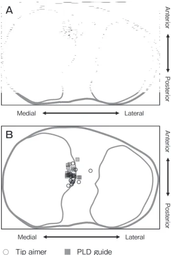
Tibial tunnel position. The tibial AM tunnel position was 28.8±4.7% for the anteroposterior (AP) direction and 44.8±4.4% for the mediolateral (ML) direction in the conventional guide group, and 28.8±3.8% for the AP and 42.7±2.6% for the ML in the PLD guide group. The PL tunnel position was 48.4±5.5% for the AP and 45.1±3.4% for the ML in the conventional guide group and 45.8±4.5% for the AP and 44.2±1.4% for the ML in the PLD guide group (Table 2, Fig.3).
Tunnel edge distance. The mean tunnel edge dis-tance was 5.2±2.0 mm in the conventional guide group and 2.3±1.0 mm in the PLD guide group (p<0.01)
A
B
5 mm
Fig. 1 (A) PLD guide for a left guide (black arrowhead) and right guide (white arrowhead) on both sides of the center of the shaft. The center shaft is inserted into the AM tunnel, and the guide pin is inserted from the side guides (white or black arrowhead). The side guides are 20 degrees from the center shaft. (B) The guide pin is offset 2 mm from the tip of the shaft hook. Hook length is 3 mm. The PL tunnel can be created with 5-mm offset from the AM tunnel edge.
b
a
c
A
B
C
Fig. 2 (A) The location of the tunnel position on the tibia was expressed. (a) The distance between the edge of the AM tunnel and PL tunnel. (B) The angle at the coronal view of CT. (b) The angle between the center of the tibial tunnels and tibial axis. (C) The angle at the sagittal view of CT. (c) The angle between the center of the tibial tunnels and tibial axis.
(Table 3). A tunnel edge distance below 0 mm (tunnel coalition) was noted in 5 knees in the conventional guide group, and no knees in the PLD guide group. Conversely, tunnel edge distance greater than 5 mm was observed in 9 knees in the conventional guide
group, but no knees in the PLD guide group (Table 3). The tunnel coalition rate was 25% for the conventional guide group and 0% for the PLD guide group.
Tunnel angle. The coronal section tunnel angle was 6.2±5.5° for the AM and 19.6±7.5° for the PL in the conventional guide group, and 8.2±4.2° for the AM and 29.8±5.1° for the PL in the PLD guide group. The sagittal section tunnel angle was 32.6±4.1° for the AM and 34.7±6.2° for the PL in the conventional guide group, and 26.9±5.1° for the AM and 30.9±5.3° for the PL in the PLD guide group (Table 4). A significant dif-ference was only noted for the coronal section PL tunnel angle.
Clinical outcomes. Joint range of motion at 6 months postoperatively was 1.6° for extension and 138.2° for flexion in the conventional guide group and 1.5° for extension and 136.3° for flexion in the PLD guide group. No positive results were noted for the Lachman test or pivot-shift test. The Lysholm score was 98.8 in the conventional guide group and 97.9 in the PLD guide group. No significant differences in clinical outcomes were noted between the 2 groups.
Discussion
The center of the ACL anatomical tibial enthesis (X/ Table 4 Tibial tunnel angles
Tip aimer PLD guide p value
Coronal AM (°) 6.2±5.5 8.2±4.2 n.s.
Coronal PL (°) 19.6±7.5 29.8±5.1 <0.01 Sagittal AM (°) 32.6±4.1 26.9±5.1 n.s. Sagittal PL (°) 34.7±6.2 30.9±5.3 n.s. Date are presented as a mean ± standard deviation
PLD, PL divergence; AM, anteromedial; PL, posterolateral. Table 3 Tibial tunnel edge distances between the AM and PL bundles
Tip aimer PLD guide p value Tunnel edge distance (mm) 5.2±2.0 2.3±1.0 <0.01
≦0 mm (cases) 5 0
>0 mm and ≦5 mm (cases) 6 23
>5 mm (cases) 9 0
Tunnel coalition rate (%) 25 0
Date are presented as a mean ± standard deviation PLD, PL divergence; AM, anteromedial; PL, posterolateral.
○ Tip aimer ■ PLD guide
Medial Lateral Anterior Posterior Medial Lateral Anterior Posterior
A
B
Fig. 3 Evaluation of tibial tunnel position. (A) Tunnel positions of the AM bundle. (B) Tunnel positions of the PL bundle.
Table 2 Tibial tunnel positions in the Tip aimer and PLD guide groups
Tip aimer PLD guide p value AM tunnel AP direction (%) 28.8±4.7 28.8±3.8 n.s.
ML direction (%) 44.8±4.4 42.7±2.6 n.s. PL tunnel AP direction (%) 48.4±5.5 45.8±4.5 n.s. ML direction (%) 45.1±3.4 44.2±1.4 n.s. Date are presented as a mean ± standard deviation
PLD, PL divergence; AM, anteromedial; AP, anteroposterior; ML, mediolateral; PL, posterolateral.
Y) was 53%/36% for the AM and 49%/51% for the PL [15]. It has been reported that tunnel position after double-bundle ACL reconstruction is 54%/42% for the AM and 54%/56% for the PL [16]. Recent anatomical studies have provided evidence that positioning the tib-ial tunnel at the center of the ACL footprint may dam-age the anterior meniscal roots [17-19]. In the present study, both the AM and PL were located more antero-medially than is anatomically normal. At our facility, when creating AM tunnels, we use the Parsons’ knob for the anterior side and an L-shaped ridge comprising the medial intercondylar ridge for the medial side [20]. The reconstructed graft is actually positioned postero-laterally from the center of the created tibial tunnel as a result of influence of the femoral tunnel. This is why we try to place the tunnel center anteromedially from the marking as much as possible. This also appears to be the case in previous reports.
The anatomical distance between the tibial AM bun-dle and PL bunbun-dle edges is 1-2 mm [21]. Lee et al. reported that a tunnel edge distance of 2 mm is required to prevent tunnel coalition in double-bundle ACL reconstruction [22]. In the present study, the mean tunnel edge distance in the PLD guide group was 2.3 mm. Thus, we were able to create a positional rela-tionship that was optimal both anatomically and in terms of preventing tunnel coalition.
Tunnel coalition is not a rare occurrence, with reports indicating that it occurred as a result of drilling in over 50%, in 27%, and in 23.8% of cases [23-25]. Tunnel coalition affects the knee stability and function of the reconstructed ligament [7]. It has been reported that tunnel coalition correlates with tunnel expansion [8]. Expansion of the tunnel can also contribute to knee stability [9]. The tunnel coalition rate in this study was 25% in the conventional guide group, which was mostly consistent with past reports. However, no cases of tun-nel coalition were observed in the PLD guide group.
Creation of the PL tunnel tended to involve insertion from a more medial aspect for the PLD guide group than the conventional guide group. Caution is required to create the AM tunnel drilling site on the tibia. An excessively medial positioning of the AM drilling site can cause the PL drilling site to become more postero-medially positioned on the tibia when using the PLD guide. No significant differences in clinical outcomes were observed between the conventional guide group and the PLD guide group. As these were only
short-term outcomes, a follow-up survey will be needed in the future.
There were 2 main limitations in this study. First, the sample size was small. Second, evaluation was only performed over the short-term postoperatively, mean-ing that long-term tunnel expansion must be assessed going forward.
In conclusion, we investigated and compared the position of tibial tunnels created with a conventional guide and the PLD guide. While the mean tibial tunnel position was similar, significant differences were noted for tunnel edge distance. Although tunnel coalition was observed in 5 knees in the conventional guide group, not a single knee showed tunnel coalition in the PLD guide group. This suggests that the PLD guide can be used to create anatomically positioned PL tunnels, reduce the probability of tunnel coalition, and achieve more stable outcomes. This device could be a beneficial aid for many surgeons.
Acknowledgments. We would like to thank Professor Yasuyuki Ishibashi and his colleagues (Hirosaki University Graduate School of Medicine) for instructing us in the handling of the PL divergence guide (Arthrex).
References
1. Louboutin H, Debarge R, Richou J, Selmi TA, Donell ST, Neyret P and Dubrana F: Osteoarthritisin patientswith anterior cruciate ligament rupture: a review ofriskfactors. Knee (2009) 16: 239-244. 2. Gabriel MT, Wong EK, Woo SL, Yagi M and Debski RE:
Distribution of in situ forces in the anterior cruciate ligament in response to rotatory loads. J Orthop Res (2004) 22: 85-89. 3. Yasuda K, Kondo E, Ichiyama H, Kitamura N, Tanabe Y,
Tohyama H and Minami A: Anatomic reconstruction of the antero-medial and posterolateral bundles of the anterior cruciate ligament using hamstring tendon grafts. Arthroscopy (2004) 20: 1015-1025. 4. Muneta T, Koga H, Morito T, Yagishita K and Sekiya I: A retro-spective study of the midterm outcome of two-bundle anterior cru-ciate ligament reconstruction using quadrupled semitendinosus tendon in comparison with one-bundle reconstruction. Arthroscopy (2006) 22: 252-258.
5. Cha PS, Brucker PU, West RV, Zelle BA, Yagi M, Kurosaka M and Fu FH: Arthroscopic double-bundle anterior cruciate ligament reconstruction: an anatomic approach. Arthroscopy (2005) 21: 1275. 6. Yasuda K, Kondo E, Ichiyama H, Tanabe Y and Tohyama H: Clinical
evaluation of anatomic double-bundle anterior cruciate ligament reconstruction procedure using hamstring tendon grafts: compari-sons among 3 different procedures. Arthroscopy (2006) 22: 240-251.
7. Hantes ME, Liantsis AK, Basdekis GK, Karantanas AH, Christel P and Malizos KN: Evaluation of the bone bridge between the bone tunnels after anatomic double-bundle anterior cruciate liga-ment reconstruction: a multidetector computed tomography study. Am J Sports Med (2010) 38: 1618-1625.
8. Samuelsson K, Andersson D and Karlsson J: Treatment of ante-rior cruciate ligament injuries with special reference to graft type and surgical technique: an assessment of randomized controlled trials. Arthroscopy (2009) 25: 1139-1174.
9. Kawaguchi Y, Kondo E, Kitamura N, Kai S, Inoue M and Yasuda K: Comparisons of femoral tunnel enlargement in 169 patients between single-bundle and anatomic double-bundle anterior cruci-ate ligament reconstructions with hamstring tendon grafts. Knee Surg Sports Traumatol Arthrosc (2011) 19: 1249-1257.
10. Fujii M, Furumatsu T, Miyazawa S, Tanaka T, Inoue H, Kodama Y, Masuda K, Seno N and Ozaki T: Features of human autologous hamstring graft elongation after pre-tensioning in anterior cruciate ligament reconstruction. Int Orthop (2016) 40: 2553-2558. 11. Shino K, Suzuki T, Iwahashi T, Mae T, Nakamura N, Nakata K
and Nakagawa S: The residentʼs ridge as an arthroscopic
land-mark for anatomical femoral tunnel drilling in ACL reconstruction. Knee Surg Sports Traumatol Arthrosc (2010) 18: 1164-1168. 12. Tensho K, Shimodaira H, Aoki T, Narita N, Kato H, Kakegawa A,
Fukushima N, Moriizumi T, Fujii M, Fujinaga Y and Saito N: Bony Landmarks of the Anterior Cruciate Ligament Tibial Footprint: A Detailed Analysis Comparing 3-Dimensional Computed Tomography Images to Visual and Histological Evaluations. Am J Sports Med (2014) 42: 1433-1440.
13. Berg EE: Parsonsʼ knob (tuberculum intercondylare tertium): a
guide to tibial anterior cruciate ligament insertion. Clin Orthop Relat Res (1993) 292: 229-231.
14. Tsukada H, Ishibashi Y, Tsuda E, Fukuda A and Toh S: Anatomical analysis of the anterior cruciate ligament femoral and tibial foot-prints. J Orthop Sci (2008) 13: 122-1229.
15. Forsythe B, Kopf S, Wong AK, Martins CA, Anderst W, Tashman S and Fu FH: The location of femoral and tibial tunnels in tomic double-bundle anterior cruciate ligament reconstruction ana-lyzed by three-dimensional computed tomography models. J Bone Joint Surg Am (2010) 92: 1418-26.
16. Amis AA and Dawkins GP: Functional anatomy of the anterior cru-ciate ligament. Fibre bundle actions related to ligament replace-ments and injuries. J Bone Joint Surg Br (1991) 73: 260-267.
17. Furumatsu T, Kodama Y, Maehara A, Miyazawa S, Fujii M, Tanaka T, Inoue H and Ozaki T: The anterior cruciate ligamentlat-eral meniscus complex: a histological study. Connect Tissue Res (2016) 57: 91-98.
18. Kodama Y, Furumatsu T, Miyazawa S, Fujii M, Tanaka T, Inoue H and Ozaki T: Location of the tibial tunnel aperture affects extru-sion of the lateral meniscus following reconstruction of the anterior cruciate ligament. J Orthop Res (2017) 35: 1625-1633.
19. LaPrade CM, Ellman MB, Rasmussen MT, James EW, Wijdicks CA, Engebretsen L and LaPrade RF: Anatomy of the anterior root attachments of the medial and lateral menisci: a quantitative anal-ysis. Am J Sports Med (2014) 42: 2386-2392.
20. Kodama Y, Furumatsu T, Hino T, Kamatsuki Y and Ozaki T: Minimal Ablation of the Tibial Stump Using Bony Landmarks Improved Stability and Synovial Coverage Following Double-Bundle Anterior Cruciate Ligament Reconstruction. Knee Surg Relat Res (2018) 30: 348-355.
21. Lee SH, Choi JY, Kim DH, Kang BJ, Nam DC, Yoon HK and Hwang SC: Correlation between femoral guidewire position and tunnel communication in double bundle anterior cruciate ligament reconstruction. Yonsei Med J (2014) 55: 1592-1599.
22. Kiekara T, Järvelä T, Huhtala H and Paakkala A: MRI evaluation of the fourvtunnels of double-bundle ACL reconstruction. Acta Radiol (2014) 55: 579-588.
23. Shimodaira H, Tensho K, Akaoka Y, Takanashi S, Kato H and Saito N: Remnant-Preserving Tibial Tunnel Positioning Using Anatomic Landmarks in Double-Bundle Anterior Cruciate Ligament Reconstruction. Arthroscopy (2016) 32: 1822-1830.
24. Kiekara T, Järvelä T, Huhtala H, Moisala AS, Suomalainen P and Paakkala A: Tunnel communication and increased graft signal intensity on magnetic resonance imaging of double-bundle anterior cruciate ligament reconstruction. Arthroscopy (2014) 30: 1595-1601.
25. Siebold R and Cafaltzis K: Differentiation between intraoperative and postoperative bone tunnel widening and communication in double-bundle anterior cruciate ligament reconstruction: a prospec-tive study. Arthroscopy (2010) 26: 1066-1073.