Contents lists available atScienceDirect
Pathology - Research and Practice
journal homepage:www.elsevier.com/locate/prp
PD-L1 expression in pancreatic adenosquamous carcinoma: PD-L1 expression is limited to the squamous component
Masahiko Tanigawa
a, Yoshiki Naito
a,b,⁎, Jun Akiba
b, Akihiko Kawahara
b, Yoshinobu Okabe
c, Yusuke Ishida
c, Hiroto Ishikawa
d, Toru Hisaka
d, Fumihiko Fujita
d, Masafumi Yasunaga
d, Takahiro Shigaki
d, Tomoya Sudo
d, Yutaro Mihara
a, Masamichi Nakayama
a, Reiichiro Kondo
a, Hironori Kusano
a, Kazuhide Shimamatsu
e, Koji Okuda
d, Yoshito Akagi
d, Hirohisa Yano
aaDepartment of Pathology, Kurume University School of Medicine, Kurume, Japan
bDepartment of Diagnostic Pathology, Kurume University Hospital, Kurume, Japan
cDivision of Gastroenterology, Department of Medicine, Kurume University School of Medicine, Kurume, Japan
dDepartment of Surgery, Kurume University School of Medicine, Kurume, Japan
eDepartment of Pathology, Omuta City Hospital, Omuta, Japan
A R T I C L E I N F O
Keywords:
Keyword Pancreas Pancreatic cancer
Pancreatic adenosquamous carcinoma PD-L1, EUS-FNA
Immunohistochemistry
A B S T R A C T
Aim: We examined the programmed death-ligand 1 (PD-L1) expression in surgically resected pancreatic ade- nosquamous carcinoma (PASC) samples. Furthermore, the detection rate was also assessed using biopsy cases obtained from endoscopic ultrasound-guidedfine needle aspiration (EUS-FNA).
Methods:Fifteen cases of PASC (six resected and nine EUS-FNA biopsied) from the Kurume University Hospital between 2009 and 2016 were used for the evaluation of PD-L1 expression. As a control group, 34 cases of pancreatic ductal adenocarcinomas (PDACs) were selected. To compare the positivity and intensity of PD-L1, two types of clones (SP263, E1L3N) were examined for immunostaining. Only the membrane expression of PD- L1 was regarded as positive. The PD-L1 expressions in the squamous cell carcinoma component (SCc), adeno- carcinoma component (ACc), and immune cells were assessed separately. The ratio of PD-L1 expression was calculated by counting the positive tumor cells, and tumor proportion score (TPS) was applied (TPS; Null < 1%, low expression; 1≤TPS≤49% and high expression;≥50%).
Results:PD-L1 expression was observed infive surgical PASC samples (83%). This shows that SCc presented a high expression in these cases. However, the overall TPS indicated a low expression. In contrast, only one case (3%) was positive for PD-L1 in PDACs, and the TPS indicated a low expression. No differences in PD-L1 ex- pression were observed between the two clones, SP263 and E1L3N. High PD-L1 expression in the EUS-FNA sample was found in only one case (11%).
Discussion: Although assessment using the tumor cells of PASC samples obtained from EUS-FNA was difficult, this study suggests the selective expression of PD-L1 in the SCc of PASC. Furthermore, it was considered that immune checkpoint inhibitors could provide therapeutic effects selectively on the SCc for the entire range of TPSs, though the PD-L1 expression was low.
1. Introduction
Pancreatic cancer is a malignant tumor with poor prognosis. As many cases are inoperable, thefive-year survival rates are generally below 30% [1–4]. Pancreatic ductal adenocarcinoma (PDAC) is a pre- dominant histological type of pancreatic cancer [5], with a malignant potential higher than that of other pancreatic cancers such as pan- creatic neuroendocrine tumor and acinar cell carcinoma [6,7]. Among
these diseases, mixed-type PDAC has a particularly poor prognosis;
histological features are thought to have a significant impact on this type of PDAC [7–11].
Pancreatic adenosquamous carcinoma (PASC) was classified as a PDAC subtype, according to the 2010 WHO classification [12]; histo- logically, PDAC is defined as a mixture of the adenocarcinoma com- ponent (ACc) and squamous cell carcinoma component (SCc) [4,5,13,14]. Like the case for PDAC, driver gene mutations, such as
https://doi.org/10.1016/j.prp.2018.10.006
Received 7 June 2018; Received in revised form 14 September 2018; Accepted 17 October 2018
⁎Corresponding author at: Department of Diagnostic Pathology, Kurume University Hospital, 67 Asahimachi, Kurume, 830-0011 Japan.
E-mail address:[email protected](Y. Naito).
0344-0338/ © 2018 Elsevier GmbH. All rights reserved.
T
those in KRAS, p53, p16 and smad4, are characteristic genetic dis- orders; thus, PASC is classified as a subtype of PDAC [14]. From the histological point of view, PASC is considered as a metaplastic cancer represented by the transition between the ACc and SCc [15,16]. In general, squamous metaplasia is a histological change reflected as an inflammatory response and is commonly seen in the respiratory and gynecological organs [17–21]. With regards to the pancreas, patho- genesis of squamous metaplasia is related to chronic pancreatitis [13], so inflammation could influence the oncogenesis of PASC.
Recent studies have found that tumor cells proliferate by avoiding immune responses [22]. After antigen presentation by dendritic cells, which recognize tumor cell surface antigens, activated cytotoxic T cells cause the apoptosis of tumor cells. Tumor-associated antigens released from apoptotic cells instigate further immune responses and enhance additional antitumor effects. During these immune responses, pro- grammed cell death 1 (PD-1) and programmed death-ligand 1 (PD-L1) suppress the T cell response, and the binding action of these two factors decreases cytokine production by T cells. Tumor cells expressing PD-L1 are thought to evade the T cell immune response.
In this study, we conducted pathomorphologic analysis by evalu- ating the expression of two different PD-L1 clones over the ACc and SCc of PASC; this may clarify the role of the PD-1 pathway associated with tumor formation. In addition, using tissue samples obtained from en- doscopic ultrasound-guided fine need aspiration (EUS-FNA), PD-L1 expression was examined to explore the capability of PD-1 ligand pathway-inhibitory drugs for treating inoperable PASCs.
2. Methods 2.1. Patients
We examined 6 resected PASCs and 9 EUS-FNA biopsied cases from the Kurume University Hospital during the period between 2009 and 2016. As a control, 34 cases of conventional PDAC that did not receive preoperative neoadjuvant therapy from 2009 to 2014 were selected (Table 1). Histologically, EUS-FNA cases were PDAC cases with squa- mous differentiation, which is highly suggestive of PASC; all these cases were inoperable. Cases of resected PASCs consisted of four men and two women, with an average age of 64.7 ± 9.7 years (range 50–80 years).
EUS-FNA cases consisted of seven men and two women, with an
average age of 66.4 ± 6.2 years. These specimens were fixed using 10% buffered formalin and cut into 4μm-thick sections, followed by hematoxylin and eosin staining. Final diagnoses were performed ac- cording to the WHO classification of the Digestive system [12]. Histo- pathological evaluations were performed by two pathologists (M.T. and Y.N). This study was approved by the ethics committee of Kurume University (No. 18116).
2.2. Immunohistochemistry
Paraffin-embedded tissue samples, which were 4-μm thick, were placed on a coated glass slide and treated with PD-L1, DPC4, p40, MLH- 1, MSH-2, PMS-2, MSH-6 antibodies using BenchMark ULTRA (Ventana Automated Systems, Inc., Tucson, AZ, USA), Bond-III autostainer (Leica Microsystems, Newcastle, UK), or Autostainer Link 48 (Agilent Technologies, Santa Clara, CA). The former was used for PD-L1 (Code 07494190001, ready to use, clone SP263, Ventana Automated Systems, Inc.), and the latter, for PD-L1 (Code #13684, 1:200, clone E1L3N, Cell Signaling Technology, Inc., Danvers, MA, USA) immunostaining.
Briefly, for clone SP263 and p40 (clone BC28, Ventana Automated Systems, Inc., Tucson, AZ, USA), each slide was heat-treated using Ventana’s ULTRA cell conditioning 1 for 64 min and incubated with each antibody for 16 min. Consecutively, the chromogenic reaction was performed automatically by the Ventana OptiVIEW DAB detection kit (catalog no. 760-700; Ventana Automated Systems, Inc.). Similarly, immunostaining for cloneE1L3N and DPC4 (clone B-8, Santa Cruz Biotechnology Inc. CA, USA) was performed using the fully automated Bond-III system (Leica Microsystems), using onboard heat-induced an- tigen retrieval with epitope retrieval solution 2 (ER2, EDTA-based buffer, pH 9.0, Leica Microsystems) for 30 min and a Refine polymer detection kit (catalog no. TA9145; Leica Microsystems).
Analysis using a four-antibody panel of mismatch repair proteins including MLH1 (clone ES05), MSH2 (clone FE11), MSH6 (clone EP49), and PMS2 (clone EP51) was performed by the DAKO EnVision method (DakoCytomation, Glostrup, Denmark). The tissue samples were pre- treated with heat-induced epitope retrieval (HIER) at 97 °C for 20 min at high pH (50×). The slides were then incubated with the following antibodies: MLH1, MSH2, MSH6, and PMS2. Immunohistochemistry was performed manually. All immunohistochemical analyses were evaluated by two experienced pathologists (M.T. and Y.N.). Only the membrane expressions of each PD-L1 clone were considered as positive.
We used the tumor proportion score (TPS) for the staining evalua- tion of the tumor areas, in accordance with previous reports [23–26].
We classified PD-L1 expression into 3 ranks, and set the following cri- teria for the staining evaluation: TPS≥1% as positive, null; TPS <
1%, low expression; TPS 1–49%, and high expression; TPS≥50%.
The scores of PD-L1-positive immune cells including lymphocytes and macrophages were evaluated as the percentage of the tumor:
IC3≥10%, IC2≥5% and < 10%, IC1≥1% and < 5%, and IC0 <
1%.
2.3. Statistical analysis
PD-L1 expression was examined statistically according to the TPS values (resected groups and EUS-FNA samples). The analysis was per- formed using a chi-squared test, and P values < 0.05 were determined to be statistically significant for all analyses. The entire analysis was performed using JMP Pro version 13.0.0 (SAS Institute, Inc. Cary, North Carolina).
3. Results
3.1. Pathologicalfindings in the resected PASC samples
A typical gross image of PASC exhibited a hemorrhagic area with an unclear border (Fig. 1a). Histologically, PASC comprised more than Table 1
Clinicopathologicalfindings in the PASC and PDAC samples.
PASC (n = 15) PDAC (n =
34) Resection (n =
6)
EUS-FNA (n
= 9)
Age 64.0 ± 11.5 66.4 ± 6.2 65.9 ± 10.4
Gender Male 4 7 25
Female 2 2 9
Location Head 3 1 32
Body/Tail 3 8 2
Tumor size (mm)
45 ± 15 27 ± 8
T 1 3 0 3
2 0 0 0
3 6 4 31
4 0 5 0
N 0 1 3 11
1 5 6 23
M 0 5 2 34
1 1 7 0
ly 6 28
v 6 24
ne 6 32
ASC; pancreatic adenosquamous carcinoma, PDAC; pancreatic ductal adeno- carcinoma, EUS-FNA; endoscopic ultrasound-guidedfine needle aspiration, ly;
lymphatic invasion, v; venous invasion, ne; nerve invasion.
30% of the SCc, some of which showed infiltration into the adjacent ACc or a transition between the two components (Fig. 1b). The ex- pression of p40 was found infive (83%) cases (Fig.1c). With regards to the grade of the SCc, four cases (67%) were of the well-differentiated
type and two cases (33%) were of the poorly differentiated type. Con- versely, the ACc in all cases was of the moderately to poorly differ- entiated type.
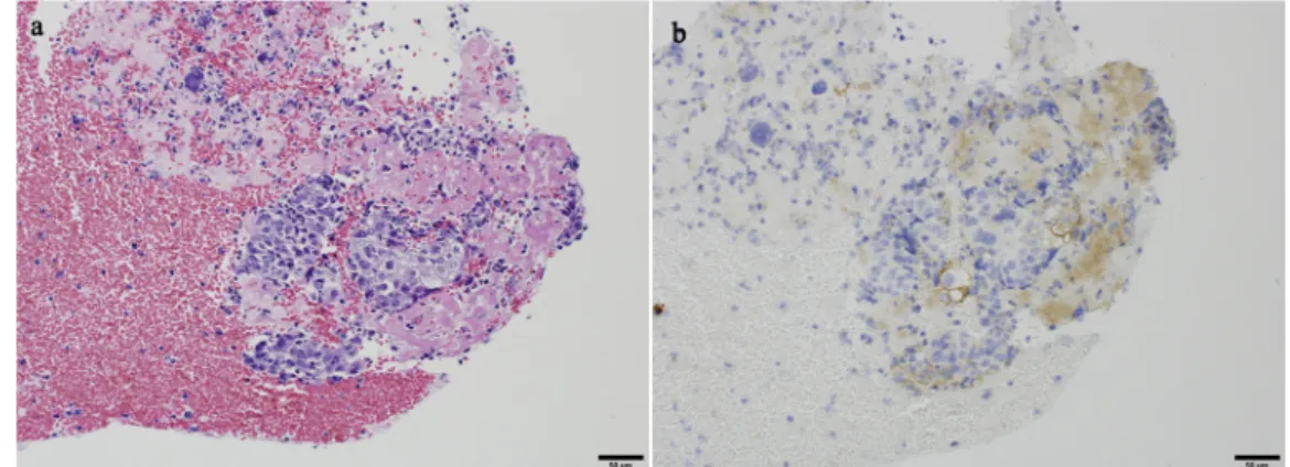
Fig. 1.Pathologicalfindings and PD-L1 expression in the resected PASC samples.
Grossly, tumor showed hemorrhage and necrosis, with an unclear border (a).
Histologically, the SCc and ACc were mixed, with the former occupying more than 30% of the tumor. (b) The SCc was confirmed by p40 staining (c).
PD-L1 expression was observed on the membrane of the SCc (d).
Conversely, PD-L1 expression was not identified in the ACc (f,★).
Table 2
Comparison of PD-L1 expression in the adenocarcinoma and squamous cell carcinoma components of the resected PASC samples.
Case No. PD-L1 p40 Smad4 MSH2 MSH6 MLH1 PMS2
TPS ACc SCc IC
1 Low Null High 3 N.Da loss + + + +
2 Low Null High 3 + loss + + + +
3 Null Null Null 0 + loss + + + +
4 Low Null High 3 + loss + + + +
5 Low Null High 1 + intact + + + +
6 Low Null High 1 + loss + + + +
PASC; pancreatic adenosquamous cell carcinoma, TPS; tumor proportion score, ACc; adenocarcinoma component, SCc; squamous cell carcinoma component, IC;
immune cells, TPS score; high≥50%, low 1–49%, null < 1%, Immune cell score; IC3≥10%, IC2≥5% and < 10%, IC1≥1% and < 5%, and IC 0 < 1%.
a There were abundant cancer pearls observed in the morphopathological analysis.
Fig. 2. PD-L1 expression in the resected PDAC samples.
PD-L1 membrane expression was observed in only one case of PDAC.
Thefigure shows a positive (TPS = 24%) and negative case (a, b).
3.2. PD-L1 expression and immunohistochemicalfindings in the resected PASC and PDAC samples
PD-L1 expressions of the resected PASC and PDAC samples are displayed inTable 2. Positive PD-L1 expression at the invasive front of the tumor and in the center of the tumor was observed infive patients (83%) with PASC. Furthermore, PD-L1 expression was observed only in the SCc (Fig. 1c); there was no expression of PD-L1 in the ACc. (Fig. 1d).
Limited to the SCc, the TPS indicated a high expression, but showed a low expression for the entire tumor. In contrast, PD-L1 expression was observed in one PDAC (2.9%) case (Fig. 2a, b), and the TPS was 24%.
For the PD-L1-positive immune cells, IC3 was observed in three cases (50%). MLH-1, MSH-2, PMS-2, and MSH-6 were expressed in all cases (Fig. 3c–f) (Table 2). On the contrary,five of six cases (83%) showed loss of DPC4 expression (Table 2) (Fig. 3g). With regards to the im- munostaining for each PD-L1 clone, no significant difference was ob- served in the PD-L1 expression (Fig. 3a, b) (Table 3).
3.3. PD-L1 expression in the EUS-FNA tissue samples
Nine EUS-FNA biopsied cases are shown inTable 4. PD-L1 expres- sion was found in one case (11%), and the TPS was 35% (Fig. 4a, b).
Fig. 3.Comparison of PD-L1 expression between the two clones, and immunohistologicalfindings in the PASC samples.
No difference was observed between Clone E1L3N (a) and Clone SP263 (b).
A normal pancreatic duct was partially positive for PD-L1.
Loss of mismatch repair gene (MMR) associated proteins, MLH1, MSH2, MSH6, and PSM6 (c–f, respectively) was not observed in both SC and AC components.
Deletion of DPC4 was confirmed infive cases (g).
PD-L1 expression was identified only in the SCc with p40 expression, but not in the ACc.
4. Discussion
PD-L1 expression has been studied across variousfields; it has been confirmed in squamous cell carcinoma of the lung, esophagus, and thymus. Pathways contributing to the evasion of T cell immune re- sponses by PD-1 or PD-L1 expression have been identified [27–29]. By inhibiting such pathways, immune-based antitumor effects can be es- tablished and used as treatment options [30]. In colorectal and gastric cancers, PD-L1 expression is observed at frequencies of around 30–40%
[31–33]; however, PD-L1 expression for PDAC is extremely low, com- pared to the case for other adenocarcinomas [34]. This study presented interesting results, that is, while PD-L1 expression was rarely found in the PDAC control group, high PD-L1 expression was found selectively in the SCc, but not in the ACc, of PASC cases.
In the past, few studies have investigated PD-1 pathway mechan- isms with regards to pancreatic disease. Considering the role of in- flammation on PD-1 pathway, we can speculate the mechanisms of PASC. One of the features of the pathogenesis of PASC is squamous metaplasia [13,35]. A report using bile duct cell lines proved that the histologic change of adenocarcinoma into squamous cell carcinoma
occurs under the influence of inflammatory cytokines [35]. Expression of selective PD-L1 in the SCc has also been observed in adenosquamous cell carcinoma in the lung, but the mechanism of its expression has not yet been elucidated [36,37]. Moreover, some reports have stated that roughly 4% of adenocarcinomas with chronic pancreatitis showed squamous metaplasia in the pancreatic duct epithelium [13]. From these results, it is hypothesized that the PD-1-mediated“cancer immune escape mechanism” may arise in cases of squamous metaplasia and cause tissue change through inflammation. This study confirmed that PD-L1 expression was observed in the SCc but not in the ACc; ad- ditionally, low PD-L1 expression was observed in cases of PDAC. PD-L1 expression in the SCc, which is induced by chemical mediators related to adenocarcinoma invasion, may play a role in the evasion of immune checkpoints. From a clinical point of view, we can attribute poor prognosis of PASC to invasion and metastasis of cancer cells that evade immune checkpoints by PD-L1 expression.
For cases of unresectable PDAC, evaluation of PD-L1 expression using EUS-FNA samples will be necessary in the future [38]. However, in this study, only a few EUS-FNA cases exhibited PD-L1 expression, although PD-L1 expression was seen in the SCc in the resected PDAC samples. An advantage of EUS-FNA is high diagnostic accuracy [39];
however, limited sample number is a drawback, especially in case of a mixed tumor, which requires sample tissue for immunohistochemistry.
In the future, while evaluating PD-1 pathway inhibitors for patients with unresectable PASC, we will encounter EUS-FNA cases with no PD- L1 expression in the SCc. Nevertheless, PD-L1 inhibitors will be actively introduced into the treatment strategy, considering the high rates of PD-L1 expression in the resected PASCs.
Currently, pembrolizumab is used as a new immune checkpoint inhibitor treatment strategy [30,40–44]. Immune checkpoint inhibitor treatment has been introduced as a strategy for treating MMR-deficient pancreatic cancer; it has actually achieved several successful outcomes.
Although TPS evaluation is required at the time of use [26], this study has demonstrated that there were no differences in the PD-L1 expres- sion of the two clones. As PASC cases with the TPS indicating a high expression of PD-L1 were not observed, it is unlikely that PD-L1/PD-1 pathway-mediated immune checkpoint inhibitors will be effective against all tumor cells. In addition, there were no cases of MMR-defi- cient PASC in this study. Unfortunately, the correlation of PD-L1 posi- tivity with the MMR gene protein status of PASC is not definite. This study suggests the selective expression of PD-L1 in the SCc of PASC, and the use of immune checkpoint inhibitors providing therapeutic effects selectively on the SCc were considered with regards to the entire TPS, though the PD-L1 expression was low.
Table 3
Comparison of the PD-L1 expression of the clones E1L3N and SP263 in the resected PASC samples.
Clone TPS PASC (n = 6) PDAC (n = 34) P-value
E1L3N High 0 0 < 0.001
Low 5 1
Null 1 33
SP263 High 0 0 < 0.001
Low 5 1
Null 1 33
PASC; Pancreatic adenosquamous, PDAC; pancreatic ductal adenocarcinoma, TPS score; high≥50%, low 1–49%, null < 1%.
Table 4
PD-L1 expression in the EUS-FNA samples of PASC.
Clone TPS SCc ACc P-value
E1L3N High 0 0 N.S
Low 1 0
Null 8 0
TPS; tumor proportion score, SCc; squamous cell carcinoma component, ACc;
adenocarcinoma component, TPS score; high≥50%, low 1–49%, null < 1%.
Fig. 4.PD-L1 expression in the EUS-FNA samples.
The only positive case of PASC obtained by EUS-FNA (a).
PD-L1 expression was observed in the SCc (b).
5. Conclusion
PASC represents a group of diseases with poor prognosis and their TPSs represent a low PD-L1 expression. Furthermore, as PD-L1 ex- pression is restricted to the SCc, immune checkpoint inhibitors may be an effective strategy for treating PASC, as they exert therapeutic effects selectively on the SCc.
Conflicts of interest
All authors have declared no conflicts of interest.
References
[1] L. Rahib, J.M. Fleshman, L.M. Matrisian, J.D. Berlin, Evaluation of pancreatic Cancer Clinical trials and benchmarks for clinically meaningful future trials: a systematic review, JAMA Oncol. 2 (2016) 1209–1216.
[2] B. Tessier-Cloutier, S.E. Kalloger, M. Al-Kandari, K. Milne, D. Gao, B.H. Nelson, D.J. Renouf, B.S. Sheffield, D.F. Schaeffer, Programmed cell death ligand 1 cut- point is associated with reduced disease specific survival in resected pancreatic ductal adenocarcinoma, BMC Cancer 17 (2017) 618.
[3] M. Kieler, M. Unseld, D. Bianconi, G. Prager, Challenges and perspectives for im- munotherapy in adenocarcinoma of the pancreas: the Cancer immunity cycle, Pancreas 47 (2018) 142–157.
[4] R. Marcus, A. Maitra, J. Roszik, Recent advances in genomic profiling of ade- nosquamous carcinoma of the pancreas, J. Pathol. 243 (2017) 271–272.
[5] Y. Fang, Z. Su, J. Xie, R. Xue, Q. Ma, Y. Li, Y. Zhao, Z. Song, X. Lu, H. Li, C. Peng, F. Bai, B. Shen, Genomic signatures of pancreatic adenosquamous carcinoma (PASC), J. Pathol. 243 (2017) 155–159.
[6] A. Al-Hader, R.N. Al-Rohil, H. Han, D. Von Hoff, Pancreatic acinar cell carcinoma: a review on molecular profiling of patient tumors, World J. Gastroenterol. 23 (2017) 7945–7951.
[7] N. Silvestris, K. Danza, V. Longo, O. Brunetti, L. Fucci, A. Argentiero, A. Calabrese, I. Cataldo, R. Tamma, D. Ribatti, S. Tommasi, Angiogenesis in adenosquamous cancer of pancreas, Oncotarget 8 (2017) 95773–95779.
[8] Y. Naito, A. Kawahara, T. Taira, Y. Takase, K. Murata, Y. Ishida, Y. Okabe, M. Tanigawa, Y. Mihara, M. Nakayama, K. Shimamatsu, H. Yano, J. Akiba, Cytopathological and immunocytochemicalfindings of pancreatic anaplastic car- cinoma with ZEB1 expression by means of touch imprint cytology, Diagn.
Cytopathol. 46 (2018) 198–203.
[9] Y. Naito, H. Kinoshita, Y. Okabe, S. Arikawa, K. Higaki, Y. Morimitsu, F. Yamasaki, K. Suda, M. Yasumoto, H. Kusano, O. Nakashima, H. Yano, Pathomorphologic study of undifferentiated carcinoma in seven cases: relationship between tumor and pancreatic duct epithelium, J. Hepatobiliary. Surg. 16 (2009) 478–484.
[10] C.A. Boyd, J. Benarroch-Gampel, K.M. Sheffield, C.D. Cooksley, T.S. Riall, 415 patients with adenosquamous carcinoma of the pancreas: a population-based ana- lysis of prognosis and survival, J. Surg. Res. 174 (2012) 12–19.
[11] C.G. Simone, T. Zuluaga Toro, E. Chan, M.M. Feely, J.G. Trevino, T.J. George Jr., Characteristics and outcomes of adenosquamous carcinoma of the pancreas, Gastrointest. Cancer Res.: GCR 6 (2013) 75–79.
[12] N. Fukushima, R.H. Hruban, N. Kato, D.S. Klimstra, G. Klöppel, M. Shimizu, B. Terris, WHO Classification of Tumors of the Digestive Systems; Ductal Adenocarcinoma Variants and Mixed Neoplasm of the Pancreas, 4th ed., IARC, Lyon, 2010.
[13] D.E. Kardon, L.D. Thompson, R.M. Przygodzki, C.S. Heffess, Adenosquamous car- cinoma of the pancreas: a clinicopathologic series of 25 cases, Mod. Pathol. 14 (2001) 443–451.
[14] J.R. Brody, C.L. Costantino, M. Potoczek, J. Cozzitorto, P. McCue, C.J. Yeo, R.H. Hruban, A.K. Witkiewicz, Adenosquamous carcinoma of the pancreas harbors KRAS2, DPC4 and TP53 molecular alterations similar to pancreatic ductal adeno- carcinoma, Mod. Pathol. 22 (2009) 651–659.
[15] O. Basturk, F. Khanani, F. Sarkar, E. Levi, J.D. Cheng, N.V. Adsay, DeltaNp63 ex- pression in pancreas and pancreatic neoplasia, Mod. Pathol. 18 (2005) 1193–1198.
[16] L.J. Layfield, H. Cramer, J. Madden, E.V. Gopez, K. Liu, Atypical squamous epi- thelium in cytologic specimens from the pancreas: cytological differential diagnosis and clinical implications, Diagn. Cytopathol. 25 (2001) 38–42.
[17] M. Herfs, P. Hubert, A.L. Poirrier, P. Vandevenne, V. Renoux, Y. Habraken, D. Cataldo, J. Boniver, P. Delvenne, Proinflammatory cytokines induce bronchial hyperplasia and squamous metaplasia in smokers: implications for chronic ob- structive pulmonary disease therapy, Am. J. Respir. Cell Mol. Biol. 47 (2012) 67–79.
[18] C. Du, J. Lu, L. Zhou, B. Wu, F. Zhou, L. Gu, D. Xu, Y. Sun, MAPK/FoxA2-mediated cigarette smoke-induced squamous metaplasia of bronchial epithelial cells, Int. J.
Chron. Obstruct. Pulmon. Dis. 12 (2017) 3341–3351.
[19] S.J. Bolton, K. Pinnion, V. Oreffo, M. Foster, K.E. Pinkerton, Characterisation of the proximal airway squamous metaplasia induced by chronic tobacco smoke exposure in spontaneously hypertensive rats, Respir. Res. 10 (2009) 118.
[20] L. Hirschowitz, C. Sen, J. Murdoch, Primary endometrioid adenocarcinoma of the cervix with widespread squamous metaplasia–a potential diagnostic pitfall, Diagn.
Pathol. 2 (2007) 40.
[21] R.H. Young, P.B. Clement, Endocervical adenocarcinoma and its variants: their morphology and differential diagnosis, Histopathology 41 (2002) 185–207.
[22] D.S. Chen, I. Mellman, Oncology meets immunology: the cancer-immunity cycle,
Immunity 39 (2013) 1–10.
[23] L. Fehrenbacher, A. Spira, M. Ballinger, M. Kowanetz, J. Vansteenkiste, J. Mazieres, K. Park, D. Smith, A. Artal-Cortes, C. Lewanski, F. Braiteh, D. Waterkamp, P. He, W. Zou, D.S. Chen, J. Yi, A. Sandler, A. Rittmeyer, Atezolizumab versus docetaxel for patients with previously treated non-small-cell lung cancer (POPLAR): a mul- ticentre, open-label, phase 2 randomised controlled trial, Lancet (Lond., Engl.) 387 (2016) 1837–1846.
[24] F. Sato, J. Akiba, A. Kawahara, Y. Naito, T. Ono, Y. Takase, K. Murata, H. Abe, T. Yamaguchi, H. Miyoshi, Y. Abe, Y. Mihara, M. Tanikawa, M. Akashi, H. Kurose, H. Umeno, H. Yano, The expression of programed death ligand-1 could be related with unfavorable prognosis in salivary duct carcinoma, J. Oral Pathol. Med. 47 (2018) 683–690.
[25] G. Lin, X. Fan, W. Zhu, C. Huang, W. Zhuang, H. Xu, X. Lin, D. Hu, Y. Huang, K. Jiang, Q. Miao, C. Li, Prognostic significance of PD-L1 expression and tumor infiltrating lymphocyte in surgically resectable non-small cell lung cancer, Oncotarget 8 (2017) 83986–83994.
[26] E.B. Garon, N.A. Rizvi, R. Hui, N. Leighl, A.S. Balmanoukian, J.P. Eder, A. Patnaik, C. Aggarwal, M. Gubens, L. Horn, E. Carcereny, M.J. Ahn, E. Felip, J.S. Lee, M.D. Hellmann, O. Hamid, J.W. Goldman, J.C. Soria, M. Dolled-Filhart, R.Z. Rutledge, J. Zhang, J.K. Lunceford, R. Rangwala, G.M. Lubiniecki, C. Roach, K. Emancipator, L. Gandhi, Pembrolizumab for the treatment of non-small-cell lung cancer, N. Engl. J. Med. 372 (2015) 2018–2028.
[27] Y. Ohigashi, M. Sho, Y. Yamada, Y. Tsurui, K. Hamada, N. Ikeda, T. Mizuno, R. Yoriki, H. Kashizuka, K. Yane, F. Tsushima, N. Otsuki, H. Yagita, M. Azuma, Y. Nakajima, Clinical significance of programmed death-1 ligand-1 and pro- grammed death-1 ligand-2 expression in human esophageal cancer, Clin. Cancer Res. 11 (2005) 2947–2953.
[28] V. Velcheti, K.A. Schalper, D.E. Carvajal, V.K. Anagnostou, K.N. Syrigos, M. Sznol, R.S. Herbst, S.N. Gettinger, L. Chen, D.L. Rimm, Programmed death ligand-1 ex- pression in non-small cell lung cancer, Lab. Invest. 94 (2014) 107–116.
[29] S. Yokoyama, H. Miyoshi, K. Nakashima, J. Shimono, T. Hashiguchi, M. Mitsuoka, S. Takamori, Y. Akagi, K. Ohshima, Prognostic Value of Programmed Death Ligand 1 and Programmed Death 1 Expression in Thymic Carcinoma, Clin. Cancer Res. 22 (2016) 4727–4734.
[30] J. Gong, A. Chehrazi-Raffle, S. Reddi, R. Salgia, Development of PD-1 and PD-L1 inhibitors as a form of cancer immunotherapy: a comprehensive review of regis- tration trials and future considerations, J. Immunother. Cancer 6 (2018) 8.
[31] B.H. O’Neil, J.M. Wallmark, D. Lorente, E. Elez, J. Raimbourg, C. Gomez-Roca, S. Ejadi, S.A. Piha-Paul, M.N. Stein, A.R. Abdul Razak, K. Dotti, A. Santoro, R.B. Cohen, M. Gould, S. Saraf, K. Stein, S.W. Han, Safety and antitumor activity of the anti-PD-1 antibody pembrolizumab in patients with advanced colorectal car- cinoma, PLoS One 12 (2017) e0189848.
[32] M. Amatatsu, T. Arigami, Y. Uenosono, S. Yanagita, Y. Uchikado, Y. Kijima, H. Kurahara, Y. Kita, S. Mori, K. Sasaki, I. Omoto, K. Maemura, S. Ishigami, S. Natsugoe, PD-L1 is a promising blood marker for predicting tumor progression and prognosis in patients with gastric cancer, Cancer Sci. (2018).
[33] Y. Wu, D. Cao, L. Qu, X. Cao, Z. Jia, T. Zhao, Q. Wang, J. Jiang, PD-1 and PD-L1 co- expression predicts favorable prognosis in gastric cancer, Oncotarget 8 (2017) 64066–64082.
[34] T. Nomi, M. Sho, T. Akahori, K. Hamada, A. Kubo, H. Kanehiro, S. Nakamura, K. Enomoto, H. Yagita, M. Azuma, Y. Nakajima, Clinical significance and ther- apeutic potential of the programmed death-1 ligand/programmed death-1 pathway in human pancreatic cancer, Clin. Cancer Res. 13 (2007) 2151–2157.
[35] A. Iemura, H. Yano, A. Mizoguchi, M. Kojiro, A cholangiocellular carcinoma nude mouse strain showing histologic alteration from adenocarcinoma to squamous cell carcinoma, Cancer 70 (1992) 415–422.
[36] X. Shi, S. Wu, J. Sun, Y. Liu, X. Zeng, Z. Liang, PD-L1 expression in lung ade- nosquamous carcinomas compared with the more common variants of non-small cell lung cancer, Sci. Rep. 7 (2017) 46209.
[37] A. Wang, H.Y. Wang, Y. Liu, M.C. Zhao, H.J. Zhang, Z.Y. Lu, Y.C. Fang, X.F. Chen, G.T. Liu, The prognostic value of PD-L1 expression for non-small cell lung cancer patients: a meta-analysis, Eur. J. Surg. Oncol. 41 (2015) 450–456.
[38] Q.M. Wu, Y.N. Guo, Y.Q. Xu, Diagnostic performance of endoscopic ultrasound- guidedfine-needle aspiration in pancreatic lesions, Eur. Rev. Med. Pharmacol. Sci.
22 (2018) 1397–1401.
[39] T. Ushijima, Y. Okabe, Y. Ishida, G. Sugiyama, Y. Sasaki, K. Kuraoka, M. Yasumoto, T. Taira, Y. Naito, M. Nakayama, O. Tsuruta, M. Sata, Evaluation of endoscopic cytological diagnosis of unresectable pancreatic cancer prior to and after the in- troduction of endoscopic ultrasound-guidedfine-needle aspiration, Mol. Clin.
Oncol. 2 (2014) 599–603.
[40] M.Y. Teo, J.E. Rosenberg, Nivolumab for the treatment of urothelial cancers, Expert Rev. Anticancer Ther. 18 (2018) 215–221.
[41] K. Sakurai, S. Niitsuma, R. Sato, K. Takahashi, Z. Arihara, Painless Thyroiditis and Fulminant Type 1 Diabetes Mellitus in a Patient Treated with an Immune Checkpoint Inhibitor, Nivolumab, Tohoku J. Exp. Med. 244 (2018) 33–40.
[42] M.J. Overman, S. Lonardi, K.Y.M. Wong, H.J. Lenz, F. Gelsomino, M. Aglietta, M.A. Morse, E. Van Cutsem, R. McDermott, A. Hill, M.B. Sawyer, A. Hendlisz, B. Neyns, M. Svrcek, R.A. Moss, J.M. Ledeine, Z.A. Cao, S. Kamble, S. Kopetz, T. Andre, Durable clinical benefit with nivolumab plus ipilimumab in DNA mis- match Repair-Deficient/Microsatellite instability-high metastatic colorectal Cancer, J. Clin. Oncol. (2018) Jco2017769901.
[43] M. Horiguchi, H. Uno, L.J. Wei, Patients with advanced melanoma who dis- continued treatment with nivolumab and ipilimumab as a result of adverse events lived significantly longer than patients who continued treatment, J. Clin. Oncol.
(2018) Jco2017760983.
[44] V. Blum, B. Muller, S. Hofer, E. Pardo, K. Zeidler, J. Diebold, K. Strobel, C. Brand, S. Aebi, O. Gautschi, Nivolumab for recurrent cutaneous squamous cell carcinoma:
three cases, Eur. J. Dermatol. 28 (2018) 78–81.