• 検索結果がありません。

High-speed single-molecule imaging reveals signal transduction by induced transbilayer raft phases

N/A
N/A
Protected

Academic year: 2021

シェア "High-speed single-molecule imaging reveals signal transduction by induced transbilayer raft phases"

Copied!
22
0
0

読み込み中.... (全文を見る)

全文

(1)

High‑speed single‑molecule imaging reveals signal transduction by induced transbilayer raft phases

Author Ikuko Koyama‑Honda, Takahiro K. Fujiwara, Rinshi S. Kasai, Kenichi G.N. Suzuki, Eriko Kajikawa, Hisae Tsuboi, Taka A. Tsunoyama, Akihiro Kusumi

journal or

publication title

Journal of Cell Biology

volume 219

number 12

page range e202006125

year 2020‑12‑07

Publisher Rockefeller University Press Rights (C) 2020 Koyama‑Honda et al.

Author's flag publisher

URL http://id.nii.ac.jp/1394/00001708/

doi: info:doi/10.1083/jcb.202006125

Creative Commons Attribution 4.0 International(https://creativecommons.org/licenses/by/4.0/)

(2)

ARTICLE

High-speed single-molecule imaging reveals signal transduction by induced transbilayer raft phases

Ikuko Koyama-Honda

1

, Takahiro K. Fujiwara

2

, Rinshi S. Kasai

3

, Kenichi G.N. Suzuki

2,4,5

, Eriko Kajikawa

6

, Hisae Tsuboi

7

, Taka A. Tsunoyama

7

, and Akihiro Kusumi

7

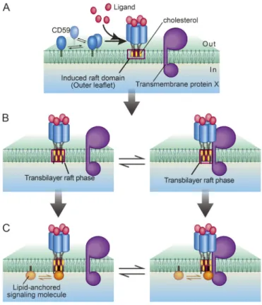
Using single-molecule imaging with enhanced time resolutions down to 5 ms, we found that CD59 cluster rafts and GM1 cluster rafts were stably induced in the outer leaflet of the plasma membrane (PM), which triggered the activation of Lyn, H-Ras, and ERK and continually recruited Lyn and H-Ras right beneath them in the inner leaflet with dwell lifetimes <0.1 s.

The detection was possible due to the enhanced time resolutions employed here. The recruitment depended on the PM cholesterol and saturated alkyl chains of Lyn and H-Ras, whereas it was blocked by the nonraftophilic transmembrane protein moiety and unsaturated alkyl chains linked to the inner-leaflet molecules. Because GM1 cluster rafts recruited Lyn and H-Ras as efficiently as CD59 cluster rafts, and because the protein moieties of Lyn and H-Ras were not required for the recruitment, we conclude that the transbilayer raft phases induced by the outer-leaflet stabilized rafts recruit lipid-anchored signaling molecules by lateral raft – lipid interactions and thus serve as a key signal transduction platform.

Introduction

In the human genome, >150 protein species have been identified as glycosylphosphatidylinositol (GPI)-anchored proteins, in which the protein moieties located at the extracellular surface of the plasma membrane (PM) are anchored to the PM by way of GPI, a phospholipid (Kinoshita and Fujita, 2016). Many GPI- anchored proteins are receptors and thus are referred to as GPI-anchored receptors (GPI-ARs). A GPI-anchored structure appears paradoxical for receptors because it spans only halfway through the membrane; yet, to function as a receptor, it has to relay the signal from the outside environment to the inside of the cell (Fig. 1 A). “Raft domains” are PM domains on the space scales from a few nanometers up to several hundred nanometers that are built by cooperative interactions of cholesterol and molecules with saturated alkyl chains of C16 or longer, as well as by their exclusion from the bulk unsaturated chain–enriched domains (Kusumi et al., 2020; Levental et al., 2020), have been implied in the signaling process of GPI-ARs across the PM (Omidvar et al., 2006; Suzuki et al., 2007b, 2012; Paulick and Bertozzi, 2008; Eisenberg et al., 2011; Fessler and Parks, 2011;

Lingwood et al., 2011; Kusumi et al., 2014; Raghupathy et al., 2015). Nevertheless, exactly how raft domains or raft-based lipid interactions participate in the transbilayer signal trans- duction of GPI-ARs remains unknown. Indeed, raft-based

interactions might even be involved in the signal transduc- tion by transmembrane (TM) receptors (Coskun et al., 2011;

Chung et al., 2016; Shelby et al., 2016).

In giant unilamellar vesicles undergoing liquid-ordered (Lo)/

liquid-disordered (Ld) phase separation, the Lo/Ld phase do- mains in the outer leaflet spatially match the same domains in the inner leaflet, indicating strong interbilayer coupling due to phase separation across the bilayer (Collins and Keller, 2008;

Blosser et al., 2015). In living cells, the long-chain phosphati- dylserine present in the PM inner leaflet was proposed to play key roles in the transbilayer coupling (Raghupathy et al., 2015).

However, the mechanisms of transbilayer coupling in the PM for the induction of signal transduction are not well understood.

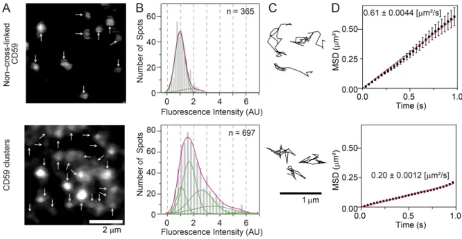
Using CD59 as a prototypical GPI-AR, our previous single–

fluorescent molecule imaging showed that nanoparticle-induced CD59 clusters form stabilized raft domains with diameters on the order of 10 nm in the PM outer leaflet, which in turn con- tinually recruit intracellular signaling molecules Gi α , Lyn, and PLC γ 2 one after another in a manner dependent on raft – lipid interactions, triggering the inositol triphosphate/Ca

2+

signaling pathway. Namely, artificially induced CD59 clusters behaved like CD59 clusters induced by the addition of the complement component C8 or the membrane attack complement complexes ...

1Department of Biochemistry and Molecular Biology, Graduate School and Faculty of Medicine, University of Tokyo, Tokyo, Japan; 2Institute for Integrated Cell-Material Sciences, Kyoto University, Kyoto, Japan; 3Institute for Frontier Life and Medical Sciences, Kyoto University, Kyoto, Japan; 4Institute for Glyco-core Research, Gifu University, Nagoya, Japan; 5Center for Highly Advanced Integration of Nano and Life Sciences, Gifu University, Gifu, Japan; 6Laboratory for Organismal Patterning, Center for Biosystems Dynamics Research, RIKEN Kobe, Kobe, Japan; 7Membrane Cooperativity Unit, Okinawa Institute of Science and Technology Graduate University, Onna-son, Okinawa, Japan.

Correspondence to Akihiro Kusumi:[email protected].

© 2020 Koyama-Honda et al. This article is available under a Creative Commons License (Attribution 4.0 International, as described athttps://creativecommons.org/

licenses/by/4.0/).

(3)

(MACCs; Suzuki et al., 2007a, 2007b, 2012). Therefore, the CD59 clusters were termed “CD59 cluster signaling rafts” or simply

“CD59 cluster rafts” (Stefanov´a et al., 1991; Suzuki et al., 2007a, 2007b, 2012; Simons and Gerl, 2010; Zurzolo and Simons, 2016).

Importantly, the recruitment of cytoplasmic signaling molecules at the CD59 signaling rafts occurred transiently, in a time scale on the order of fractions of a second (in the following text, we use the expression “recruitment of signaling molecules ‘at’ CD59 clusters ” rather than “ the recruitment ‘ to ’ CD59 clusters ” be- cause our imaging method could not directly show the binding of the signaling molecules located in the inner leaflet to the CD59 clusters located in the outer leaflet). Raftlike properties of the artificial antibody (Ab)-induced CD59 clusters were confirmed by the finding that fluorescently labeled gangliosides and sphingomyelins are colocalized with the artificial CD59 clusters (Komura et al., 2016; Kinoshita et al., 2017). CD59-TM, in which the GPI anchor was replaced by the TM domain of a prototypical nonraft molecule, low-density lipoprotein receptor (LDLR), failed to exhibit the raftlike behaviors and to trigger the downstream signal, in ways similar to the CD59 clusters after cholesterol depletion (Suzuki et al., 2007a, 2007b, 2012). The

present research was designed on the basis of these previous research results. Furthermore, our previous single-molecule studies revealed that, although gangliosides and sphingomye- lins are always present in the CD59 cluster signaling rafts, each lipid molecule associates with the CD59 cluster raft for only 50–100 ms (Komura et al., 2016; Kinoshita et al., 2017), like signaling molecules Giα, Lyn, and PLCγ2.

Meanwhile, the time resolution of the single-molecule

imaging method used to detect such transient colocalization

events was only 33 ms. In the present study, we greatly en-

hanced the imaging time resolutions down to 5.0 and 6.45 ms, an

improvement by factors of 6.7 and 5.2, respectively, and thus

substantially refined the detection of cytoplasmic signaling

molecule colocalizations with CD59 cluster rafts (and GM1

cluster rafts). To the best of our knowledge, these are likely to

be the fastest simultaneous, two-color, single-molecule ob-

servations ever performed. We previously found Lyn recruit-

ment at CD59 cluster rafts, but in the present research, by

applying single-molecule imaging at enhanced time resolutions

and using various lipid-anchored cytoplasmic molecules, in-

cluding Lyn, H-Ras, and four artificially designed molecules, as

Figure 1. Outer- and inner-leaflet lipid-

anchored molecules employed in this study

and their cross-linking schemes. (A) The

outer-leaflet molecules employed in this work

were a prototypical GPI-AR, CD59; a prototypical

ganglioside, GM1; and a prototypical nonraft

phospholipid, DNP-DOPE. The inner-leaflet

molecules examined here were (G and GFP

represent EGFP) the following: Lyn-FG, Lyn

conjugated at its C-terminus to two molecules of

FKBP in series and then to GFP; Myrpal-N20Lyn-

GFP, myristoyl, palmitoyl-anchored Lyn peptide

conjugated to GFP, where the peptide was the

20-aa N-terminal sequence of Lyn, which con-

tains the conjugation sites for both myristoyl and

palmitoyl chains; TM-Lyn-GFP, the TM mutant of

Lyn-GFP, in which the TM domain of a proto-

typical nonraft molecule LDLR was conjugated to

the N-terminus of the full-length Lyn-GFP (which

cannot be fatty acylated); Palpal-N16GAP43-GFP,

palmitoyl, palmitoyl-anchored GAP43 peptide

conjugated to GFP, in which the peptide was the

16-aa N-terminal sequence of GAP43 containing

two palmitoylation sites (likely to be raft asso-

ciated); GFP-C5Rho-geranylgeranyl, GFP an-

chored by a geranylgeranyl chain, in which GFP

was conjugated at its C-terminus to the five-aa

C-terminal sequence of Rho, which contains a

site for attaching an unsaturated geranylgeranyl

chain (likely to be non

raft associated); FGH-Ras,

H-Ras chimera molecule in which two tandem

FKBP molecules linked to GFP were then conju-

gated to H-Ras; and GFP-tH, GFP linked to the

10-aa C-terminal sequence of H-Ras containing

two sites for palmitoylation and a site for far-

nesylation. These molecules were expressed and

observed in live HeLa cells. (B–D) The schemes for clustering (cross-linking) CD59 (B), GM1 (C), and FGH-Ras (D). CD59 was clustered by the sequential additions

of anti-CD59 mAb IgG labeled with the fluorescent dye A633 and secondary Abs (+2°-antibodies; B). GM1 was clustered by the sequential additions of CTXB

conjugated with A633 and anti-CTXB Abs (C). FGH-Ras (as well as Lyn-FG) was clustered by the addition of AP20187 (cross-linker for FKBP; D). After the

induction of clustering of these molecules, the possible recruitment of lipid-anchored molecules in the other leaflet of the PM at these clusters was examined.

(4)

well as by using the stabilized ganglioside GM1 cluster rafts in addition to the CD59 cluster rafts, we sought to unravel the mechanisms by which cytoplasmic lipid-anchored signaling molecules in the PM inner leaflet are recruited at CD59 cluster rafts and GM1 cluster rafts formed in the PM outer leaflet.

In addition to the well-known function of CD59 to protect normal cells in the body against self-attack by MACCs, CD59 is involved in tumor growth. First, CD59 renders autologous car- cinoma cells insensitive to the MACC action, providing tumor cells with a key strategy to evade the immune system (Morgan et al., 1998; Carter and Lieber, 2014). Second, the MACC-induced CD59 clusters activate the extracellular signal-regulated kinase (ERK) signaling pathway, thus enhancing tumor cell prolifera- tion (Jurianz et al., 1999). Therefore, the basic understanding of CD59 signaling, particularly the Lyn (Src family kinase) signal- ing to trigger the inositol triphosphate/Ca

2+

pathway for pro- tection against MACC binding, as well as the signaling cascades for ERK activation by way of Lyn and Ras (Bertotti et al., 2006;

Harita et al., 2008; Wang et al., 2011; Suzuki et al., 2012;

Croucher et al., 2013; Dorard et al., 2017), would be useful for developing methods to regulate CD59 function, eventually leading to better therapeutic outcomes in oncology by sup- pressing ERK activities and reversing complement resistance (Carter and Lieber, 2014).

In the present research, we first aimed to unravel how the CD59 cluster rafts in the PM outer leaflet recruit the down- stream intracellular lipid-anchored signaling molecules Lyn and H-Ras, located in the PM inner leaflet. Lyn is anchored to the PM inner leaflet by a myristoyl chain and a palmitoyl chain (myrpal), whereas H-Ras is anchored by two palmitoyl chains and a farnesyl chain (Fig. 1 A). Because CD59 cannot directly interact with and activate Lyn and H-Ras, and because Lyn and H-Ras are proposed to be raft domain associated in the PM inner leaflet (Field et al., 1997; Sheets et al., 1999; Prior et al., 2001, 2003), we paid special attention to raft – lipid in- teractions as a recruiting mechanism (Wang et al., 2005) while also considering protein – protein interactions (Fig. 1 B;

Douglass and Vale, 2005).

Second, to directly examine the possibility that the signal transfer from the PM outer leaflet to the inner leaflet is mediated by raft–lipid interactions, we cross-linked the prototypical raft lipid ganglioside GM1 in the outer leaflet to examine whether GM1 clusters could recruit Lyn and H-Ras in the inner leaflet (Fig. 1, A and C). Many studies have examined the cytoplasmic signals triggered by GPI-AR stimulation and GM1 clustering in a raft-dependent manner (Pyenta et al., 2001; McKerracher and Winton, 2002; Wang et al., 2005; Todeschini et al., 2008; Fujita et al., 2009; Um and Ko, 2017), although the results varied considerably. In contrast, very few studies have investigated the actual recruitment of cytoplasmic lipid-anchored signaling molecules at the stabilized nanoraft domains formed in the PM outer leaflet (Harder et al., 1998; Suzuki et al., 2007a, 2007b, 2012), and particularly the molecular dynamics of the recruit- ment in live cells. In the present study, as a control, we induced the clustering of lipid-anchored Lyn or H-Ras in the PM inner leaflet and observed whether this could induce the recruitment of CD59 and GM1 in the PM outer leaflet (Fig. 1, A and D).

Results

Ab-induced CD59 clusters in the PM and their ERK activation First, we improved the time resolution of our home-built single- molecule imaging station, described previously (Koyama-Honda et al., 2005; Komura et al., 2016; Kinoshita et al., 2017). The improvements were accomplished by using two kinds of camera systems that can operate at higher frame rates (see Materials and methods) and modifying the single-molecule imaging sta- tion by using lasers with higher outputs and tuning the excita- tion optics. As a result, the time resolution was enhanced from 33.3 ms (30 Hz) to 5.0 or 6.45 ms (200 or 155 Hz, respectively, which is faster than normal video rate by factors of 6.7 and 5.2, respectively), with frame sizes of 640 × 160 pixels and 653 × 75 pixels, respectively. We employed the same two cameras for performing simultaneous, two-color, single-molecule imaging (see Materials and methods). Throughout this work, all of the microscopic observations of CD59 cluster rafts (Alexa Fluor 633 [A633] tagged) and the downstream cytoplasmic signaling mol- ecules (fused to EGFP, which is simply called “ GFP ” for con- ciseness) were performed simultaneously in the bottom (basal) PM of HeLa cells.

CD59 cluster signaling rafts were formed by the addition of the primary (anti-CD59 IgG mAb conjugated with A633) and secondary Abs, according to previous reports (Field et al., 1997;

Janes et al., 1999; Chen and Williams, 2013). Using this method, CD59 clusters could be formed in both the apical and basal PMs, whereas in our previous method of using nanoparticles to in- duce CD59 clusters, due to the nonaccessibility of the particles in the space between the basal PM and the coverslip, CD59 clusters were formed only in the apical PM. Therefore, in this study, we observed the CD59 clusters and signaling molecules in the basal PM, which enabled observations with improved signal-to-noise ratios. These observations were conducted within 10 min after the addition of the secondary Abs, when more than 92% of the CD59 clusters were located outside caveolae (Fig. S1 A).

To better observe the short-term colocalizations of lipid- anchored signaling molecules with CD59 cluster rafts, we ho- ped to slow down the colocalization processes, and therefore all microscopic observations were performed at 27°C, which is 10°C lower than the physiological temperature of 37°C. It is known that raft formation is temperature dependent, but in all the cell lines examined thus far, the temperature-dependent changes are pronounced below ∼15°C, at which large Lo phase–like raft do- mains are induced and become visible by fluorescence micros- copy (for visualization, actin-based membrane skeleton meshes must be removed from the PM cytoplasmic surface); this would not occur at 27°C (Holowka and Baird, 1983; Gidwani et al., 2001;

Veatch and Keller, 2003; Baumgart et al., 2007; Lingwood et al., 2008; Sengupta et al., 2008; Levental et al., 2009; Kusumi et al., 2020). Namely, the changes found in the PM when the tem- perature is lowered from 37°C to 27°C would be quantitative rather than qualitative. For example, the diffusion coefficients of various lipids and GPI-ARs in two very different cell types, CHO and rat basophilic leukemia (RBL)-2H3 cells, were reported to decrease only by a factor of at most 1.4 when the temperature was lowered from 37°C to 27°C (Lee et al., 2015; Saha et al., 2015).

Meanwhile, the diffusion coefficients of both the prototypical

(5)

nonraft phospholipid L- α -dioleoylphosphatidylcholine (DOPE) and the prototypical raft-associated phospholipids C18- sphingomyelin and

L

- α -distearoylphosphatidylcholine (all of them fluorescently labeled) would be reduced by a factor of approximately 2 when the temperature was lowered from 37°C to 27°C (assuming that the activation energy for diffusion is the same between 37°C and 23°C; Kinoshita et al., 2017, where we used T24 and PtK2 cells; on the basis of these results, we de- cided to perform all of the microscopic observations at 27°C to better detect the colocalization processes). Therefore, we be- lieve that the conclusions obtained in the present work based on the observations performed at 27°C are essentially correct.

The number of CD59 molecules located in a CD59 cluster was estimated to be ∼10 (molecules) on average (the variations would be quite large; Fig. 2, A and B; Materials and methods).

Because CD59 is anchored to the PM outer leaflet by way of two saturated, long alkyl chains, the CD59 clusters employed here would contain an average of 20 saturated long alkyl chains of CD59 in the small cross-sectional area of the CD59 cluster. The CD59 clusters diffused at a threefold slower rate than mono- meric CD59 (labeled with anti-CF59 – antigen-binding fragment [Fab]-A633; Fig. 2, C and D). Because we used the dye (A633)- conjugated Ab (and the secondary Abs) to induce CD59 clusters, the recording periods were quite limited due to photobleaching (∼0.51 s), and signal-to-noise ratios for observing the CD59 clusters were worse than with our previous observations using fluorescent nanoparticles. In the present study, we could not detect stimulation-induced temporary arrest of lateral diffu- sion, and the CD59 clusters appeared to simply undergo slow diffusion.

CD59 clustering triggered the signaling cascade to activate the ERK1/2 kinases (performed at 37°C instead of 27°C; Fig. 3), in agreement with a previous finding (Jurianz et al., 1999). The signaling pathways leading to ERK activation could involve the small G-protein H-Ras, as well as Lyn (Bertotti et al., 2006;

Harita et al., 2008; Porat-Shliom et al., 2008; Wang et al., 2011;

Croucher et al., 2013; Dorard et al., 2017). Therefore, we per- formed direct single-molecule observations of the recruitment of both Lyn kinase and H-Ras to the CD59 cluster signaling rafts.

We had previously detected Lyn recruitment at CD59 clusters (Suzuki et al., 2007a, 2007b), but in the present research we focused on understanding the recruitment mechanism by using other related molecules and H-Ras, as well as by using single- molecule observations with improved time resolutions.

Lyn is continually and transiently recruited at CD59 cluster rafts one molecule after another, but not at

nonclustered CD59

Lyn is anchored to the PM inner leaflet by myristoyl and pal- mitoyl chains conjugated to its N-terminus (Fig. 1 A). The Lyn conjugated at its C-terminus to two molecules of FK506-binding protein (FKBP) in series and then to GFP (Lyn-FG) used here for single-molecule observations would be functional because it could be phosphorylated in RBL-2H3 cells after antigen (DNP) stimulation (Fig. S2 A). Virtually all of the Lyn-FG molecules on the PM inner leaflet were monomers (undergoing a single-step photobleaching like GFP molecules sparsely adsorbed on the

glass; Fig. S3) and underwent thermal diffusion, with a mean diffusion coefficient (in the time scale of 124 ms) of 0.76 ± 0.0019 µm

2

/s (Fig. 4 A).

Simultaneous two-color single-molecule observations re- vealed that Lyn-FG molecules diffusing in the inner leaflet were continually recruited at CD59 clusters located in the PM outer leaflet, one molecule after another. Importantly, the dwell time of each Lyn-FG molecule at the CD59 cluster was on the order of 0.1 s (Fig. 5 and Video 1). Quantitative detection of colocaliza- tions was performed by using our previously developed defini- tion, in which fluorescent spots with two different colors are located within 150 nm (Koyama-Honda et al., 2005). Although the colocalization distance of 150 nm is clearly much greater than the sizes of the interacting molecules, which would gen- erally be on the order of several nanometers, the colocalization analysis is still useful for detecting molecular interactions for the following reason. Unassociated molecules may track together by chance over short periods of time for short distances, but the probability of this occurring for multiple frames is small.

Therefore, longer colocalization durations imply the presence of molecular interactions between the two molecules rather than incidental encounters (although molecular interactions are ini- tiated by incidental encounters; see Materials and methods).

Each time we detected a colocalization event of an Lyn-FG molecule with a CD59 cluster, we measured its duration, and after observing sufficient numbers of colocalization events, we obtained a histogram showing the distribution of colocalized durations for Lyn-FG and CD59 clusters (Fig. 6 A, a; Materials and methods). However, this duration histogram must also contain the colocalization events due to incidental close en- counters of molecules within 150 nm, without any molecular interactions. To obtain the histogram of incidental colocalization durations, the image obtained in the longer-wavelength channel (A633) was shifted toward the right by 20 pixels (1.0 and 1.19 µm, depending on the camera) and then overlaid on the image obtained in the GFP channel ( “ shifted overlay ” ). The duration histogram for incidental colocalization, called h(incidental-by- shift), could effectively be fitted with a single exponential function with a decay time constant τ

1

of 15 ± 0.93 ms (Throughout this report, the SEM of the dwell lifetime is pro- vided by the fitting error of the 68.3% confidence limit for the decay time constant).

The distribution of the durations obtained for correctly

overlaying the Lyn-FG movies and CD59 cluster movies was

significantly different from that for the shifted overlay (P =

0.00076 using the Brunner-Munzel test; Brunner and Munzel,

2000; throughout this report, the Brunner-Munzel test was used

for the statistical analysis, and all statistical parameters are

summarized in Table S1, Table S2, and Table S3). The histogram

of colocalization durations for Lyn-FG at CD59 clusters could be

fitted with the sum of two exponential functions with decay

time constants of τ

1

and τ

2

. In the fitting, τ

1

was preset as the

decay time constant determined from h(incidental-by-shift), and

τ

2

was determined as a free parameter. In the previous studies

using normal video rate (30 Hz; 33-ms resolution), due to in-

sufficient time resolutions, such distinct components could not

be observed in the colocalization duration histogram.

(6)

As described in Materials and methods, τ

2

directly represents the binding duration (inverse off-rate assuming simple zero- order dissociation kinetics for Lyn-FG from the CD59 cluster).

Although the errors involved in the determinations of τ

2

are

quite large due to the problem of signal-to-noise ratios of the images, we emphasize that, within the scope of this report, the presence or absence of the τ

2

components in the colocalization duration histograms would already be of key importance.

In the case of the colocalizations of Lyn-FG with CD59 cluster rafts, the fitting provided a τ

2

of 80 ± 25 ms (Table S1). Both τ

1

and τ

2

for all of the molecules investigated here were much shorter than the photobleaching lifetimes of GFP and A633 (>400 ms; i.e., 62 or 80 image frames), and therefore no cor- rections for photobleaching were performed in this research.

The 80-ms dwell lifetime of Lyn at CD59 clusters is shorter than that observed previously (median, 200 ms; Suzuki et al., 2007a), probably due to the improved time resolutions and signal-to- noise ratios (previously, shorter colocalizations were likely missed) as well as the different ways of forming CD59 clusters.

Therefore, this result indicates that Lyn is recruited at CD59 cluster rafts more transiently than we previously evaluated.

Next, the colocalizations of Lyn-FG with nonclustered CD59 (labeled with anti-CD59 Fab-A633) were examined. The dura- tion histogram obtained by the correct overlay was almost the same as h(incidental-by-shift) (P = 0.86; τ

1

= 19 ms; Fig. 6 A, b, and Table S1), and it was significantly different from the histo- gram with CD59 clusters (P = 0.018).

Lyn recruitment at CD59 clusters requires raft–lipid interactions

Next, we asked whether raft–lipid interactions and protein–

protein interactions are required for recruiting Lyn-FG at CD59 Figure 2. CD59 clusters in the PM outer leaflet contained an average of

∼10 CD59 molecules and diffused slowly. (A)

Fluorescence images of non–cross-linked CD59 bound by A633–anti-CD59 Fab (D/P, 0.27; top) and CD59 clusters induced by the sequential additions of A633–anti-CD59 IgG (D/P, 0.63) and the secondary Abs (bottom), obtained at single-molecule sensitivities. Arrows indicate all of the detected fluorescence spots in each image.

(B) Histograms showing the distributions of the signal intensities of individual fluorescence spots of non–cross-linked CD59 (Fab-A633 probe; top,

n

= 355) and CD59 clusters (bottom,

n

= 697). On the basis of these histograms, we concluded that each CD59 cluster contained an average of

10 CD59 molecules (see Materials and methods), although the number distributions would be quite broad. (C) Typical trajectories of non

cross-linked CD59 (top) and CD59 clusters (bottom) for 0.2 s, obtained at a time resolution of 6.45 ms. (D) Ensemble-averaged mean-square displacements (MSDs) plotted against time, suggesting that in the time scale of 1 s, both non

cross-linked and clustered CD59 (68 and 119 trajectories, respectively) undergo effective simple Brownian diffusion, and the diffusion is slowed by a factor of about 3 after Ab-induced clustering. All error bars represent SEM.

Figure 3. Both CD59 clusters and GM1 clusters induced by the se-

quential additions of CTXB and its polyclonal Abs (Ab-CTXB-GM1 clus-

ters) induced Erk phosphorylation (activation). Note that the simple

clustering of five GM1 molecules by CTXB (CTXB-5-GM1) failed to trigger ERK

activation. Western blotting was performed by using antiphosphorylated Erk

Abs (top) with anti-H-Ras Abs as the loading controls (bottom). The addition

of 20 nM EGF was used as a positive control for Erk activation.

(7)

clusters. First, we examined the recruitment of myrpal- N20(Lyn)-GFP (Fig. 1 A), which was proposed to be associated with raft domains (Pyenta et al., 2001). The duration histogram for the colocalizations of myrpal-N20(Lyn)-GFP molecules with CD59 clusters clearly exhibited two components (significant difference from h(incidental-by-shift), P = 0.025), with a sta- tistically nonsignificant (P < 0.068) 18% reduction in τ

2

com- pared with the duration histogram for Lyn-FG (Fig. 6 B, a, and Table S1). Second, we found that the TM mutant of Lyn-GFP (TM-Lyn-GFP; Fig. 1 A) did not exhibit any detectable longer- lifetime component in the colocalization duration histogram (Fig. 6 B, b, and Table S1; P = 0.46 against h(incidental-by- shift)). These results suggest that (1) the protein moiety of Lyn by itself cannot induce the recruitment; (2) the raft–lipid in- teraction by itself can induce Lyn recruitment at CD59 clusters;

and (3) when both the Lyn protein moiety and raftophilic myristoyl + palmitoyl chains exist, the lifetime at the CD59 cluster raft appears to be prolonged (could be proved in the future when single-molecule imaging is further improved).

To further examine whether the raft – lipid interaction alone can recruit cytoplasmic saturated chain – anchored proteins at CD59 clusters, we examined the recruitment of two more arti- ficial molecules with large deletions in their protein moieties, but with preserved lipid-binding sites: Palpal-N16 growth- associated protein 43 (GAP43)-GFP (raftophilic) and GFP-C5 Rho-gerger (nonraftophilic; Fig. 1 A). Palpal-N16 GAP43-GFP exhibited a clear two-component histogram (significant differ- ence from h(incidental-by-shift); P = 0.0023), with a τ

2

(71 ms) quite comparable to the τ

2

values for Lyn-FG and myrpal- N20(Lyn)-GFP with CD59 clusters (Fig. 6 B, c, and Table S1).

Meanwhile, GFP-C5 Rho-gerger did not exhibit any detectable τ

2

component (Fig. 6 B, d, and Table S1; P = 0.97 against h(in- cidental-by-shift)). Taken together, the results obtained with these four designed molecules (Fig. 6 B) suggest that a raft – lipid interaction without a specific protein – protein interaction could induce the recruitment of cytoplasmic proteins with two satu- rated chains at CD59 clusters. However, if the protein – protein interaction does exist (Lyn-FG; τ

2

= 80 ms), then it could slightly prolong the colocalization lifetime (myrpal-N20(Lyn)-GFP; τ

2

= 66 ms). In short, the outside-in interlayer coupling occurs when stabilized CD59 cluster rafts are induced in the outer leaflet, and the outside-in transbilayer coupling mechanism is predomi- nantly lipid based.

H-Ras is continually and transiently recruited at CD59 clusters in a manner dependent on raft–lipid interactions

Next, we examined the recruitment of fluorescently labeled H-Ras (FKBP2-GFP-H-Ras [FGH-Ras]), which is anchored to the PM inner leaflet via two saturated (palmitoyl) chains and an unsaturated (farnesyl) chain covalently conjugated to the C-terminal domain of H-Ras (Fig. 1 A). Virtually all of the FGH- Ras molecules underwent thermal diffusion, with a diffusion coefficient (in the time scale of 124 ms) of 1.12 ± 0.0017 µm

2

/s (Fig. 4 B). The FGH-Ras was functional because it was activated by EGF stimulation (Fig. S2 B).

The histogram of the colocalization durations of FGH-Ras at CD59 clusters exhibited two clear components (Fig. 6 C, a, and Table S1; P = 0.029 against h(incidental-by-shift); τ

2

= 91 ms), whereas no significant τ

2

component was detected in the his- togram for the colocalizations at nonclustered CD59 (Fig. 6 C, b, and Table S1; P = 0.52 against h(incidental-by-shift)). After mildly treating the cells with methyl- β -cyclodextrin (M β CD;

4 mM at 37°C for 30 min), the FGH-Ras colocalization with CD59 clusters was strongly suppressed (Fig. 6 C, c; P = 0.41 against h(incidental-by-shift)). The strong effect of partial cholesterol depletion supports the critical importance of raft – lipid interac- tions for the recruitment of lipid-anchored FGH-Ras at CD59 clusters.

Next, we examined the colocalization of GFP-C10H-Ras-pal- far (GFP-tH; Fig. 1 A), which lacks the majority of the H-Ras protein moiety (Prior et al., 2001, 2003), with CD59 clusters.

The colocalization duration histogram exhibited two clear components (Fig. 6 C, d, and Table S1; P = 0.027 against h(inci- dental-by-shift); τ

2

= 75 ms versus 91 ms for the full-length FGH- Ras; nonsignificant difference). Taken together, these results suggest that the two palmitoyl chains of H-Ras probably mask the effect of the unsaturated farnesyl chain, and thus FGH-Ras ’ s two palmitoyl chains might work like Lyn-FG ’ s myristoyl + palmitoyl chains. The τ

2

values are summarized in Fig. 6 D.

In the present report, we focused on the recruitment of lipid- anchored cytoplasmic signaling molecules, Lyn-FG and FGH- Ras, at CD59 cluster rafts. Because Lyn-FG and FGH-Ras are continually recruited to CD59 clusters, they are considered to be more concentrated within the nanoscale region (on the order of 10 nm) of the CD59 cluster raft. This will enhance the homo- and heterointeractions of Lyn, H-Ras, and other recruited raftophilic signaling molecules at CD59 cluster rafts. Indeed, we found that Figure 4. Lyn-FG and FGH-Ras molecules underwent simple Brownian

diffusion in/on the inner PM leaflet as observed at a 6.45-ms resolution,

when they were not colocalized with CD59 clusters or Ab-CTXB-GM1

clusters. (A and B) Representative trajectories of single Lyn-FG (A) and FGH-

Ras (B) molecules and the ensemble-averaged MSDs plotted against

Δt

for

Lyn-FG and FGH-Ras. A and B are based on 109 and 456 trajectories, re-

spectively. Their mean diffusion coefficients are shown in the figure. All error

bars represent SEM.

(8)

the homo-oligomerization of FGH-Ras by cross-linking its FKBP domain by the AP20187 addition could activate FGH-Ras (Fig.

S2). This result further suggests that the recruitment of Lyn-FG and FGH-Ras at the small cross-sectional area of the CD59 cluster raft, leading to their higher concentrations at CD59 clusters, would have important signaling consequences.

GM1 clusters formed by Ab cross-linked cholera toxin B subunit (CTXB) in the PM outer leaflet activate ERK1/2 kinases To further investigate the raft–lipid interactions across the bi- layer, we induced clusters of GM1, a prototypical raft-associated glycosphingolipid (ganglioside), in the PM outer leaflet and ex- amined whether Lyn-FG and FGH-Ras located in/on the inner leaflet could be recruited at GM1 clusters in the outer leaflet.

GM1 clusters were induced by applying CTXB conjugated with A633 (dye/protein molar ratio [D/P], 0.8), which could bind five GM1 molecules (CTXB-5-GM1; Merritt et al., 1994), and greater GM1 clusters containing an average of approximately three CTXB and 15 GM1 molecules (virtually 30 saturated acyl chains) were induced by the further addition of a goat polyclonal anti- CTXB Ab IgG (Ab-CTXB-GM1 clusters; Fig. 7 A; see the caption for Fig. 7 B and Materials and methods; the actual variation of the number of CTXB molecules in a greater GM1 cluster could be quite large). We anticipated that all five of the of the GM1 binding sites in CTXB are filled with GM1 because GM1 exists abundantly in the PM outer leaflet of HeLa cells, and the 2D

collision rate is much higher than that in 3D space (Grasberger et al., 1986).

Ab-CTXB-GM1 clusters (30 saturated alkyl chains) diffused with a mean diffusion coefficient of 0.077 µm

2

/s, 5.1 times slower than non – cross-linked CTXB-5-GM1 (five saturated alkyl chains; 0.39 µm

2

/s; Fig. 7 D), whereas they diffused 2.6 times slower than CD59 clusters (0.20 µm

2

/s; 20 saturated alkyl chains; Fig. 2 D). Namely, the average cross-sectional area of the hydrophobic region of the Ab-CTXB-GM1 cluster would be somewhat greater than that of the CD59 cluster.

The GM1 clusters slowly became entrapped in caveolae; ∼9%

of the fluorescent spots were colocalized with caveolae at 10 min after the addition of the anti-CTXB Abs at 27°C (Fig. S1 B).

Therefore, in the present investigation, all of the microscopic observations involving GM1 clusters were made within 10 min after the addition of the Abs, when most of Ab-CTXB-GM1 clusters were located outside caveolae.

CTXB binding to the cell surface did not trigger the ERK signaling cascade, but when Ab-CTXB-GM1 clusters were in- duced, the ERK signaling cascade was activated (Fig. 3), consis- tent with the previous observations (Janes et al., 1999; Kiyokawa et al., 2005). The differences found here might be induced by the larger sizes of Ab-CTXB-GM1 clusters than the size of CTXB- 5GM1. However, we suspect that this is due not simply to the differences in the sizes of the entire CTXB-5-GM1 and Ab-CTXB- GM1 clusters, but rather to those in the local densities of the Figure 5. High-speed, simultaneous, two- color, single-molecule imaging showed tran- sient recruitment of Lyn-FG in/on the inner leaflet at CD59 clusters located in/on the outer leaflet. (A) Typical single-molecule image sequences (6.45-ms resolution; every other im- age is shown) showing the colocalization of a CD59 cluster (top row and magenta spots in the bottom row) and a single molecule of Lyn-FG (green arrowheads in the middle row and green spots in the bottom row). Lyn-FG spots appear brighter during colocalization due to slower diffusion. (B) The trajectories of the CD59 cluster (magenta) and the Lyn-FG molecule (green) shown in A. These molecules became colocalized (orange circular region with a radius of 150 nm around the CD59 cluster position) between 39 and 91 ms (52 ms; orange box).

(C) Another display of the colocalization event shown in A and B, showing the displacements of an Lyn-FG molecule and a CD59 cluster along the x and y axes (left and right, respectively) from the average position of the CD59 cluster during the colocalization period, plotted against time.

Circles indicate the times employed in B.

(9)

saturated chains in the CTXB-5-GM1 and Ab-CTXB-GM1 clus- ters, based on the following reason.

A crystallographic study showed that the five GM1 binding sites in CTXB are all located ∼ 3.7 nm away from adjacent binding sites (Fig. 1 C; Merritt et al., 1994), and thus the five GM1 mol- ecules (10 saturated chains) are located on a circle with a di- ameter of ∼ 6.3 nm. Considering the size of the acyl chains (occupying a cross-section of <0.35 nm

2

; i.e., a diameter of <0.33 nm), two adjacent GM1 molecules bound to CTXB will be located

>3 nm away from each other in a space that could accommodate

>9 acyl chains and, in the middle of the GM1 binding sites, there would be a circular space with a cross-section of >5 nm in di- ameter, which could accommodate >25 acyl chains (in the outer

leaflet; Fig. 1 C). Namely, the space between two GM1 molecules with saturated acyl chains bound to a CTXB molecule is much larger than the cross-section of a few lipid molecules; that is, CTXB induces only sparse GM1 clusters. The observation that CTXB molecules simply bound to the PM cannot trigger the downstream signals is consistent with this consideration: the five GM1 molecules bound to a single CTXB molecule would not provide the threshold densities of saturated lipids necessary to create stable rafts by assembling and keeping cholesterol and saturated chains in CTXB-5-GM1 and excluding unsaturated chains. The five GM1 molecules bound to a single CTXB molecule would not serve as a nucleus to induce raft domains beneath the CTXB molecule (in the outer leaflet of the PM) and hence would Figure 6. Lyn-FG, FGH-Ras, and other lipid- anchored raftophilic molecules were recruited at CD59 clusters but not at non–cross-linked CD59.

The distributions (histograms) of the colocalization durations for the

correct

and

shifted

overlays, shown in semilog plots. The histograms for shifted overlays were fitted by a single exponential function (dashed line), and those for the correct overlays were fitted by the sum of two exponential functions (solid line), with the shorter time constant set to

τ1

ob- tained from the histogram of the shifted overlay. The boxes highlighted in orange contain histograms that could be better fitted with the sum of two expo- nential decay functions rather than a single expo- nential function. The values of

τ1

and

τ2

are indicated in each box. See Table S1 for statistical parameters.

(A) Lyn-FG was recruited at CD59 clusters but not

at non–cross-linked CD59 (a, b). (B) Recruitment of

Lyn-related molecules and other lipid-anchored cy-

toplasmic model proteins at CD59 clusters: myrpal-

N20LynGFP (a) and palpal-N16GAP43-GFP (c) were

recruited, but TM-Lyn-GFP (b) and GFP-C5Rho-

gerger (d) were not. (C) FGH-Ras was recruited at

CD59 clusters but not at non

cross-linked CD59 (a,

b), and FGH-Ras recruitment at CD59 clusters de-

pended on the PM cholesterol (c). Meanwhile, GFP-

tH was recruited at CD59 clusters. (D) Summary of

the bound lifetimes (

τ2

) of Lyn-FG, FGH-Ras, and

other cytoplasmic lipid-anchored signaling molecules

at CD59 clusters. The differences in

τ2

values are

nonsignificant. ND, not detected. The M

β

CD treat-

ments (4 mM at 37°C for 30 min; see part C, c) have

been controversial. However, the involvement of

raft domains was examined in a variety of methods in

the present research, including the use of various

lipid-anchoring chains and the TM domain of a pro-

totypical nonraft molecule, LDLR, and a prototypical

nonraft phospholipid DOPE. In the past, we employed

the MβCD treatments together with other control

experiments (using TM artificial mutants of GPI-ARs,

saponin treatment, cholesterol repletion after the

M

β

CD treatment) and found that the M

β

CD treat-

ment with 4 mM M

β

CD at 37°C for 30 min repro-

ducibly gave the results consistent with the results

obtained by using other methods of testing the raft

involvement.

(10)

fail to recruit signaling molecules that trigger the ERK signaling cascade. This possibility was directly examined in the present study (see the next section; in the case of CD59 clusters, we suspect that due to the long flexible glycochain of GPI, CD59 has reorientation freedom, and thus the saturated chains of CD59 in the cluster and the cholesterol, sphingomyelin, and gangliosides recruited from the bulk PM can form a tighter complex beneath the cluster of CD59 protein moieties).

Meanwhile, when the CTXB molecules were cross-linked by anti-CTXB Abs, because the GM1 molecules are bound near the outer edges of CTXB (Merritt et al., 1994), they would be located very close to the GM1 molecules bound to other CTXB molecules in the Ab- CTXB-GM1 cluster (Fig. 1 C). These closely associated GM1 molecules could form the stable raft nucleus for recruiting cholesterol and lipids with saturated alkyl chains, recruiting raftophilic signaling molecules and thus triggering the ERK signaling pathways.

The stabilization and enlargement of raft domains induced by CTXB and its Abs as well as signaling by the enhanced raft do- mains have been established quite well in the literature, al- though the data have been quite qualitative (reviewed by Kusumi et al., 2020). For example, using the T cell line E6.1 Jurkat, Janes et al. (1999) reported that the addition of CTXB and its Ab-induced membrane patches contained lymphocyte- specific protein tyrosine kinase (Lck), linker for activation of T cells (LAT), and the T cell receptor, but excluded CD45.

These patches were considered to be enhanced raft domains because they were colocalized by CD59, used as a prototyp- ical raft marker. Therefore, we next investigated whether

CTXB-5-GM1 or Ab-CTXB-GM1 clusters could recruit Lyn-FG and FGH-Ras.

Lyn and H-Ras are continually and transiently recruited at Ab- CTXB-GM1 clusters in a manner dependent on raft – lipid interactions, but not at CTXB-5-GM1

We directly examined whether single molecules of Lyn-FG and FGH-Ras were recruited at CTXB-5-GM1 and Ab-CTXB-GM1 clusters located in/on the PM outer leaflet. As described in the previous section, CTXB-5-GM1 failed to trigger ERK activation, in contrast to Ab-CTXB-GM1 clusters.

The histogram of the colocalization durations of FGH-Ras at Ab-CTXB-GM1 exhibited two clear components, indicating that Lyn-FG was recruited at Ab-CTXB-GM1 (Fig. 8 A, a, and Table S2;

P = 0.013 against h(incidental-by-shift); τ

2

= 110 ms). Meanwhile, no significant τ

2

component was detectable for the colocaliza- tions at CTXB-5-GM1 (Fig. 8 A, b, and Table S2; P = 0.24 against h(incidental-by-shift)). Similarly, FGH-Ras was recruited at Ab- CTXB-GM1 (Fig. 8 B, a, Table S2; P = 0.025 against h(incidental- by-shift); τ

2

= 97 ms), but not at CTXB-5-GM1 (Fig. 8 B, b, and Table S2; P = 0.52 against h(incidental-by-shift)).

Partial cholesterol depletion eliminated the τ

2

component for the FGH-Ras colocalization with Ab-CTXB-GM1 (Fig. 8 B, c, and Table S2; P = 0.96 against h(incidental-by-shift)). Furthermore, when DNP-DOPE, a nonraft reference unsaturated phospholipid, was clustered in the outer leaflet by the addition of anti-DNP Abs and secondary Abs (the Ab concentrations were adjusted so that

>90% of DNP-DOPE clusters became immobile; i.e., the cross-section

Figure 7. Ab-CTXB-GM1 clusters generated in the PM outer leaflet contained an average of

15 GM1 molecules and diffused 2.6 times slower than

CD59 clusters. (A) Fluorescence images of non

cross-linked fluorescently labeled CTXB (which could bind up to five GM1 molecules; A633 conjugated with a

D/P of 0.80; called CTXB-5-GM1; top) and CTXB clusters induced by the further addition of anti-CTXB Abs (Ab-CTXG-GM1 cluster; bottom), obtained at single-

molecule sensitivities. Arrows indicate all of the detected fluorescence spots in each image. (B) Histograms showing the distributions of the signal intensities of

individual fluorescent spots of A633-labeled CTXB-5-GM1 (top) and Ab-CTXB-GM1 clusters (bottom). On the basis of these histograms, we concluded that each

Ab-CTXB-GM1 cluster contained an average of

15 GM1 molecules (see Materials and methods), although the distribution would be quite broad. (C) Typical

trajectories of CTXB-5-GM1 (top) and Ab-CTXB-GM1 clusters (bottom) for 0.2 s, obtained at a time resolution of 6.45 ms. (D) Ensemble-averaged MSDs plotted

against time, suggesting that in the time scale of 1 s, both CTXB-5-GM1 (top; 154 trajectories) and Ab-CTXB-GM1 clusters (bottom; 91 trajectories) undergo

effective simple Brownian diffusion, and the diffusion is slowed by a factor of about 5 after Ab-induced clustering. All error bars represent SEM.

(11)

of the DNP-DOPE cluster would be substantially greater than that of Ab-CTXB-GM1), no significant τ

2

component was detected for the FGH-Ras colocalization with DNP-DOPE clusters (Fig. 8 C and Table S2; P = 0.39 against h(incidental-by-shift)). These results further support the proposal that raft – lipid interactions are essential for the recruitment of cytoplasmic lipid-anchored signaling molecules at Ab-CTXB-GM1 and therefore that the GM1 molecules closely ap- posed to each other inside the Ab-CTXB-GM1 cluster induce stable raft nuclei by recruiting cholesterol and other raftophilic molecules.

The results for τ

2

are summarized in Fig. 8 D.

Small clusters of inner-leaflet signaling molecules did not recruit CD59 or GM1 in the outer leaflet

The homo-oligomerization of Lyn-FG and FGH-Ras in the cyto- plasm was induced by the addition of AP20187 (dimerizer sys- tem developed by Schreiber and then ARIAD Pharmaceuticals;

Schreiber, 1991; Clackson et al., 1998). The presence of a single FKBP molecule in a protein could only create dimers but not oligomers greater than dimers upon AP20187 addition, but the presence of two FKBP molecules in a single protein could induce oligomers (Fig. 1 B, bottom). The average number of Lyn-FG or FGH-Ras molecules in a single cluster was estimated to be ap- proximately three (Fig. S2, C and D, and Materials and methods).

The oligomerized FGH-Ras triggered the downstream sig- naling, as shown by the pull-down assay using the Ras-binding

domain of the downstream kinase Raf-1 (Fig. S2 B), consistent with previous observations (Inouye et al., 2000; Nan et al., 2015).

Meanwhile, the oligomerization-induced self-phosphorylation of Lyn-FG was not detected (Fig. S2 A).

We examined whether CD59 and CTXB-5GM1 located in the PM outer leaflet could be recruited at FGH-Ras or Lyn-FG oligomers induced in the PM inner leaflet by the addition of AP20187 (Fig. 9 and Table S3). No significant recruitment was detectable, indicating that the oligomers of the inner-leaflet lipid- anchored signaling molecules cannot recruit the outer-leaflet raft- associated molecules. This result suggests that although FGH-Ras and Lyn-FG could be transiently recruited to stabilized raft do- mains, they would only be passengers and not the main molecules for inducing raft domains, probably due to their shorter saturated chains (palmitoyl) and the presence of unsaturated chains. Fur- thermore, FGH-Ras and Lyn-FG could only be recruited to the outer edges of the raft domains or perhaps the interfaces of the raft and bulk domains. Meanwhile, the lack of CD59 and GM1 recruitment might be due to the smaller sizes (an average of approximately three molecules) of the FGH-Ras and Lyn-FG oligomers.

Discussion

The recruitment of cytoplasmic signaling molecules to small regions in the PM after stimulation is considered to be important Figure 8. Lyn-FG and FGH-Ras were recruited at Ab-CTXB-GM1 clusters but not at CTXB-5-GM1.

The distributions (histograms) for the colocalization durations are shown. See the

Fig. 6

legend for details and keys. See Table S2 for statistical parameters.

(A) Lyn-FG was recruited at Ab-CTXB-GM1 clusters but not at CTXB-5-GM1 (a, b). (B) FGH-Ras was re- cruited at Ab-CTXB-GM1 clusters but not at CTXB-5- GM1 (a, b), and its recruitment at Ab-CTXB-GM1 clusters depended on the PM cholesterol (c).

(C) FGH-Ras was not recruited to DNP-DOPE clus-

ters. (D) Summary of the bound lifetimes (

τ2

) of Lyn-

FG and FGH-Ras at Ab-CTXB-GM1 clusters. ND,

τ2

component not detected.

(12)

for inducing the downstream signaling, because higher con- centrations of signaling molecules in small regions would en- hance homo- and heterointeractions and possibly the formation of transient dimers and oligomers. We indeed found that the homo-oligomerization of FGH-Ras induced by AP20187 activated the downstream signaling of FGH-Ras and H-Ras (Fig. S2 B).

Therefore, in the present research, we extensively studied the recruitment of Lyn, H-Ras, and other lipid-anchored cytoplas- mic molecules at CD59 cluster rafts and Ab-CTXB-GM1 clusters.

Our results clearly showed that Ab-CTXB-GM1 clusters of a raftophilic lipid (GM1) formed in the PM outer leaflet can recruit the cytoplasmic lipid-anchored signaling molecules Lyn and H-Ras to the inner-leaflet region apposed to the outer-leaflet Ab- CTXB-GM1 clusters. This recruitment was not induced after the PM cholesterol was mildly depleted or when the unsaturated lipid (DNP)-DOPE was clustered in the outer leaflet. These re- sults unequivocally demonstrate that the cytoplasmic lipid- anchored signaling molecules Lyn and H-Ras can be assembled at the stabilized raft–lipid clusters formed in the outer leaflet by raft–lipid interactions. The involvement of TM proteins in the recruitment process would be quite limited, because (1) even cytoplasmic lipid-anchored molecules after the deletions of the majorities of their protein moieties were recruited at Ab-CTXB- GM1 clusters, (2) their dwell lifetimes at CD59 clusters and Ab- CTXB-GM1 clusters were very similar to those of Lyn-FG and FGH-Ras, and (3) the recruitment of FGH-Ras depended on the PM cholesterol level. Of course, this does not rule out the specific interactions of GPI-ARs with TM proteins as coreceptors (Klein et al., 1997; Wang et al., 2002; Zhou, 2019). The results showing that Ab-CTXB-GM1 clusters, but not CTXB-5GM1, can recruit Lyn-FG and FGH-Ras (Fig. 8) would suggest that critical con- centrations (number densities) of saturated chains would probably exist for generating the outer-leaflet raft domains that can recruit raftophilic molecules in the inner leaflet. However, the concentration effect might further be compounded by the larger sizes of the Ab-CTXB-GM1 cluster-induced raft domains compared with the CTXB-5GM1–induced raft domains.

To summarize the sequence of events in CD59 signaling (Fig. 10), first, the stable CD59 cluster rafts in the outer leaflet are induced by the clustering of raftophilic CD59 molecules by extracellular stimulation, such as MACC binding. The stabilized raft domains tend to last for durations on the order of tens of minutes (Suzuki et al., 2007b, 2012), whereas their constituent molecules, such as the gangliosides, tend to stay there only for 50 ms and turn over quickly, continually exchanging with those located in the bulk PM region (Komura et al., 2016). Second, at the signal-induced stabilized CD59 cluster raft domains, the raftophilic cytoplasmic signaling molecules, Lyn and H-Ras, are recruited by raft – lipid interactions with lifetimes on the order of 0.1 s (Figs. 6 and 8); that is, each molecule stays at the CD59 cluster raft quite transiently. However, because many molecules would continually arrive one after another, and because each raft domain can accommodate several hundred lipid molecules (when the raft radius is 10 nm, each leaflet within the raft can accommodate ∼500 phospholipids), many cytoplasmic rafto- philic signaling molecules could be dynamically concentrated in the small cross-sectional area beneath the CD59 cluster raft, leading to locally enhanced molecular interactions.

Let us assume that the sizes of the stabilized CD59 cluster rafts and Ab-CTXB-GM1 cluster rafts are in the range of 20–100 nm in diameter (Figs. 1, B and C; 2; and 7) and the diffusion coefficient of the lipid-anchored signaling molecules is ∼ 1 µm

2

/s (Fig. 4). Then, these signaling molecules would stay in the 20 – 100-nm region in the bulk PM for only 0.03 – 0.63 ms.

However, they remained in the stabilized raft domains for 80 – 110 ms (Figs. 6 and 8); that is, the dwell lifetimes were prolonged by a factor of 200 – 2,000, which is a large factor.

Namely, the dwell lifetimes in the range of 80 – 110 ms might appear to be short, but in fact, Lyn-FG, FGH-Ras, and other raftophilic lipid-anchored molecules exhibited extremely pro- longed dwell lifetimes beneath the stabilized raft domains in the outer leaflet. Such extreme prolongation would not be possible by simple interactions of the lipids in the inner leaflet with the lipids in stabilized raft domains in the outer leaflet.

Figure 9. FGH-Ras oligomers and Lyn-FG oligom- ers induced by AP20187 addition failed to recruit non–cross-linked CD59 and CTXB-5-GM1. Shown here are the histograms for the durations in which non

cross-linked CD59 and CTXB-5-GM1 located in/

on the PM outer leaflet are colocalized with FGH-Ras oligomers and Lyn-FG oligomers artificially induced in the PM inner leaflet by the addition of AP20187. See the

Fig. 6

legend for details and keys. See Table S3 for statistical parameters. (A) Recruitment of non

cross- linked CD59 located in the outer leaflet at the induced FGH-Ras oligomers located in the inner leaflet.

(B) Recruitment of non

cross-linked CD59 located in the outer leaflet at the induced Lyn-FG oligomers located in the inner leaflet. (C) Recruitment of CTXB- 5-GM1 located in the outer leaflet at the induced FGH-Ras oligomers located in the inner leaflet.

(D) Recruitment of CTXB-5-GM1 located in the outer

leaflet at the induced Lyn-FG oligomers located in

the inner leaflet.

(13)

Because the micrometer-scale transbilayer raft phases have been detected in artificial bilayer membranes (Collins and Keller, 2008; Blosser et al., 2015), we propose that nanoscale transbilayer raft phases are induced in both leaflets by the sta- bilized raft domains initially formed in the outer leaflet and that when cytoplasmic raftophilic lipid-anchored signaling mole- cules arrive at the transbilayer raft phases, they tend to be trapped in the inner-leaflet part of the transbilayer raft phase, exhibiting dwell lifetimes on the 0.1-s order. Namely, trans- bilayer raft–lipid interactions would only make sense when

cooperative lipid interactions occur due to the formation of the transbilayer raft phase. Indeed, raft domains are generally considered to form by the cooperative interactions of saturated alkyl chains and cholesterol, as well as by their cooperative exclusions from the bulk PM enriched in unsaturated alkyl chains. Therefore, we propose that the nanoscale transbilayer raft phases would be induced at stabilized rafts initially formed in the outer leaflet. We further propose that the transbilayer raft phases induced by the stimulation-triggered GPI-AR cluster rafts would act as a key signaling platform for the engaged GPI-AR clusters, recruiting inner-leaflet raftophilic signaling molecules, and that the formation of the transbilayer raft phase would be a general mechanism for GPI-AR signal transduction (however, this does not rule out the possibility that some TM proteins with raft affinities or those that can be concentrated at the interface between the raft and bulk domains are involved in the recruit- ment of Lyn and H-Ras, as depicted in Fig. 10; compare the result shown in Fig. 6 A, a, with that shown in Fig. 6 B, a).

This recruitment mechanism based on the transbilayer raft phase appears to suggest the lack of specificity in the cytoplas- mic signaling without any dependence on the GPI-AR species.

However, because different GPI-ARs would form signaling cluster rafts with a variety of sizes, because of closeness of the saturated acyl chains within the cluster (as found here for GM1 clusters induced by CTXB), and because the TM protein species with which different GPI-ARs interact would vary (Suzuki et al., 2012; Zhou, 2019), the GPI-AR cluster rafts formed in the outer leaflet could induce transbilayer raft phases with distinct properties. These transbilayer raft phases could recruit a variety of lipid-anchored signaling molecules with differing efficiencies, thus triggering various downstream signaling cascades with different strengths; that is, the relative activation levels among the many intracellular signaling cascades triggered by GPI-ARs would vary depending on the GPI-AR species.

Materials and methods

Improved camera systems for simultaneous, dual-color, single-molecule imaging in living cells at enhanced time resolutions of 5 and 6.45 ms

The major improvement of our single-molecule imaging station from the previously published version (Koyama-Honda et al., 2005; Komura et al., 2016; Kinoshita et al., 2017) was the em- ployment of two camera systems that allow higher frame rates.

With an increase in the frame rate of the camera system, we employed lasers with higher outputs (see the next paragraph).

The two camera systems both employed two-stage microchannel plate intensifiers (C8600-03; Hamamatsu). In one camera system, the image intensifier was lens coupled to an electron multiplying charge-coupled device camera (Cascade 650; Photometrics), which was operated at 155 Hz (6.45 ms/frame), with a frame size of 653 × 75 pixels (38.9 × 4.46 µm

2

for a total of 240× magnifi- cation). In the other camera system, the image intensifier was fiber coupled, with a 1.6:1 tapering, to a charge-coupled device camera (XR/MEGA-10ZR; Stanford Photonics) cooled to −20°C and operated at 200 Hz (5 ms/frame), with a frame size of 640 × 160 pixels (27.1 × 6.75 µm

2

for a total of 240× magnification).

Figure 10. Schematic model showing the CD59 signal transduction

mediated by the transbilayer raft phase, which recruits lipid-anchored

signaling molecules at the ligated, stabilized CD59 cluster domains in

the PM outer leaflet, inducing enhanced interactions of recruited mol-

ecules. (A) First, the ligand binding triggers the conformational changes of

CD59, which in turn induce CD59 clustering, creating stable CD59 cluster

signaling rafts. If GM1 is clustered closely, then stable GM1 cluster rafts will

be produced. (B) Then, the transbilayer raft phase is induced by the CD59

cluster raft by involving molecules in the inner leaflet, recruiting cholesterol

and molecules with saturated alkyl chains (left) and also excluding molecules

with unsaturated alkyl chains. An as yet unknown TM protein(s) X, which has

affinities to raft domains, might also be recruited to the transbilayer raft

phase (right; recruitment of X could be enhanced by specific protein–protein

interactions with the ligated CD59 exoplasmic protein domain). (C) Finally,

cytoplasmic lipid-anchored signaling molecules, such as H-Ras and Lyn, are

recruited to the transbilayer raft phase in the inner leaflet by the raft

lipid

interaction (left). This could be enhanced by the protein

protein interaction

with the TM protein X (right). Although the residency times of the inner-

leaflet signaling molecules beneath the CD59 cluster may be limited, because

many molecules will be recruited there one molecule after another, inter-

actions of two or more species of cytoplasmic signaling molecules will occur

efficiently beneath the CD59 cluster raft. This way, the transbilayer raft phase

induced by the stabilized CD59 cluster raft would function as an important

signaling platform.

(14)

Right before microscopic observations of the cells, the culture medium was replaced by HBSS buffered with 2 mM Pipes at pH 7.4 (P-HBSS), and the bottom PMs of the cells growing on glass- bottom dishes were observed by a homebuilt objective lens – type total internal reflection fluorescence microscope constructed on an inverted microscope (IX-70; Olympus) with a 60× objec- tive lens (NA, 1.4) with two detection arms for simultaneous two-color single-molecule imaging, as described previously (Koyama-Honda et al., 2005). The temperature of the sample and the microscope was maintained at 27 ± 1°C. The cells were illuminated simultaneously by a 488-nm laser (for GFP, Sap- phire 488-20; Coherent) and a 594-nm laser (for A633, 05-LYR- 173; Melles Griot/IDEX Health & Science). Fluorescence signals from GFP and A633 were split into the two detection arms by using a dichroic mirror at 600 nm (600DCXR; Chroma) and further isolated by interference filters (HQ535/70 for GFP and HQ655/100 for A633; Chroma). The fluorescence image in each arm was projected onto the photocathode of the image intensi- fier in the camera system described above (the same cameras were employed for the two channels). MetaMorph software (Molecular Devices) was used for image acquisition and pre- processing, and the obtained images were further processed using ImageJ software.

Determining the positions of fluorescence spots of single molecules and molecular clusters in the image

The positions (x and y coordinates) of individual fluorescence spots were determined by using an in-house computer program (Koyama-Honda et al., 2005; Hiramoto-Yamaki et al., 2014;

Fujiwara et al., 2016), based on a spatial cross-correlation matrix (Gelles et al., 1988). For each frame, the entire image was cor- related with a symmetric 2D Gaussian point spread function with an SD of 150 nm (kernel). The resulting 2D cross-correlation function for each molecule and each molecular cluster was thresholded, and their positions were determined as the center of mass of the thresholded correlation intensity.

Colocalization detection and evaluation of colocalization lifetimes

For the colocalization analysis, GFP trajectories longer than 19 frames and A633 trajectories longer than 29 frames were used.

The colocalization of an A633 spot with a GFP spot was defined as the event in which the two fluorescence spots, representing A633 and GFP molecules, became localized within 150 nm of each other. This is a distance at which an exactly colocalized molecule is detected as colocalized at probabilities >90%, using the Cascade 650 camera operated at 155 Hz, and higher proba- bility was achieved using the XR/MEGA-10ZR camera operated at 200 Hz (Koyama-Honda et al., 2005).

A colocalization distance of 150 nm is much greater than the molecular scale, and therefore, in addition to colocalization due to specific molecular binding, events in which molecules inci- dentally encounter each other within a distance of 150 nm, termed “incidental colocalizations,” can occur. However, as de- scribed in the Results section, nonassociated molecules may track together by chance over a short distance, but the proba- bility of moving together for multiple frames is small, and

therefore longer colocalizations imply the binding of two molecules.

In the analysis of colocalization durations, those as short as one or two frames were neglected to avoid higher-frequency noise. Likewise, if two colocalization events are separated by a gap of one or two frames, then they are linked and counted as a single longer colocalization event. To obtain the histogram of incidental colocalization durations, the image obtained in the longer-wavelength channel (A633) was shifted toward the right by 20 pixels (1.0 and 1.19 µm, depending on the camera) and then overlaid on the image obtained in the GFP channel (“shifted overlay”). The histogram of the incidental colocalization dura- tions was called h(incidental-by-shift). We found h(incidental- by-shift) could effectively be fitted by a single exponential decay function, using nonlinear least-squares fitting by the Levenberg- Marquardt algorithm provided in OriginPro software, and the decay time constant was called the “ incidental colocalization lifetime, ” τ

1

(Figs. 6 and 8).

Meanwhile, the distribution of the colocalization durations for correctly overlaid A633 and GFP images ( “ correct overlay ” ) was obtained, and we found that some of the histograms (such as that for Lyn-FG versus CD59 clusters) could be fitted with the sum of two exponential functions with a decay time constant τ

1

9 and the other, longer time constant τ

2

(Figs. 6 and 8). The τ

1

9 component was considered to represent the duration of inci- dental colocalization, and thus τ

1

9 = τ

1

. Therefore, in the fol- lowing discussion, we describe τ

1

9 simply as τ

1

.

The τ

2

component of the histogram was considered to de- scribe the colocalization durations, including the durations of true molecular interactions ( τ

B

). Here, we propose that the binding duration τ

B

can be approximated by τ

2

, which can be directly determined from the histogram, based on the following argument. As described previously (Kasai et al., 2018), in the simplest and probably most primary case in which the binding occurs only once during a single colocalization event, the du- ration τ

2

would be the sum of (1) the duration between the in- cidental encounter and actual molecular binding, (2) the duration of molecular binding (τ

B

), and (3) the duration between the dissociation of two molecules and separation by >150 nm.

Therefore, the mathematical function to describe the histogram for the colocalization durations including the molecular binding would be exp(−t/τ

B

) convoluted with the histogram h(inciden- tal-by-shift), which is proportional to exp(−t/τ

1

; t = time) at the present experimental accuracies (see, e.g., Sungkaworn et al., 2017; Figs. 6 and 8). Here, we are assuming simple zero-order kinetics for the release of lipid-anchored cytoplasmic molecules from the CD59 cluster rafts (and thus the binding duration distribution is proportional to exp( − t/ τ

B

)). The result of the convolution of an exponential function with another exponen- tial function is well known, and the convoluted function is the sum of these two exponential functions (exp( − t/ τ

1

) and exp( − t/

τ

B

)). Therefore, the entire histogram is the sum of the histogram

for simple close encounters, h(incidental-by-shift), which has

the form of exp(t/τ

1

), and the histogram for the colocalization

events that include molecular interactions and is expressed by

the sum of exp(−t/τ

1

) and exp(−t/τ

B

). Meanwhile, as described,

some of the experimentally obtained histograms (such as that

Figure 3. Both CD59 clusters and GM1 clusters induced by the se- se-quential additions of CTXB and its polyclonal Abs (Ab-CTXB-GM1  clus-ters) induced Erk phosphorylation (activation)
Figure 9. FGH-Ras oligomers and Lyn-FG oligom- oligom-ers induced by AP20187 addition failed to recruit non–cross-linked CD59 and CTXB-5-GM1
Figure 10. Schematic model showing the CD59 signal transduction mediated by the transbilayer raft phase, which recruits lipid-anchored signaling molecules at the ligated, stabilized CD59 cluster domains in the PM outer leaflet, inducing enhanced interactio
Figure S1. Less than 10% of CD59 cluster rafts and Ab-CTXB-GM1 clusters became trapped in caveolae within 10 min after their induction
+2

参照

関連したドキュメント

Conjecture 5.4, concerning the existence of disordered sphere packings, is plausible for a number of reasons: (i) the decorrelation principle of Section 4.3; (ii) the neces- sary

Standard domino tableaux have already been considered by many authors [33], [6], [34], [8], [1], but, to the best of our knowledge, the expression of the

Let Q be an acyclic quiver, Q the corresponding framed quiver and Q = Q op. Let mod-k Q be the category of finite dimensional right modules over k Q considered in [13].

In this section we generalize some of the results of Sommers [16] on bounded dominant regions of Cat and positive filters in + to bounded dominant regions of A m and

Our work provides insight into how cluster sizes and number of subtrees in a cluster are impacted by the value of α, the maximum degree d in the tree, the relationship between c and

If Φ is a finite root system, and if we take H 0 to be the category of representations of an alternating quiver corresponding to Φ , then our generalized cluster complex is the

Furuta, Two extensions of Ky Fan generalization and Mond-Pecaric matrix version generalization of Kantorovich inequality, preprint.

We show that a discrete fixed point theorem of Eilenberg is equivalent to the restriction of the contraction principle to the class of non-Archimedean bounded metric spaces.. We