RELATIONSHIP BETWEEN GLENOHUMERAL JOINT STABILITY AND GLENOID DEPTH : A CADAVERIC STUDY
Hidekazu Abe1), Yoichi Shimada1), Nobuyuki Yamamoto1), Hiroshi Minagawa1), Nobutoshi Seki1), Kazuma Kikuchi1) and Eiji Itoi2)
(Received 25 December 2008, Accepted 16 January 2009)
1)Division of Orthopaedic Surgery, Department of Neuro and Locomotor Science, Akita University School of Medicine, Hondo 1-1-1, Akita 010-8543, Japan
2)Department of Orthopaedic Surgery, Tohoku University School of Medicine, Seiryo-machi 1-1, Aobaku, Sendai 980-8574, Japan
Abstract
The purpose is to directly measure the glenoid depth and clarify its relationship with the stability ratio. We used 9 fresh-frozen cadaveric shoulders (mean age : 66 years, 5 males and 4 females).
Naked glenohumeral joint was investigated. The shoulder was fixed to the custom designed mea- surement device with the glenoid surface horizontal to the floor and the humerus with 45° abduc- tion and 35° external rotation. The stability ratio was 27.1 ± 6.5% (mean ± standard deviation) at 90° in the anterior, 26.2 ± 7.9% at 280° in the posterior (the minimum), 48.3 ± 10.4 at 10° in the superior, and 52.9 ± 12.5% at 180° in the inferior (the maximum) direction. The mean glenoid depth was 1.4 ± 0.3 mm at 90° and 270°, and 3.4 ± 0.9 and 3.3 ± 0.9 mm at 0° and 180°, respective- ly, showing that both the stability ratio and glenoid depth were minimal in the anterior 3 o’clock direction. The stability ratio increased as the glenoid depth increased, and the correlation coeffi- cient was 0.92 (p=0.01), showing a markedly strong positive correlation. The glenohumeral joint is most unstable in the anterior direction in terms of the bony stability.
Key words : biomechanics, stability ratio, glenoid depth
Introduction
It is well known the shoulder has the largest range of motion in the body and is the most frequently dislocated joint among human joints. Most traumatic shoulder dis- locations are anterior dislocations. Previous studies re- ported that bone defects and erosion of the glenoid oc- curred in the anteroinferior direction at an incidence of 8-90%1-9), but Saito et al. recently performed an analysis using CT, and found that bone defects most frequently
occurred at the 3 o’clock position, i.e., the anterior direc- tion10). Various stabilizing mechanism are active during movement of the shoulder joint. In the hanging position, muscles and ligaments are relaxed, for which intra-artic- ular pressure is considered important for stability in the inferior direction11), concavity compression, which kept the humeral head centered in the glenoid fossa, at the mid-range position12), and restriction by the glenohumer- al ligament at the end-range of motion13). The stability ratio is an index proposed by Fukuda et al. in 1988, com- presenting joint stability as a value calculated by dividing the force causing humeral head dislocation by the force pressing the humeral head to the glenoid14). Studies us- ing this index clarified that the humerus-glenoid mor- phology, particularly the glenoid depth, is important for the concavity compression in the mid-range of motion Correspondence : Hidekazu Abe
Division of Orthopedic Surgery, Department of Neuro and Locomotor Science, Akita University School of Medicine, 1-1-1 Hondo, Akita 010-8543, Japan
Tel : 81-18-884-6148 Fax : 81-18-836-2617
E-mail : hidekazu@doc.med.akita-u.ac.jp
during movement . However, all these studies inves- tigated the glenoid in only 8 directions, and the direction with bone instability involved in shoulder joint stability was not closely investigated. Moreover, it has not yet been demonstrated whether dislocation occurs at an un- stable position and produces a bone defect. The purpose of this study was to closely measure the stability ratio in multiple directions, and evaluate bone instability of the glenoid. In addition, the glenoid depth was directly mea- sured using a high-precision laser, and its relationship with the stability ratio was clarified.
Materials and Methods
We used 9 fresh-frozen cadaveric shoulders (mean age at the time of death : 66 years, 5 males and 4 females).
Shoulders with macroscopic joint deformity and degener- ation of the cartilage surface were excluded. All soft tis- sues including the supra- and infraspinatus muscles, teres minor muscle, subscapularis, the long head of the biceps brachii muscle, joint capsule, and glenohumeral ligament were removed from the shoulders, and only the skeletal structure was investigated. To avoid the drying of samples, the cartilage surface was moistened to main- tain the wet condition with a spray of saline solution from its preparation to the end of measurement.
The scapula was cut at its body, leaving the glenoid, and the specimen was fixed to a custom designed mea- surement device using bone cement (Fig. 1). The sample was set in the position used in previous reports12,16). The specimen was set with the glenoid horizontal to the floor, and fixed to the table with bone cement so as to adjust the 0 and 6 o’clock positions of the glenoid to an identical level, using a laser displacement sensor (RV3-55B/R, KEYENCE, Osaka, Japan). The specimen was set at 45°
abduction and 35° external rotation. To set the rotation, a screw was inserted perpendicular to the humeral shaft, 10° internally rotated from the plane including the hu- meral axis and the bicipital groove.
The humeral head was adjusted using a force transduc- er (Force moment sensor, NITTA, Osaka, Japan) so as to set the ‘compressive load’ representing the force press- ing the humeral head to the glenoid to 50 N, following previous reports. The force causing humeral head dislo-
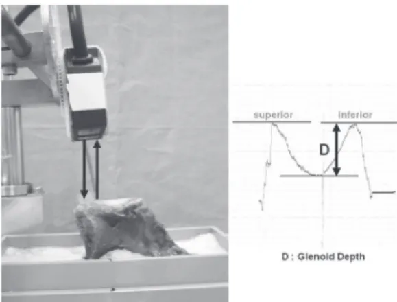
cation, ‘translational force’, was measured using a force gauge (Digital Force Gauge DPS-20, IMADA, Aichi, Ja- pan). The force gauge was connected to an amplifier (Polygraph 366, Nippon Denki Sanei, Yokohama, Japan), and the data were recorded (OMNICORDER 8M15, Nip- pon Denki Sanei, Yokohama, Japan) during the experi- ment. The glenoid depth was also measured using the laser displacement sensor (Fig. 2).
Fig. 1. A specimen and the custom designed measure- ment device.
The specimen was placed with the glenoid surface horizontal to the floor, and the humerus was fixed to the device at 45° abduction and 35° external rotation.
The stability ratio was calculated by dividing the translation force by the compressive load and multiplying by 100 (%).
Fig. 2. Measurement of the glenoid depth using a laser displacement sensor
D : Glenoid depth
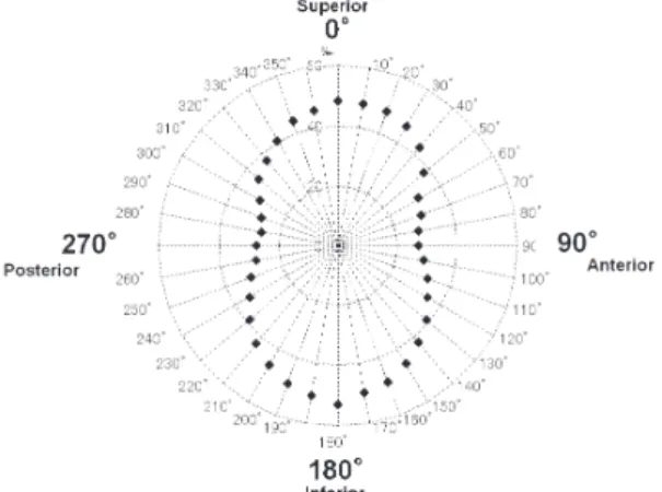
The mechanical parameter was measured every 10 de- grees in 36 directions, as well as every 45 degrees in 4 directions to compare with preceding reports, with a total of 40 directions. The parameter was measured twice, and the mean was adopted. The glenoid depth was mea- sured every 45 degrees in 8 directions.
The stability ratio was calculated from the measured value in each direction. The calculated value was multi- plied by 100(%) following the previous reports for ease of understanding. The relationship of the value with the glenoid depth was investigated using Pearson’s correla- tion coefficient. A significance level of 5% or less was regarded as significant.
Results
The mean stability ratio is shown in Fig. 3. The stabil- ity ratio is presented as those in the right shoulder fol- lowing the conventional method. The stability ratio was low in the antero-posterior and high in the superio-infe- rior direction. The stability ratio was 27.1 ± 6.5% (mean
± standard deviation) at 90° in the anterior, 26.2 ± 7.9% at 280° in the posterior (the minimum), 48.3 ± 10.4 at 10° in the superior, and 52.9 ± 12.5% at 180° in the inferior (the maximum) direction.
The glenoid depth was shallow in the antero-posterior direction and deep in the superio-inferior direction, and
the value was 1.4 ± 0.3 mm at 90° and 270°, and 3.4 ± 0.9 and 3.3 ± 0.9 mm at 0° and 180°, respectively (Fig. 4).
Regarding the relationship between the stability ratio and glenoid depth, the stability ratio increased as the gle- noid depth increased, showing a high positive correlation, with a correlation coefficient of 0.92 (p=0.01) (Fig. 5).
Discussion
Regarding glenohumeral joint stability, instability was stronger in the antero-posterior than in the superio-infe- rior direction when only the bony structures were con- sidered, showing that, experimentally, the glenohumeral joint structure is least stable in the anterior direction, not the antero-inferior direction, which reportedly causes clinically problematic anterior dislocation. The stability
Fig. 3. Stability ratio
The stability ratio was high in the vertical and low in the antero-posterior direction, showing an almost symmetric peanut-like shape.
Fig. 5. Relationship between the stability ratio and glenoid depth
A strong correlation was noted.
Fig. 4. Glenoid depth
ratio representing the joint stability was strongly corre- lated with the glenoid depth, showing that the glenoid depth is important in glenohumeral joint stability.
Several biomechanical studies using the stability ratio have been reported12,15,16), but all these investigated the glenoid in only 8 directions. The values measured in our study were low in the antero-posterior and high in the superior-inferior direction, similar to those in previous reports. When the directions with minimum and maxi- mum values were investigated in detail, the values in the anterior and inferior directions were similar to those in the previous reports, but the maximum value was detect- ed at 10° in the superior direction, and the minimum at 280° in the posterior direction, showing that the skeletal component is the least stable at 90°, i.e., 3 o’clock direc- tion within the clinically important anterior to anteroinfe- rior direction, when instability of only the skeletal com- ponents was considered.
Lippitt and Halder et al.12,15) measured the stability ratio of the glenoid in 8 directions before and after resection of the glenoid labrum, and found that the rates of its contri- bution were about 20 and 10%, respectively. Although we did not include the glenoid labrum in the evaluation, based on the similar tendency of the measured stability ratio, the influence of the glenoid labrum may also be similar.
Halder et al.15) changed the abduction position of the forearm from 0 to 30°, 60°, and 90°, and compared the stability ratio, in which the ratio was the highest in the dropped position but no significant difference was noted within a range of 30-90° in the presence of the glenoid la- brum, whereas the stability ratio was not affected by the abduction position when the glenoid labrum was resect- ed. Based on these, the glenohumeral joint is unstable in the anterior direction in the middle of the range of mo- tion regardless of the presence or absence of the glenoid labrum.
Regarding the measurement of the glenoid depth, Lip- pitt et al.12) measured the glenoid concavity as the effec- tive depth based on the displacement of the humeral head, and identified a linear correlation with the stability ratio, but the glenoid depth was indirectly measured based on the movement of the humeral head. We com- pared the stability rate with the directly measured gle-
noid depth, and observed a strong positive correlation between these parameters.
To demonstrate the importance of the glenoid depth, Lazarus et al.16) prepared a bone defect in the antero-infe- rior region of the glenoid, and measured the stability ratio before and after repair of the defect by tendon grafting.
They reported that decreased concavity due to a bone defect served as a destabilizing factor, and its repair is important for acquiring stability based on the findings that the stability ratio was decreased in the presence of a bone defect, and increased after repair by tendon graft- ing. Although it is difficult to directly measure the gle- noid depth in actual clinical cases, Itoi et al. performed a study using cadaver shoulders, and found that a bone de- fect covering 21% or more of the glenoid length signifi- cantly induced instability17).
It was previously considered that bony defects occur anteroinferiorly, but Saito et al.10) performed 3D-CT mea- surement in 123 shoulders of patients with recurrent shoulder dislocation, and found that bony defects were located at 3 : 01. This value was mostly consistent with our finding that the minimum stability ratio was detected in the 3 o’clock direction, experimentally demonstrating that the humeral head dislocated in the anterior direction in which bone instability was the most marked, and formed a bone defect.
It was clarified that the stability ratio is strongly corre- lated with the glenoid depth, and the instability in the an- terior direction and glenoid depth are important for gle- nohumeral joint stability when only the bony structures are considered.
Conclusion
Both the stability ratio and glenoid depth were low in the anterior 3 o’clock direction. The stability ratio was strongly positively correlated with the glenoid depth, showing the importance of the glenoid depth in joint sta- bility.
References
1) Bankart, A.S.B. (1923) Recurrent or habitual disloca- tion of the shoulder joint. BMJ, 2, 1132-1133.
2) Aston, J.W. Jr. and Gregory, C.F. (1973) Dislocation of the shoulder with significant fracture of the glenoid.
J. Bone Joint Surg. Am., 55, 1531-1533.
3) Kummel, B.M. (1970) Fracture of the glenoid caus- ing chronic dislocation of the shoulder. Clin. Orthop., 69, 189-191.
4) Henry, J.H. and Genung, J.A. (1982) Natural History of glenohumeral dislocation revisited. Am. J. Sports Med., 10, 135-137.
5) Hovelius, L. (1978) Shoulder dislocation in Swedish ice hockey players. Am. J. Sport Med., 6, 373-377.
6) Hovelius, L., Eriksson, K., Fredin, H., Hagberg, G., Hussenius, A., Lind, B.,Thorling, J. and Weckstrom, J. (1983) Recurrences after initial dislocation of the shoulder : results of a prospective study of a treat- ment. J. Bone Joint Surg. Am., 65, 343-349.
7) Kralinger, F.S., Golser, K., Wischatta, R., Wambacher, M. and Sperner, G. (2002) Predicting recurrence af- ter primary anterior shoulder dislocation. Am. J.
Sport Med., 30, 116-120.
8) Rowe, C.R., Patel, D. and Southmayd, W.W. (1978) The Bankart procedure : a long term end-result study. J. Bone Joint Surg. Am., 60, 1-16.
9) Sugaya, H., Moriishi, J., Dohi, M., Kon, Y. and Tsuchi- ya, A. (2003) Glenoid rim morphology in recurrent anterior glenohumeral instability. J. Bone Joint Surg.
Am., 85, 878-884.
10) Saito, H., Itoi, E., Sugaya, H., Minagawa, H., Yama- moto, N. and Tuoheti, Y. (2005) Location of the gle- noid defect in shoulders with recurrent anterior dis- location. Am. J. Sports Med., 33, 889-893.
11) Itoi, E., Motzkin, N.E., Browne, A.O., Hoffmeyer, P., Morrer, B.F. and An, K.N. (1993) Intraarticular pres- sure of the shoulder. Arthroscopy, 9, 406-413.
12) Lippitt, S.B., Vanderhooft, E., Harris, S.L., Sidles, J.A., Harryman, D.T. and Matsen, F.A. (1993) Gleno- humeral stability from concavity-compression : a quantitative analysis. J. Shoulder Elbow Surg., 2, 27- 35.
13) Turkel, S.J., Ithaca, M.A., Panio, M.W., Marshall, J.L.
and Girgis, F.G. (1981) Stabilizing mechanisms pre- venting anterior dislocation of the glenohumeral joint. J. Bone Joint Surg. Am., 63, 1208-1217.
14) Fukuda, K., Chen, C.M., Cofield, R.H. and Chao, E.Y.
(1988) Biomechanical analysis of stability and fixa- tion strength of total shoulder prosthesis. Orthope- dics, 11, 141-149.
15) Halder, A.M., Kuhl, S.G., Zobitz, M.E., Larson, D.
and An, K.N. (2001) Effects of the glenoid labrum and glenohumeral abduction on stability of the shoul- der joint through concavity-compression. J. Bone Joint Surg., 83A, 1062-1069.
16) Lazarus, M.D., Sidles, J.A., Harryman, D.T.and Mat- sen, F.A. (1996) Effect of a chondral-labral defect on glenoid concavity and glenohumeral stability. J. Bone Joint Surg., 178A, 94-102.
17) Itoi, E., Lee, S.B., Berglund, L.J., Berge, L.L. and An, K.N. (2000) The effect of a glenoid defect on antero- inferior stability of the shoulder After Bankart repair ; a cadaveric study. J. Bone Joint Surg. Am., 82, 35-46.