Acta Med. Nagasaki 30 :276-284
A Case Report of Hepatic Artery-portal Vein Fistula with Portal Hypertension
Tosiomi KUSANO, Noboru HARADA, Nobuyuki MINAMI, Yoshiteru KUSANO, Ryoichi TSUCHIYA
The Second Department of Surgery, Nagasaki University School of Medicine,
Nagasaki, Japan
Received for publication, June 30, 1985
A rare case of portal hypertension secondary to traumatic hepatic artery portal vein arteriovenous fistula (A-P fistula) due to liver needle biopsy was reported. Successfull resection of the A-P fistula in this patient was made by a limited partial hepatectomy under guidance of intraoperative ultrasonic evaluation of the site of the fistula. In high risk cirrhotic patient, mass resection of the liver parenchyma is usually fatal. Therefore, the reported cases in the literature were treated by conservative such as ligation of the artery, and the result was almost not acceptable. In this report, wee mphasized the use- fullness of intraoperative orientation of the A-P fistula by ultrasonic examination which allowed a limited partial hepatectomy for complete removal of the affected area without any postoperative complication.
INTRODUCTION
Since SACKS7) reported a case of hepatic artery-portal vein arteriovenous fistula (A-P fistula) who died of bleeding from esophageal varices due to portal hypertension in 1982, 47 patients with hepatic A-P fistula have been appeared in the literature. In Japan, there have been only a few cases such as by OKUDA'), and HARADA3) from our de- partment. Almost all cases in the literature') resulted in secondary portal hypertension, and ligation of the hepatic artery or additional systemic portacaval shunting was used as a treatment for these patients. The results of these treatments were unrecommendable be- cause of not only palliative but also additional creation of morbid condition by systemic shunting. Direct intervention to A-P fistula should be the choice for the treatment. How- ever, it needs wide resection of liver parenchyma because of blind situation of A-V fistula during surgery, and this operation is great burden for the patient especially with liver
草 野 敏 臣,原 田 昇,南 宣 行,草 野 義 輝,土 屋 涼 一
dysfunction such as liver cirrhosis, causing to hepatic failure. In this report, a case of a bulky A-V fistula in the anterosuperior region of the right hepatic lobe was presented.
In this case, A-V fistula in the cirrhotic liver was successfully resected by a limited partial hepatectomy under using intraoperative ultrasonographic guidance. We emphasize the usefullness of intraoperative ultrasonographic evaluation in this disease.
Case Report
A 51-year-old woman, housewife, was admitted on 14th June, 1980 to our clinic with the chief complaints of severe hematemesis and melena. Her past history included that abnormal liver function was noted on examination in October 1980 for after-effect of expousure to the atomic bomb, and chronic hapatitis was diagnosed by SILVERMAN's needle liver biopsy on 8 th October 1980. Ascites began appearing from June 1983, upon which the patient was given a diuretic. Starting in about October 1983, the patient began noticing tarry stool, and clinical laboratory tests showed mild iron deficiency anemia. On January 14th of the year, the patient had severe hematemesis and melena during the night, and was brought to our hospital.
Clinical Record
Although the patient was in shock, but she was conscious and not jaundiced. Rapid fluid replacement restored her blood pressure to 128/78mmHg and pulse rate to 96/min.
An emergency endoscopy revealed a round, bulging blood clot on the posterior wall of the stomach immediately below the cardiac orifice. It seemed to be the focus of the hemo- rrhage. Esophageal varix was not detected. There were no ulcerative changes in the both of the stomach and esophagus.
The abdomen was swollen. The liver and spleen were palpable below the costal margin in three and four fingerbreadths, respectively. Ascites and edema of the lower extremities were also present.
Laboratory Findings on Admission
Blood examination gave RBC 1.87 X 10⁴, hemoglobin 4.4g/dl, hematocrit 17.3%, WBC 5,200, and platelets 10.8 X 10³ ; serum biochemistry showed total bilirubin of 0.7mg%, GOT 38 SFE, GPT 26 SFE, alkali phosphatase 8.4 KA, cholinesterase 0.394 pH, BUN 123 mg/dl, and T.P. 5.6 g/dl. Table 1 represents the results of preoperative laboratory data after ten bottles of blood transfusion.
Radiological and Endoscopic Studies
Upper digestive tract series showed rosette-like varices surrounding the cardiac orifice (Fig. 1). Repeated endoscopy with GTF-P3 disclosed a low bulge at a spot on the anterior wall of the esophagus about 30cm from the incisors, but the color was the same as the surrounding mucosa. Beyond the E-C junction, a typical gastric varices were seen in the lesser curvature. However, this time it was not clear where the hemorrhagic focus was. The varix was blue, without R-C signs.
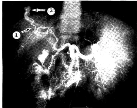
On the selective celiac angiography, the liver was almost in normal size. The right
anterosuperior branch of the hepatic artery was slightly enlarged, and communicated with
a blanch of the portal vein which showed a cystically enlarged termination. Hepatic artery-portal vein fistula was diagnosed (Fig. 2). In venous phase, splenomegaly was apparent but without demonstrable splenic vein. A huge varicous-like venous dilatation
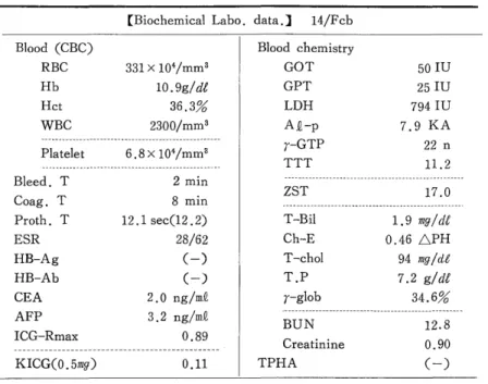
Table 1. Preoperative laboratory data after ten bottles of blood transfusion
[Biochemical Labo. data.] 14/Fcb
Blood (CBC) Blood chemistry
RBC 331 x 104/mm3 GOT 50 IU
Hb 10.9g/d& GPT 25 IU
Hct 36.3% LDH 794 IU
WBC 2300/mm3 A2-p 7.9 KA
---
r-GTP 22 n
Pl atelet 6.8 x 10⁴/mm3
--- ---- - -- -- - - --- TTT 11.2
--- -- ---
B eed. T 2 min ZST
17.0 C
oag. T 8 min --__
Proth. T 12.1 sec(12.2) T-Bil 1,9 mg/df
ESR 28/62 Ch-E 0.46 APH
HB-Ag (-) T-chol 94 mg/dZ
HB-Ab (-) T.P 7.2 g/dl
CEA 2.0 ng/m4 r-glob 34.6%
AFP 3.2 ng/mQ BUN --- 12 --- .8
ICG-Rmax 0.89 C
reatinine 0.90
---