1
Protease-Activated Receptor-2 Plays a Critical Role in Vascular Inflammation and
Atherosclerosis in Apolipoprotein E-deficient Mice
Tomoya Hara, MD. PhD1*, Pham Tran Phuong, MSc1*, Daiju Fukuda, MD. PhD1, 2, Koji Yamaguchi,
MD. PhD1, Chie Murata, PhD3, Sachiko Nishimoto, PhD1, Shusuke Yagi, MD. PhD1, Kenya
Kusunose, MD. PhD1, Hirotsugu Yamada, MD. PhD1, Takeshi Soeki, MD. PhD1, Tetsuzo Wakatsuki,
MD. PhD1, Issei Imoto, MD. PhD3, Michio Shimabukuro, MD. PhD2, Masataka Sata, MD. PhD1
*; equal contribution
1. Department of Cardiovascular Medicine, Tokushima University Graduate School of Biomedical Sciences, Tokushima 770-8503, Japan.
2. Department of Cardio-Diabetes Medicine, Tokushima University Graduate School of Biomedical Sciences, Tokushima 770-8503, Japan.
3. Department of Human Genetics, Tokushima University Graduate School of Biomedical Sciences, Tokushima 770-8503, Japan.
First author’s surname and short title: Hara and Roles of PAR-2 in atherogenesis Total word count: 6908 words
Total number of figure and table: 8 figures and 4 tables
Total number of supplementary figure and table: 2 figures and 1 table
2
Daiju Fukuda, MD, PhD
Department of Cardio-Diabetes Medicine,
Tokushima University Graduate School of Biomedical Sciences 3-18-15, Kuramoto-cho, Tokushima 770-8503, Japan
Phone: +81-88-633-7859, Fax: +81-88-633-7894 E-mail: [email protected]
3
Abstract
Background: The coagulation system is closely linked with vascular inflammation, although the underlying mechanisms are still obscure. Recent studies show that protease-activated receptor (PAR)-2, a major receptor of activated factor X (FXa), are expressed in both vascular cells and leukocytes, suggesting that PAR-2 may contribute to the pathogenesis of inflammatory diseases. Here we investigated the role of PAR-2 in vascular inflammation and atherogenesis.
Methods: We generated apolipoprotein E-deficient (ApoE-/-) mice lacking systemic PAR-2
expression (PAR-2-/-ApoE-/-). ApoE-/- mice which lack or express PAR-2 only in bone-marrow (BM)
cells were also generated by BM transplantation. Atherosclerotic lesions were investigated after 20 weeks on a western-type diet (WTD) by histological analyses, quantitative RT-PCR, and western blotting. In vitro experiments using BM-derived macrophages were performed to confirm pro-inflammatory roles of PAR-2. The association between plasma FXa level and the severity of coronary atherosclerosis was also examined in humans who underwent coronary intervention. Results: PAR-2-/-ApoE-/- mice showed reduced atherosclerotic lesions in the aortic arch (P<0.05)
along with features of stabilized atherosclerotic plaques such as less lipid deposition (P<0.05), collagen loss (P<0.01), macrophage accumulation (P<0.05), and inflammatory molecule
expression (P<0.05) compared with ApoE-/- mice. Systemic PAR2 deletion in ApoE-/- mice
significantly decreased the expression of inflammatory molecules in the aorta. The results of BM transplantation experiments demonstrated that PAR-2 in hematopoietic cells contributed to
atherogenesis in ApoE-/- mice. PAR-2 deletion did not alter metabolic parameters. In vitro
experiments demonstrated that FXa or a specific peptide agonist of PAR-2 significantly increased expression of inflammatory molecules and lipid uptake in BM-derived macrophages from wild-type
4
mice compared with those from PAR-2-deficient mice. Activation of NF-κB signaling was involved in PAR-2-associated vascular inflammation and macrophage activation. In humans who underwent coronary intervention, plasma FXa level independently correlated with the severity of coronary atherosclerosis as determined by Gensini score (P<0.05) and plaque volume (P<0.01).
Conclusions: PAR-2 signaling activates macrophages and promotes vascular inflammation,
increasing atherosclerosis in ApoE-/- mice. This signaling pathway may also participate in
atherogenesis in humans.
5
Clinical Perspective (1) What is new?
Systemic deletion or hematopoietic deletion of protease-activated receptor-2 (PAR-2) attenuates vascular inflammation and atherogenesis in atherosclerotic mouse model. The activation of PAR-2 promotes pro-inflammatory activation of macrophages as
determined by inflammatory molecule expression and foam cell formation.
In humans, plasma FXa level positively correlates with the severity of coronary artery disease such as Gensini score and plaque volume determined by intravascular ultrasound analysis.
In addition to classical coronary risk factors, plasma FXa level predicts the severity of coronary artery disease.
(2) What are the clinical implications?
The PAR-2 signaling plays an important role in the development of vascular inflammation and atherosclerosis.
The PAR-2 signaling pathway may provide a novel mechanism of atherogenesis and serve as a potential therapeutic target for atherosclerosis.
6
Introduction
It is a widely accepted view that atherosclerosis is a chronic inflammatory disease 1, 2. Many cellular
and molecular pathways cause vascular inflammation 3, 4. Previous reports have suggested a link
between the blood coagulation system and inflammatory diseases 5, 6. Several coagulation
proteases and protease-activated receptor (PAR) pathways serve as underlying mechanisms 7-11.
PARs are a family of G protein-coupled, seven transmembrane domain receptors that are activated by proteolytic cleavage of the receptor N-terminus by several proteases, generating a novel tethered ligand, which subsequently activates the receptor via intramolecular binding. Among the four subtypes of the PAR family, recent studies demonstrated that PAR-2, one of the major receptors of activated factor X (FXa), is expressed in vascular cells (i.e., endothelial cells and
smooth muscle cells) and leukocytes, but not in platelets 7, 10, 11, and contributes to the development
of inflammatory diseases, such as insulin resistance 12 and neointima formation after vascular injury
13. However, the role of PAR-2 signaling in the development of atherosclerosis is not fully
understood. Several studies have reported that PAR-2 in endothelial cells plays an important role
in the pathophysiology of inflammatory diseases 14-16; however, little is known about the role of
PAR-2 in macrophage activation, which is a key player in atherogenesis.
Recently, we reported that a direct FXa inhibitor, rivaroxaban, attenuates atherosclerotic
plaque progression and destabilization in apolipoprotein E-deficient (ApoE-/-) mice 17. Consistent
with the results of our animal study, recent clinical trials demonstrated that patients with acute coronary syndrome treated with rivaroxaban experienced fewer cardiovascular events compared
with the control 18-20. Furthermore, the result of the Cardiovascular Outcomes for People Using
7
rivaroxaban to aspirin reduced major adverse cardiovascular events in patients with coronary or
peripheral artery disease compared with traditional aspirin alone group 21. The anti-coagulation
effects of FXa inhibitors might play a role in these results, although these studies also suggest that the inhibition of FXa-PAR-2 signaling by a FXa inhibitor might have anti-atherosclerotic effects through attenuation of vascular inflammation.
In this study, to address the hypothesis that FXa-PAR-2 signaling, especially in macrophages, promotes vascular inflammation and atherosclerosis, we performed in vivo studies
using ApoE-/- mice that genetically lack PAR-2 (PAR-2-/-ApoE-/- mice) and bone marrow (BM)
chimeric ApoE-/- mice which lack or express PAR-2 only in hematopoietic cells. In vitro experiments
using macrophages were also performed to investigate the role of PAR-2 in pro-inflammatory activation of this cell-type. Furthermore, we examined the relationship between plasma FXa level and the severity of coronary atherosclerosis in human to obtain clinically translatable evidence that FXa-PAR-2 signaling is involved in the development of atherosclerosis. The results of our study suggest a novel mechanism of atherogenesis that could serve as a potential therapeutic target.
Methods
All data and supporting materials have been provided with the published article.
Animals
Male ApoE-/- mice and PAR-2-deficient (PAR-2-/-) mice (C57BL/6J background), originally
8
by cross-breeding PAR-2-/- mice and ApoE-/- mice. Sex-age-matched ApoE-/- mice served as the
control. We administered a western-type diet (WTD) for 20 weeks from 8 weeks of age. Some of
8-week-old male PAR-2-/-ApoE-/- mice received a WTD supplemented with rivaroxaban (5 mg/kg
body weight/day) for 20 weeks 17. Sex-age-matched non-treated PAR-2-/-ApoE-/- mice were served
as the control. Rivaroxaban was supplied by Bayer Health Care. The mice were housed in a room in which lighting was controlled (12 h on/12 h off), and room temperature was kept at 25 °C. All experimental procedures conformed to the guidelines for animal experimentation of Tokushima University. The protocol was reviewed and approved by our institutional ethics committee.
Bone marrow transplantation
To investigate the role of PAR-2 in hematopoietic cells, we performed bone marrow transplantation
(BMT) as described previously 22. For generating ApoE-/- mice which lack PAR-2 in hematopoietic
cells, BM of ApoE-/- mice was reconstituted with that of PAR-2-/-ApoE-/- mice. For generating ApoE
-/- mice which express PAR-2 only in hematopoietic cells, BM of PAR-2-/-ApoE-/- mice was
reconstituted with that of ApoE-/- mice. ApoE-/- mice which BM was replaced with that of ApoE
-/-mice and PAR-2-/-ApoE-/- mice which BM was replaced with that of PAR-2-/-ApoE-/- mice were used
for the control respectively. BM cells were harvested from the femurs and tibias of female donor mice. Eight-week-old male recipient mice were lethally X-irradiated with a total dose of 9.0 Gy. One day later, recipient mice received unfractionated BM cells by injection into the tail vein. At 8 weeks after BMT, all animals were started on a WTD. The replacement rate of BM cells was determined by fluorescence in-situ hybridization (FISH) of the X and Y chromosomes in male recipient mice
air-9
drying method. FISH analysis was performed using mouse X and Y chromosome probes (Chromosome Science Labo) following the manufacturer’s protocol, and the signals were observed under an all-in-one fluorescence microscope BZ-9000 (KEYENCE Japan). We used only BM chimeric mice in which more than 90% of BM had been replaced by donor BM.
Blood pressure and plasma lipid level measurement
The blood pressure of each mouse was measured using a tail-cuff system (BP-98A, Softron) in conscious animals. The average value of three measurements was used for comparison. At the time of sacrifice, blood was collected from the heart into K2-EDTA-containing tubes. After centrifuged, plasma was stored at -80°C until required. Plasma lipid levels were measured using commercially available kits (Wako Diagnostics).
Preparation of aortas and atherosclerotic lesion analysis
Atherosclerotic lesion analysis was performed as described previously 17. Mice were sacrificed by
administration of an overdose of pentobarbital, and perfused with 0.9% sodium chloride solution at a constant pressure via the left ventricle. Both the heart and whole aorta were immediately removed.
The thoracic aorta was excised, opened longitudinally, and fixed with 10% neutral buffered formalin.
To quantify atherosclerotic lesions in the aortic arch, en face Sudan IV staining was performed. The percentage of the Sudan IV-positive area was measured. The abdominal aorta was removed and snap-frozen in liquid nitrogen for gene or protein expression analysis.
10
Histological and immunohistochemical staining were performed as described previously 17. Each
heart was cut along a horizontal plane between the lower tips of the left and right atria. The upper portion was snap-frozen in OCT compound (TissueTeck). Then, the aortic root was sectioned
serially (5 μm intervals) from the point where the aortic valves appeared to the ascending aorta,
until the valve cusps were no longer visible. Lipid and collagen content within atherosclerotic plaques were determined by Oil red O staining and picrosirius red staining, respectively. Macrophage antigen-3 (Mac-3), monocyte chemotactic protein-1 (MCP-1) and matrix metallopeptidase (MMP)-9 expression were detected using anti-macrophage antigen-3 (BD Biosciences), anti-MCP-1 (Santa-Cruz) or anti-MMP-9 (R&D Systems Inc.) antibody followed by the avidin-biotin complex technique, and stained using a Vector Red substrate kit (Vector). Each section was counterstained with hematoxylin.
Cell culture
BM-derived macrophages were used in this study. BM cells were obtained from femurs and tibias
of 8-week-old wild-type mice and PAR-2-/- mice. Cells were cultured in DMEM supplemented with
10 ng/ml macrophage colony-stimulating factor (R&D Systems) for 7 days. To assess the role of
PAR-2 in macrophage activation, BM-derived macrophages from wild-type mice and PAR-2-/- mice
were treated with 50 nM activated factor X (FXa) (Haematologic Technologies, Inc.) or 10 nM PAR-2 specific agonist peptide (AP-PAR-2; Sigma-Aldrich) for PAR-24 h after PAR-24-h serum starvation. In addition, to assess the role of FXa-PAR-2 signaling in foam cell formation, BM-derived macrophages from
wild-type mice and PAR-2-/- mice were pre-treated with 50 nM FXa or 10 nM AP-2 for 24 h, and
11
Intracellular lipids were stained by Oil red O staining after fixation with 10% neutral buffered formalin. The ratio of Oil red O-positive area/cellular area (%) was calculated in 100 cells and averaged. All cells were cultured in DMEM supplemented with 10% fetal bovine serum, 100 U/ml penicillin, and
100 mg/ml streptomycin in a 5% CO2humidified atmosphere.
Reverse transcription, real-time polymerase chain reaction
Total RNA was extracted from tissues and cells using an illustra RNAspin RNA Isolation Kit (GE Healthcare). Reverse transcription was performed using a QuantiTect Reverse Transcription kit
(Qiagen) from 1 μg of the extracted total RNA. Quantitative real-time PCR (qPCR) was performed
on an Mx3000P (Agilent Technologies) using gene-specific primers (Supplemental Table) and
Power SYBR Green PCR Master Mix (Applied Biosystems). Data are expressed in arbitrary units
that were normalized by β-actin.
Western blot
Protein were isolated from cells or tissue, and separated by SDS–polyacrylamide gel
electrophoresis gels as we described previously23. The following antibodies were used: IκBα, and
β-actin (Sigma). β-actin was used as an internal control. Antibody distribution was visualized with
enhanced chemiluminescence-plus reagent (GE Healthcare) using a luminescent image analyzer (LAS-1000, Fuji Film).
Clinical research
12
patients underwent elective percutaneous coronary intervention (PCI) and intravascular ultrasound (IVUS) study of the vessel undergoing PCI. We excluded patients with acute coronary syndrome, renal insufficiency, history of PCI or coronary artery bypass surgery, and those with inadequate IVUS imaging. Blood samples were collected into Na-EDTA-containing tubes just before PCI. After blood samples were centrifuged, plasma was stored at -80°C until required. Plasma FXa level was measured using a commercially available kit (Human Coagulation factor Xa ELISA Kit; MBS721605, MyBioSource). All experimental procedures were approved by the Institutional Review Board of Tokushima University Hospital and all subjects were gave informed consent.
We calculated Gensini scores 24 from initial angiograms as a global severity marker of
coronary atherosclerosis. Our IVUS imaging protocol was described previously 25. In brief, we
selected plaques of intermediate stenotic lesions (25% < percent diameter stenosis < 50% on quantitative coronary angiography) > 5 mm from the intervention site. We used imaging catheters (Atlantis; Boston Scientific/Cardiovascular System) and a console (Galaxy; Boston Scientific/Cardiovascular System). We captured 10 continuous slice images (total length 5 mm) at intervals of 0.5 mm using an auto-pullback system in each plaque. Cross-sectional lumen area, cross-sectional vessel area within the external elastic membrane, and plaque area were determined using software attached to the IVUS system. Plaque volume was calculated using integration.
Statistical analysis
Numerical values are expressed as mean ± SEM. Comparison of parameters between two groups
13
Mann-Whitney U test when data did not follow a normal distribution. Univariate analysis between the severity of coronary atherosclerosis (Gensini score and plaque volume) and plasma FXa level
was performed by Spearman's rank correlation coefficient. Multiple regression analysis was
performed for prediction of the Gensini score and coronary plaque volume using metabolic
parameters and plasma FXa level. A value of P<0.05 was considered significant.
Results
Genetic deletion of PAR-2 attenuated plaque progression and destabilization in ApoE-/- mice
After 20 weeks of WTD feeding, en face Sudan IV staining of the aortic arch showed a significant
reduction of atherosclerotic lesions in PAR-2-/-ApoE-/- mice compared with ApoE-/- mice (49.0±3.1
vs. 38.9±3.7%, P<0.05) (Figure 1). Genetic deletion of PAR-2 demonstrated a significant reduction
of lipid deposition (6.98±0.21 vs. 6.23±0.21%, P<0.05) (Figure 2A) and an increase in collagen
content (66.0±1.3 vs. 72.3±1.3%, P<0.01) (Figure 2B) in atherosclerotic plaques in ApoE-/- mice.
Also, genetic deletion of PAR-2 in ApoE-/- mice significantly reduced the accumulation of
macrophages as determined by Mac-3 staining (5.35±0.58 vs. 3.02±0.58%, P<0.05) (Figure 2C) and the expression of MCP-1 (2.17±0.34 vs. 1.00±0.34%, P<0.05) (Figure 2D) and MMP-9 (2.60±0.24 vs. 1.39±0.27%, P<0.05) (Figure 2E) in atherosclerotic plaques compared with control animals. There were no differences in body weight gain, blood pressure, plasma glucose level, and
plasma lipid levels between ApoE-/- mice and PAR-2-/-ApoE-/- mice (Table 1). PAR-2 deletion did not
affect plasma FXa level. These results indicated that genetic deletion of PAR-2 in ApoE-/- mice
14
Genetic deletion of PAR-2 attenuated vascular inflammation in ApoE-/- mice
We also compared the expression of inflammatory molecules in the atherosclerotic aorta using
qPCR between ApoE-/- mice and PAR-2-/-ApoE-/- mice. Consistent with the results of
immunohistochemical study, PAR-2-/-ApoE-/- mice demonstrated reduced expression of the
macrophage marker F4/80 (P=0.05) compared with ApoE-/- mice. PAR-2-/-ApoE-/- mice also
demonstrated a reduction of inflammatory molecules, such as MCP-1, macrophage inflammatory
protein (MIP)-1α, interleukin (IL)-6 and MMP-9 (P<0.05, respectively) (Figure 3A). PAR-2 deletion attenuated the degradation of IκBα in the atherosclerotic aorta, suggesting inhibition of NF-κB signaling (Figure 3B). Genetic deletion of 2 did not change RNA expression of 1,
PAR-3, and PAR-4 (Figure 3C). These results indicated that genetic deletion of PAR-2 in ApoE-/- mice
reduces vascular inflammation.
FXa inhibition did not affect atherogenesis in PAR-2-/-ApoE-/- mice
FXa activates both PAR-1 and PAR-2. Previous studies reported the expression of PAR-1 and PAR-2 in the vasculature and contribution to the development of vascular inflammation and
atherogenesis 7, 10, 11. Therefore, to examine the role of FXa-PAR-2 signaling in atherogenesis, we
administered a specific FXa inhibitor, rivaroxaban, to PAR-2-/-ApoE-/- mice for 20 weeks.
Rivaroxaban attenuated atherogenesis in ApoE-/- mice as demonstrated in our previous study 17,
although it did not inhibit the progression of atherosclerotic lesions in the aortic arch (35.1±2.3 vs. 31.4±3.5%) and the expression of inflammatory molecules, except for IL-1β, in atherosclerotic aorta
in PAR-2-/-ApoE-/- mice, suggesting the pivotal role of FXa-PAR-2 signaling in atherogenesis
15
PAR-2 in hematopoietic cells contributed to plaque progression and destabilization in
ApoE-/- mice
To further investigate the role of PAR-2 in hematopoietic cells in the development of atherosclerosis,
we generated BM specific PAR-2 deleted or expressing ApoE-/- mice. The replacement rate of BM
cells determined by fluorescence in-situ hybridization was more than 90% in all BM chimeric mice as shown in Supplemental Figure 2.
PAR-2 deletion in BM cells reduced the development of atherosclerotic lesions in the aortic arch (10.8±1.1 vs. 5.8±1.0%, P<0.01) (Figure 4A) and lipid deposition in atherosclerotic plaques in the aortic root (5.71±0.66 vs. 3.08±0.63%, P<0.01) (Figure 5A). In contrast, PAR-2 deletion in BM cells significantly increased collagen content in plaques (15.3±2.1 vs. 24.2±2.0%,
P<0.01) (Figure 5B). Furthermore, the results of immunostaining of atherosclerotic lesions revealed
that PAR-2 deletion in BM cells in ApoE-/- mice reduced the accumulation of macrophages as
determined by Mac-3 staining (7.43±0.29 vs. 4.03±0.35%, P<0.01) (Figure 5C), and the expression of MMP-9 (6.13±0.18 vs. 4.25±0.48%, P<0.01) (Figure 5E) compared with control chimeric mice.
The expression of MCP-1 was also lower in plaques in ApoE-/- mice lacking PAR-2 in BM cells
compared with that in control mice (Figure 5D). On the other hand, BM specific PAR-2 expression promoted atherosclerotic lesion progression (Figure 4B). Lipid deposition and collagen loss in atherosclerotic lesions were greater in BM specific PAR-2 expressing mice compared to the control (Supplementary Figure 3).
There were no differences in body weight gain, blood pressure, plasma glucose level, and plasma lipid levels despite the presence or absence of PAR-2 in BM (Table 2 and Supplementary
16
Table 2). These results indicated that PAR-2 in hematopoietic cells contributes to plaque
progression and destabilization in ApoE-/- mice. Also, these results suggested that PAR-2 signaling
in macrophages contributes, at least partially, to atherogenesis.
PAR-2 in hematopoietic cells promoted vascular inflammation in ApoE-/- mice
We also examined the role of PAR-2 in hematopoietic cells in the expression of inflammatory
molecules in the atherosclerotic aorta using qPCR. PAR-2 deletion in hematopoietic cells reduced the expression of the macrophage marker F4/80 (P<0.05). PAR-2 deletion in BM cells also reduced
the expression of inflammatory molecules, such as MCP-1 (P<0.05), MIP-1α (P<0.05), IL-1β
(P<0.05), IL-6 (P<0.01), tumor necrosis factor (TNF)-α (P<0.05), and MMP-9 (P<0.01) (Figure 6A). On the other hand, PAR-2 expression in BM increased the expression of these inflammatory molecules (Figure 6B). These results indicated that PAR-2 in hematopoietic cells promotes the expression of inflammatory molecules in atherosclerotic aorta, leading to the development of vascular inflammation.
PAR-2 signal promoted pro-inflammatory activation of macrophages
To investigate the mechanism by which inhibition of PAR-2 signaling prevented the development and destabilization of atherosclerotic lesions without improvement of metabolic parameters, we performed in vitro experiments using BM-derived macrophages. We treated BM-derived macrophages with FXa, a major ligand of PAR-2, or a specific agonist peptide of PAR-2 (AP-2). The results of qPCR analysis demonstrated that the expression of inflammatory molecules (e.g., MCP-1, IL-6 and TNF-α) was significantly higher in BM-derived macrophages from wild-type mice
17
compared with those from PAR-2-/- mice after FXa or AP-2 treatment (Figure 7A). The result of
western blotting demonstrated that the activation of PAR-2 signaling by FXa increased the degradation of IκBα, suggesting the activation of NF-κB in macrophages (Figure 7B). We further examined the role of PAR-2 signaling in lipid uptake by macrophages in vitro. The activation of PAR-2 signaling by FXa or AP-2 significantly increased uptake of oxidized low-density lipoprotein
(oxLDL) by BM-derived macrophages from wild-type mice compared with those from PAR-2-/- mice
(Figure 7C). These results suggested that PAR-2 signaling promotes pro-inflammatory activation
of macrophages.
Relationship between FXa-PAR-2 signaling and atherosclerosis in clinical samples
To investigate the role of the FXa-PAR-2 signaling pathway in the development of atherosclerosis in humans, we investigated the link between plasma FXa level and the severity of coronary atherosclerosis. Blood samples, Gensini score and intravascular ultrasound (IVUS) data were obtained from 32 patients who underwent percutaneous coronary intervention. Patient characteristics at enrollment are shown in Table 3. Mean age of the participants was 69.6±1.1 years; 63% were men. Plasma FXa level positively correlated with the Gensini score (Spearman ρ=0.36, P<0.05) (Figure 8A). Plasma FXa level tended to correlate positively with coronary plaque volume determined by intravascular ultrasound study (Spearman ρ=0.42, P=0.06) (Figure 8B). Multiple regression analysis revealed that plasma FXa level remained as an independent predictor
of the Gensini score (corrected R2=0.29, P<0.05). Multiple regression analysis revealed that
plasma FXa level, as well as male sex and HbA1c level, remained as independent predictors of
18
promotes vascular inflammation through PAR-2 and is associated with the severity of atherosclerosis in coronary arteries in humans.
Discussion
Accumulating evidence suggests that PAR-2 signaling promotes pro-inflammatory responses in
many cell types 7, 10, 11, contributing to the pathogenesis of inflammatory diseases 12-16, although
little is known about the role of PAR-2, especially in hematopoietic cells, in the development of vascular inflammation and atherosclerosis. In this study, in vivo experiments using PAR-2 deficient
ApoE-/- mice and BM-specific PAR-2 expression or deletion animals demonstrated that PAR-2
deletion attenuated vascular inflammation and reduced the development and destabilization of atherosclerotic lesions without the alteration of metabolic parameters including blood lipid levels. The results of in vitro experiments using peritoneal macrophages demonstrated that PAR-2 signaling participates in pro-inflammatory activation of macrophages through NF-κB pathway. Our findings suggest that the PAR-2 signaling promotes vascular inflammation and atherogenesis, and that pro-inflammatory activation of macrophages through the PAR-2 signaling plays a pivotal role as an underlying mechanism.
Previous reports demonstrated that several proteases including trypsin, tryptase and FXa, but not thrombin, activate PAR-2. Different from other members of the PAR family, platelets do not express PAR-2, whereas vascular cells, such as endothelial cells and smooth muscle cells, and
leukocytes express PAR-2 11. Because of its unique expression pattern, previous studies
19
26, 27 and inflammatory responses 15, 16 has been reported. However, the role of PAR-2 signaling
in macrophage activation is not fully investigated. Therefore, in this study, we generated BM-chimeric mice which express or lack PAR-2 in BM. PAR-2 deletion in hematopoietic cells attenuated vascular inflammation and atherogenesis. On the contrary, PAR-2 expression only in hematopoietic
cells promoted vascular inflammation and atherogenesis. Furthermore, our in vitro experiments
demonstrated that treatment of macrophages with FXa, the major endogenous ligand of PAR-2, or AP-2, a specific agonist for PAR-2, increases inflammatory gene expression and lipid uptake, both of which are critical for atherosclerotic lesion progression and destabilization. These results suggested that PAR-2 plays a pivotal role in pro-inflammatory activation of macrophages, contributing to the development of vascular inflammation and atherogenesis.
Several previous studies have already reported a role of PAR-2 in non-hematopoietic cells
in the development of vascular inflammation in different models 28-31. Therefore, it is plausible that
PAR-2 in non-hematopoietic cells such as endothelial cells may participate in atherogenesis. In fact, in our BMT experiments, expression of PAR-2 in the vasculature enhanced lesion development despite of the presence or absence of PAR-2 in hematopoietic cells. These results indicated pivotal roles of PAR-2 of both hematopoietic cells, especially macrophages, and non-hematopoietic cells in atherogenesis, and suggested PAR-2 can be a potential therapeutic target against atherosclerosis.
Since clinical approval as an anti-coagulant, a direct oral FXa inhibitor, rivaroxaban, attracts much attention because of its beneficial effects on cardiovascular events proved by several
clinical trials 18-21. In our clinical study, plasma FXa level predicts the severity of coronary
20
and upregulates inflammation through NF-κB pathway32-34. Therefore, the results of in vivo animal
studies and in vitro studies of the present study may provide one of the possible mechanisms for recent clinical studies. Interestingly, recent studies have demonstrated that several coagulation factors such as tissue factor and factor X exist not only in the circulating blood but also in
atherosclerotic plaques in human autopsy samples 35. In our present study, plasma FXa level
between ApoE-/- mice and PAR-2-/-ApoE-/- mice did not differ, although, in our previous study, we
reported higher expression of PAR-2 in atherosclerotic aorta of ApoE-/- mice compared with the
aorta in wild-type mice 17. These results suggested that the role of FXa-PAR-2 signaling in vascular
inflammation is enhanced in the atherosclerotic plaques. In addition, in this study, we could not
observe the inhibitory effect of rivaroxaban on atherogenesis in PAR-2-/-ApoE-/- mice. This result
also may give support to the role of FXa-PAR-2 signaling in the development of atherosclerosis at least partially.
This study has several limitations. First, to investigate the role of PAR-2 in macrophages, a key cell-type in atherogenesis, we performed BMT experiments. Previous studies demonstrated that other hematopoietic cells such as neutrophils express PAR-2. Although our in vitro studies using peritoneal macrophages showed that FXa-PAR-2 signaling participates in pro-inflammatory activation of macrophages, other hematopoietic cells activated by FXa-PAR-2 signaling might have contributed to the results of in vivo studies. Second, we only examined the role of FXa-PAR-2 signaling in atherogenesis, however, FXa activates PAR-1 as well. Some of previous studies
reported that contribution of PAR-1 to inflammation36, 37. Therefore, it might also participate in the
development of atherosclerosis. Further studies are needed to clarify the role of FXa-PARs signaling in vascular inflammation and atherogenesis. Third our patient population in our clinical
21
study was small, and a large scale study is required to confirm our results. Lastly, in our clinical study, total cholesterol level did not predict the coronary atherosclerosis determined by IVUS. We speculate that because we included only patients receiving statins for at least 6 months, lipid levels were well controlled in this study population.
Our present study demonstrated proof of the concept that PAR-2 signaling in macrophages plays a pivotal role in the development of atherosclerosis. Further studies are needed to elucidate the participation of FXa-PARs signaling in the development of vascular inflammation and atherosclerosis. In conclusion, PAR-2 deletion attenuates vascular inflammation
and atherogenesis in ApoE-/- mice. PAR-2 in hematopoietic cells may contribute, at least in part, to
the development of atherosclerosis. Combined with the results of our in vitro studies, these results indicated that PAR-2 signaling in macrophages plays a pivotal role in vascular inflammation. The PAR-2 signaling pathway may provide a novel mechanism of atherogenesis and serve as a potential therapeutic target for atherosclerosis.
Acknowledgements
The authors thank Yumiko Saga and Etsuko Uematsu for their technical assistance. The authors also thank Dr. Yoshihiro Okayama, University Hospital of Tokushima Clinical Trial Center for Developmental Therapeutics, for his significant advice for statistical analyses.
Funding
This work was partially supported by Japan Society for the Promotion of Science KAKENHI Grants (Number 26860565 to T.H., Number 16K09517 to D.F., and Number 16H05299 and 26248050 to
22
M. Sata), a Sakakibara Memorial Research Grant from The Japan Research Promotion Society for Cardiovascular Diseases (D.F.), a Grant from Japan Cardiovascular Research Foundation (D.F.), a grant-in-aid from the Cardiovascular Research Fund, Tokyo, Japan (D.F.), the Takeda Science Foundation (D.F. and M.Sata), the Fugaku Trust for Medical Research (M.S.), the Vehicle Racing Commemorative Foundation (M.S.), and a Bayer Scholarship for Cardiovascular Research (D.F). M. Sata received research funding from Bayer Yakuhin, Ltd. The funders had no role in the study design, data collection, and analysis, or preparation of the manuscript.
Conflict of Interest Disclosures
The Department of Cardio-Diabetes Medicine, Tokushima University Graduate School, is supported in part by unrestricted research grants from Boehringer Ingelheim, Tanabe-Mitsubishi, Kowa, and Actelion. Dr. Sata received research funding from Bayer Yakuhin, Ltd. Other authors declare that they have no conflict of interest.
23
References
1. Aikawa M, Libby P. Atherosclerotic plaque inflammation: the final frontier? Can J Cardiol.
2004;20:631-634.
2. Ross R. Atherosclerosis--an inflammatory disease. N Engl J Med. 1999;340:115-126.
3. Hansson GK. Inflammation, atherosclerosis, and coronary artery disease. N Engl J Med.
2005;352:1685-1695.
4. Paoletti R, Gotto AM, Jr., Hajjar DP. Inflammation in atherosclerosis and implications for
therapy. Circulation. 2004;109:III20-26.
5. Esmon CT, Fukudome K, Mather T, Bode W, Regan LM, Stearns-Kurosawa DJ, Kurosawa
S. Inflammation, sepsis, and coagulation. Haematologica. 1999;84:254-259.
6. Levi M, van der Poll T, Buller HR. Bidirectional relation between inflammation and
coagulation. Circulation. 2004;109:2698-2704.
7. Borissoff JI, Spronk HM, ten Cate H. The hemostatic system as a modulator of
atherosclerosis. N Engl J Med. 2011;364:1746-1760.
8. Coughlin SR. Thrombin signalling and protease-activated receptors. Nature.
2000;407:258-264.
9. Croce K, Libby P. Intertwining of thrombosis and inflammation in atherosclerosis. Curr Opin
Hematol. 2007;14:55-61.
10. Leger AJ, Covic L, Kuliopulos A. Protease-activated receptors in cardiovascular diseases.
Circulation. 2006;114:1070-1077.
11. Major CD, Santulli RJ, Derian CK, Andrade-Gordon P. Extracellular mediators in
24 Arterioscler Thromb Vasc Biol. 2003;23:931-939.
12. Badeanlou L, Furlan-Freguia C, Yang G, Ruf W, Samad F. Tissue factor-protease-activated
receptor 2 signaling promotes diet-induced obesity and adipose inflammation. Nat Med. 2011;17:1490-1497.
13. Tennant GM, Wadsworth RM, Kennedy S. PAR-2 mediates increased inflammatory cell
adhesion and neointima formation following vascular injury in the mouse. Atherosclerosis. 2008;198:57-64.
14. Redecha P, Franzke CW, Ruf W, Mackman N, Girardi G. Neutrophil activation by the tissue
factor/Factor VIIa/PAR2 axis mediates fetal death in a mouse model of antiphospholipid syndrome. J Clin Invest. 2008;118:3453-3461.
15. Samad F, Ruf W. Inflammation, obesity, and thrombosis. Blood. 2013;122:3415-3422.
16. Spronk HM, de Jong AM, Crijns HJ, Schotten U, Van Gelder IC, Ten Cate H. Pleiotropic
effects of factor Xa and thrombin: what to expect from novel anticoagulants. Cardiovasc
Res. 2014;101:344-351.
17. Hara T, Fukuda D, Tanaka K, Higashikuni Y, Hirata Y, Nishimoto S, Yagi S, Yamada H,
Soeki T, Wakatsuki T, Shimabukuro M, Sata M. Rivaroxaban, a novel oral anticoagulant, attenuates atherosclerotic plaque progression and destabilization in ApoE-deficient mice.
Atherosclerosis. 2015;242:639-646.
18. Mega JL, Braunwald E, Mohanavelu S, Burton P, Poulter R, Misselwitz F, Hricak V,
Barnathan ES, Bordes P, Witkowski A, Markov V, Oppenheimer L, Gibson CM. Rivaroxaban versus placebo in patients with acute coronary syndromes (ATLAS ACS-TIMI 46): a randomised, double-blind, phase II trial. Lancet. 2009;374:29-38.
25
19. Mega JL, Braunwald E, Murphy SA, Plotnikov AN, Burton P, Kiss RG, Parkhomenko A,
Tendera M, Widimsky P, Gibson CM. Rivaroxaban in patients stabilized after a ST-segment elevation myocardial infarction: results from the ATLAS ACS-2-TIMI-51 trial (Anti-Xa Therapy to Lower Cardiovascular Events in Addition to Standard Therapy in Subjects with Acute Coronary Syndrome-Thrombolysis In Myocardial Infarction-51). J Am Coll Cardiol. 2013;61:1853-1859.
20. Mega JL, Braunwald E, Wiviott SD, Bassand JP, Bhatt DL, Bode C, Burton P, Cohen M,
Cook-Bruns N, Fox KA, Goto S, Murphy SA, Plotnikov AN, Schneider D, Sun X, Verheugt FW, Gibson CM. Rivaroxaban in patients with a recent acute coronary syndrome. N Engl
J Med. 2012;366:9-19.
21. Eikelboom JW, Connolly SJ, Bosch J, Dagenais GR, Hart RG, Shestakovska O, Diaz R,
Alings M, Lonn EM, Anand SS, Widimsky P, Hori M, Avezum A, Piegas LS, Branch KRH, Probstfield J, Bhatt DL, Zhu J, Liang Y, Maggioni AP, Lopez-Jaramillo P, O'Donnell M, Kakkar AK, Fox KAA, Parkhomenko AN, Ertl G, Stork S, Keltai M, Ryden L, Pogosova N, Dans AL, Lanas F, Commerford PJ, Torp-Pedersen C, Guzik TJ, Verhamme PB, Vinereanu D, Kim JH, Tonkin AM, Lewis BS, Felix C, Yusoff K, Steg PG, Metsarinne KP, Cook Bruns N, Misselwitz F, Chen E, Leong D, Yusuf S. Rivaroxaban with or without Aspirin in Stable Cardiovascular Disease. N Engl J Med. 2017;377:1319-1330.
22. Sata M, Saiura A, Kunisato A, Tojo A, Okada S, Tokuhisa T, Hirai H, Makuuchi M, Hirata Y,
Nagai R. Hematopoietic stem cells differentiate into vascular cells that participate in the pathogenesis of atherosclerosis. Nat Med. 2002;8:403-409.
26
Sato F, Bando M, Yagi S, Soeki T, Hayashi T, Imoto I, Sakaue H, Shimabukuro M, Sata M. Obesity-induced DNA released from adipocytes stimulates chronic adipose tissue inflammation and insulin resistance. Sci Adv. 2016;2:e1501332.
24. Gensini GG. A more meaningful scoring system for determining the severity of coronary
heart disease. Am J Cardiol. 1983;51:606.
25. Yamaguchi K, Wakatsuki T, Soeki T, Niki T, Taketani Y, Oeduka H, Kusunose K, Ise T, Iwase
T, Yamada H, Sata M. Effects of telmisartan on inflammatory cytokines and coronary plaque component as assessed on integrated backscatter intravascular ultrasound in hypertensive patients. Circ J. 2014;78:240-247.
26. Damiano BP, Cheung WM, Santulli RJ, Fung-Leung WP, Ngo K, Ye RD, Darrow AL, Derian
CK, de Garavilla L, Andrade-Gordon P. Cardiovascular responses mediated by protease-activated receptor-2 (PAR-2) and thrombin receptor (PAR-1) are distinguished in mice deficient in PAR-2 or PAR-1. J Pharmacol Exp Ther. 1999;288:671-678.
27. Saifeddine M, al-Ani B, Cheng CH, Wang L, Hollenberg MD. Rat proteinase-activated
receptor-2 (PAR-2): cDNA sequence and activity of receptor-derived peptides in gastric and vascular tissue. Br J Pharmacol. 1996;118:521-530.
28. Hezi-Yamit A, Wong PW, Bien-Ly N, Komuves LG, Prasad KS, Phillips DR, Sinha U.
Synergistic induction of tissue factor by coagulation factor Xa and TNF: evidence for involvement of negative regulatory signaling cascades. Proc Natl Acad Sci U S A. 2005;102:12077-12082.
29. Nystedt S, Ramakrishnan V, Sundelin J. The proteinase-activated receptor 2 is induced by
27 J Biol Chem. 1996;271:14910-14915.
30. Sparkenbaugh EM, Chantrathammachart P, Mickelson J, van Ryn J, Hebbel RP, Monroe
DM, Mackman N, Key NS, Pawlinski R. Differential contribution of FXa and thrombin to vascular inflammation in a mouse model of sickle cell disease. Blood. 2014;123:1747-1756.
31. Svensson KJ, Kucharzewska P, Christianson HC, Skold S, Lofstedt T, Johansson MC,
Morgelin M, Bengzon J, Ruf W, Belting M. Hypoxia triggers a proangiogenic pathway involving cancer cell microvesicles and PAR-2-mediated heparin-binding EGF signaling in endothelial cells. Proc Natl Acad Sci U S A. 2011;108:13147-13152.
32. Borensztajn K, Peppelenbosch MP, Spek CA. Factor Xa: at the crossroads between
coagulation and signaling in physiology and disease. Trends Mol Med. 2008;14:429-440.
33. Krupiczojc MA, Scotton CJ, Chambers RC. Coagulation signalling following tissue injury:
focus on the role of factor Xa. Int J Biochem Cell Biol. 2008;40:1228-1237.
34. Ringwala SM, Dibattiste PM, Schneider DJ. Effects on platelet function of a direct acting
antagonist of coagulation factor Xa. J Thromb Thrombolysis. 2012;34:291-296.
35. Borissoff JI, Heeneman S, Kilinc E, Kassak P, Van Oerle R, Winckers K, Govers-Riemslag
JW, Hamulyak K, Hackeng TM, Daemen MJ, ten Cate H, Spronk HM. Early atherosclerosis exhibits an enhanced procoagulant state. Circulation. 2010;122:821-830.
36. Antoniak S, Cardenas JC, Buczek LJ, Church FC, Mackman N, Pawlinski R.
Protease-Activated Receptor 1 Contributes to Angiotensin II-Induced Cardiovascular Remodeling and Inflammation. Cardiology. 2017;136:258-268.
37. Grimsey NJ, Trejo J. Integration of endothelial protease-activated receptor-1 inflammatory
29
Figure legends
Figure 1. Effect of genetic deletion of RAR-2 on atherosclerotic lesion formation in ApoE
-/-mice.
En face Sudan IV staining of the aortic arch showed that PAR-2-/-ApoE-/- mice had reduced
atherosclerotic lesions compared with ApoE-/- mice (n=9-13). Scale bar: 1 mm. *P<0.05. All values
are mean ± SEM.
Figure 2. Effect of genetic deletion of PAR-2 on characteristics of atherosclerotic plaques in ApoE-/- mice.
(A) Oil red O staining of atherosclerotic plaques in the aortic root. Lipid deposition in plaques was
decreased in PAR-2-/-ApoE-/- mice compared with that in ApoE-/- mice. Scale bar: 100 μm. (B)
Picrosirius red staining observed under polarization microscopy. Collagen content in plaques was
increased in PAR-2-/-ApoE-/- mice compared with that in ApoE-/- mice. Scale bar: 100 μm. (C-E)
Immunostaining against Mac-3 (C), MCP-1 (D), and MMP-9 (E). PAR-2-/-ApoE-/- mice showed
reduced accumulation of macrophages and less expression of MCP-1 and MMP-9 in
atherosclerotic plaques compared with ApoE-/- mice. Scale bar: 50 μm. (n=10 in A and B, n = 5 in
C through E, per group). Mac-3 indicates macrophage antigen-3; MCP-1, monocyte chemotactic protein-1; and MMP-9, matrix metallopeptidase-9. *P<0.05 and **P<0.01. All values are mean ± SEM.
Figure 3. Effect of genetic deletion of PAR-2 on vascular inflammation.
30
expression of F4/80, a macrophage marker. PAR-2 deletion also reduced the expression of
inflammatory molecules, such as MCP-1, MIP-1α, IL-6 and MMP-9, in the atherosclerotic aorta
(n=10-12). (B) The expression of IκBα was examined by western blotting. The degradation of IκBα was reduced in PAR-2 deficient animals, suggesting suppression of NF-κB activity (n=5). (C) Expression of PARs in the aorta. qPCR analyses using abdominal aorta revealed that genetic
deletion of PAR-2 did not change the expression of PAR-1, PAR-3, and PAR-4. (n=10). ApoE‒/‒
indicates apolipoprotein E–deficient; IL, interleukin; MCP-1, monocyte chemotactic protein-1; MIP-1 α, macrophage inflammatory protein α; MMP-9, matrix metallopeptidase-9; N.D., not detectable;
and TNF- α, tumor necrosis factor-α. *P<0.05. All values are mean ± SEM.
Figure 4. Role of PAR-2 in hematopoietic cells in atherosclerotic lesion formation.
(A) En face Sudan IV staining of the aortic arch demonstrated that hematopoietic deletion of
PAR-2 in ApoE-/- mice reduced atherosclerotic lesion progression in the aortic arch compared with the
control BM chimeric mice (n=9-10). (B) En face Sudan IV staining of the aortic arch demonstrated
that hematopoietic restoration of PAR-2 in PAR-2-/-ApoE-/- mice accelerated atherosclerotic lesion
progression in the aortic arch compared with the control BM chimeric mice (n=5-7). Scale bar: 1 mm. BM indicates bone marrow. *P<0.05 and **P<0.01. All values are mean ± SEM.
Figure 5. Effect of hematopoietic deletion of PAR-2 on characteristics of atherosclerotic plaques in aortic root.
(A) Oil red O staining of atherosclerotic plaques in the aortic root. PAR-2 deletion in BM cells
31
staining observed under polarization microscopy. Collagen content was increased in ApoE-/- mice
lacking PAR-2 in BM cells compared with the control group. Scale bar: 100 μm. (C-E)
Immunostaining against Mac-3 (C), MCP-1 (D), and MMP-9 (E). ApoE-/- mice lacking PAR-2 in BM
cells demonstrated reduced accumulation of macrophages and less expression of MCP-1 and
MMP-9 in atherosclerotic plaques compared with the control chimeric mice. Scale bar: 50 μm.
(n=9-10 in A and B, n=5-10 in C through E, per group). BM indicates bone marrow; Mac-3,
macrophage antigen-3; MCP-1, monocyte chemotactic protein-1; MIP-1 α, macrophage
inflammatory protein α; and MMP-9, matrix metallopeptidase-9. **P<0.01. All values are mean ±
SEM.
Figure 6. Role of PAR-2 in hematopoietic cells in expression of inflammatory molecules in
abdominal aorta.
(A) qPCR analyses using abdominal aorta revealed that hematopoietic delete of PAR-2 in ApoE -/-mice decreased the expression of F4/80, a macrophage marker, and inflammatory molecules in the atherosclerotic aorta (n=7-8). (B) qPCR analyses using abdominal aorta revealed that
hematopoietic restoration of PAR-2 in PAR-2-/-ApoE-/- mice increased inflammatory molecules in
the atherosclerotic aorta (n=4-7). IL indicates interleukin; MCP-1, monocyte chemotactic protein-1;
MIP-1 α, macrophage inflammatory protein α; MMP-9, matrix metallopeptidase-9; and TNF-α,
tumor necrosis factor-α.*P<0.05 and **P<0.01. All values are mean ± SEM.
Figure 7. Role of PAR-2 signaling in macrophage activation.
32
inflammatory molecules in BM-derived macrophages from wild-type mice compared with those
from PAR-2-/- mice (n=4, per group). (B) The expression of IκBα in BM-derived macrophages was
examined by western blotting. FXa enhanced the degradation of IκBα in wild-type macrophage but not in PAR-2-deficient macrophages (n=5-6). (C) Lipid uptake by BM-derived macrophages was examined by Oil red O staining. The activation of PAR-2 signaling by FXa or AP-2 significantly increased uptake of oxidized low-density lipoprotein (oxLDL) to BM-derived macrophages from
wild-type mice compared with those from PAR-2-/- mice. The ratio of Oil red O positive area/cell
area (%) was calculated in 100 cells under light microscopy and averaged. Scale bar: 10 μm. (n=4
per group). AP-2 indicates specific agonist peptide of PAR-2; BM, bone marrow; FXa, activated factor X; IL, interleukin; MCP-1, monocyte chemotactic protein-1; MW, molecular weight; NT,
nontreatment; TNF-α, tumor necrosis factor-α. and WT, wild-type. *P<0.05 and **P<0.01. All values
are mean ± SEM.
Figure 8. Relationship between plasma FXa level and severity of coronary atherosclerosis in humans.
(A) Plasma FXa level in patients with stable angina showed a positive correlation with Gensini score. (B) Plasma FXa level in patients with stable angina tended to correlate with coronary plaque volume as determined by IVUS study. FXa indicates activated factor X; and IVUS, intravascular ultrasound.
33
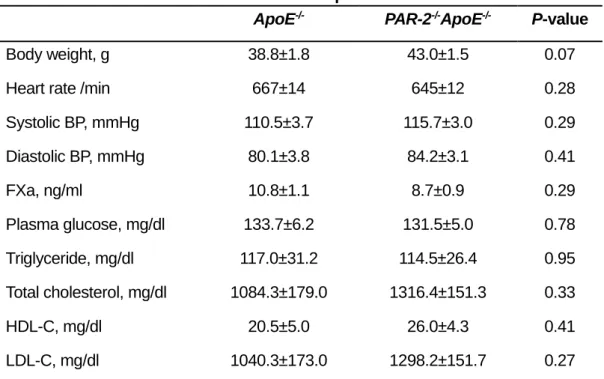
Table 1. Effects of PAR-2 deletion on metabolic parameters.
ApoE-/- PAR-2-/-ApoE-/- P-value
Body weight, g 38.8±1.8 43.0±1.5 0.07
Heart rate /min 667±14 645±12 0.28
Systolic BP, mmHg 110.5±3.7 115.7±3.0 0.29 Diastolic BP, mmHg 80.1±3.8 84.2±3.1 0.41 FXa, ng/ml 10.8±1.1 8.7±0.9 0.29 Plasma glucose, mg/dl 133.7±6.2 131.5±5.0 0.78 Triglyceride, mg/dl 117.0±31.2 114.5±26.4 0.95 Total cholesterol, mg/dl 1084.3±179.0 1316.4±151.3 0.33 HDL-C, mg/dl 20.5±5.0 26.0±4.3 0.41 LDL-C, mg/dl 1040.3±173.0 1298.2±151.7 0.27
All values are mean±SEM. ApoE‒/‒ indicates apolipoprotein E–deficient; BP, blood pressure, FXa,
activated factor X; HDL-C, high-density lipoprotein cholesterol; LDL-C, low-density lipoprotein cholesterol; and PAR-2, protease-activated receptor.
34
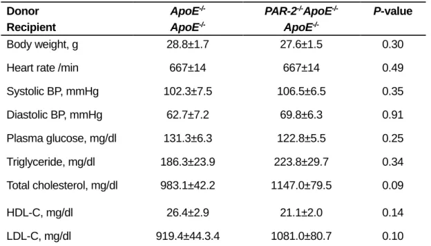
Table 2. Effects of PAR-2 deletion in BM on metabolic parameters. Donor Recipient ApoE-/- ApoE -/-PAR-2-/-ApoE-/- ApoE -/-P-value Body weight, g 28.8±1.7 27.6±1.5 0.30
Heart rate /min 667±14 667±14 0.49
Systolic BP, mmHg 102.3±7.5 106.5±6.5 0.35 Diastolic BP, mmHg 62.7±7.2 69.8±6.3 0.91 Plasma glucose, mg/dl 131.3±6.3 122.8±5.5 0.25 Triglyceride, mg/dl 186.3±23.9 223.8±29.7 0.34 Total cholesterol, mg/dl 983.1±42.2 1147.0±79.5 0.09 HDL-C, mg/dl 26.4±2.9 21.1±2.0 0.14 LDL-C, mg/dl 919.4±44.3.4 1081.0±80.7 0.10
All values are mean±SEM. ApoE‒/‒ indicates apolipoprotein E–deficient; BM, bone marrow; BP,
blood pressure; HDL-C, high-density lipoprotein cholesterol; LDL-C, low-density lipoprotein cholesterol; and PAR-2, protease-activated receptor.
35
Table 3. Patients' characteristics.
(N = 32) Age, yrs 69.6±1.1 Male/female 20 (63) / 12 (37) Smoking 19 (59) Hypertension 30 (94) HbA1c, % 6.1±0.2 Triglyceride, mg/dl 125±11 Total cholesterol, mg/dl 176±6 HDL-C, mg/dl 105±5 LDL-C, mg/dl 52±2
Data presented are mean±SEM or number (percentage). HDL-C indicates high-density lipoprotein cholesterol; and LDL-C, low-density lipoprotein cholesterol.
36
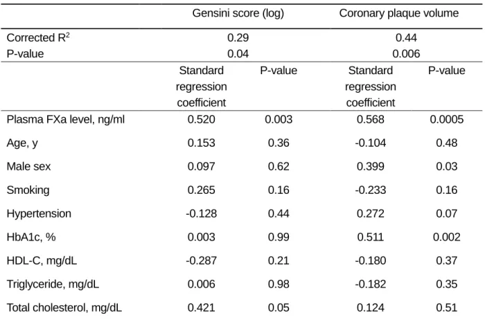
Table 4. Multivariate analysis for prediction of the severity of coronary atherosclerosis.
Gensini score (log) Coronary plaque volume
Corrected R2 P-value 0.29 0.04 0.44 0.006 Standard regression coefficient P-value Standard regression coefficient P-value
Plasma FXa level, ng/ml 0.520 0.003 0.568 0.0005
Age, y 0.153 0.36 -0.104 0.48 Male sex 0.097 0.62 0.399 0.03 Smoking 0.265 0.16 -0.233 0.16 Hypertension -0.128 0.44 0.272 0.07 HbA1c, % 0.003 0.99 0.511 0.002 HDL-C, mg/dL -0.287 0.21 -0.180 0.37 Triglyceride, mg/dL 0.006 0.98 -0.182 0.35 Total cholesterol, mg/dL 0.421 0.05 0.124 0.51