INTRODUCTION
Sp6, one of the Sp family members, is a tissue-specific transcription factor. Sp6 was previously called KLF14 (1). However, authentic KLF14 gene is localized at rat chromosome 2 and has been iden-tified to be different from Sp6 gene (2). Epiprofin is another name of Sp6 based on its gene
expres-sion pattern and function (3). In situ hybridization analysis has revealed Sp6 mRNA expression in tooth germ, limb bud, and hair follicle (3). In newborn mouse incisors, Sp6 mRNA is expressed in early stages of tooth development to the secretory stage of ameloblasts. Sp6 mRNA is also weakly expressed in mesenchymal odontoblasts of the incisor (3). Recently, it has been reported that Sp6 mRNA is strongly expressed in embryos compared with adult tissues, suggesting a functional role in development and tissue homeostasis (4). However, there is no in-formation about the levels of Sp6 protein expres-sion in various tissues in the embryo.
Sp6 has a unique domain structure, as shown
ORIGINAL
Sp6 downregulation of follistatin gene expression in
ameloblasts
Intan Ruspita, Keiko Miyoshi, Taro Muto, Kaori Abe, Taigo Horiguchi, and
Takafumi Noma
Department of Molecular Biology, Institute of Health Biosciences, The University of Tokushima Gradu-ate School, Tokushima, Japan
Abstract : Sp6 is a member of the Sp family of transcription factors that regulate a wide range of cellular functions, such as cell growth and differentiation. Sp6, also called epiprofin, is specifically expressed in tooth germ, limb bud, and hair follicle, but there is little in-formation on its function.
To investigate the possible role of Sp6 in tooth development, first we established an Sp6-overproducing clone, CHA9, and analyzed the features of the cell, including cell prolif-eration and gene expression. The parental cells of CHA9 are the ameloblast-lineage G5 cells that we previously established from rat dental epithelia of lower incisor. Sp6 over-production accelerated cell proliferation and induced the expression of ameloblastin mRNA, a marker of ameloblast differentiation. Second, we performed genome-wide screen-ing of Sp6 target genes by microarray analysis. Out of a total 20,450 genes, 448 genes were up-regulated and 500 genes were down-regulated by Sp6. We found the expression of fol-listatin, a BMP antagonist, to be 22.4-fold lower in CHA9 than in control cells. Transfec-tion of the Sp6-antisense construct into CHA9 cells restored follistatin expression back to equivalent levels seen in control cells, indicating that Sp6 regulates follistatin gene expression in ameloblasts.
Our findings demonstrate that the follistatin gene is one of the Sp6 target genes in ameloblasts and suggest that Sp6 promotes amelogenesis through inhibition of follista-tin gene expression. J. Med. Invest. 55 : 87-98, February, 2008
Keywords : ameloblast, amelogenesis, follistatin, microarray, Sp
Received for publication November 7, 2007 ; accepted Decem-ber 7, 2007.
Address correspondence and reprint requests to Takafumi Noma, M.D., Ph.D, Department of Molecular Biology, Institute of Health Biosciences, The University of Tokushima Graduate School, Kuramoto-cho, Tokushima 770-8504, Japan and Fax : +81-88-633-7326.
in Fig. 1A. It has three zinc finger motifs at the C-terminal region and two potential nuclear localiza-tion signals near the zinc finger domain. It is be-lieved to bind to GC- or GT-rich DNA sequences, sharing common binding characteristics of all Sp and KLF family proteins (5). A proline-rich domain, presumed to be a transcriptional regulatory domain, at the N-terminal region, is also a unique charac-teristic of Sp5, Sp6, and Sp7 among the Sp family members (5). This proline-rich domain may pro-vide unique functions in tooth development distinct from other Sp proteins (3). However, there is no information about the functional roles of Sp6 in transcriptional regulation involving target genes and interactive partners.
We explored the physiological roles of Sp6 in the developing tooth. We first prepared an antibody against rat Sp6 protein and confirmed the protein localization in the rat incisor. We also developed an Sp6-overproducing clone, CHA9. Next, we screened potential Sp6 target genes using microarray analy-sis by comparing expression in CHA9 with a con-trol clone, V1. Among many potential target genes, we found significant downregulation of follistatin gene, which is an extracellular glycoprotein that directly binds to bone morphogenic protein (BMP) and acts by antagonizing BMP signals (6). Trans-fection of Sp6-antisense expression plasmid into CHA9 cells restored follistatin gene expression to the same level as control. It has been reported that follistatin is responsible for the formation of an enamel-free area in the mouse incisor and molar by inhibiting ameloblast differentiation (7-10).
Taken together, our findings demonstrate that Sp6 has a role in amelogenesis by inducing ameloblastin expression and inhibiting follistatin expression.
MATERIALS AND METHODS
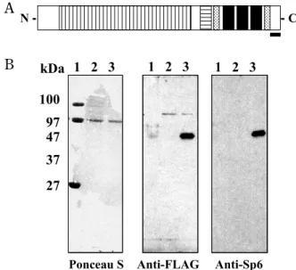
Generation of anti-Sp6 antibodyCarboxyl-terminal peptides of rat Sp6, CGKGK-REAEGSSASSN (XP_343962 ; amino acid 362-376), were conjugated with keyhole limpet hemocyanin (KLH) and used for immunization in rabbits (Fig. 1A) (Hokkaido System Sciences, Hokkaido, Japan). The specificity of antisera was analyzed by West-ern blotting of protein extracts of COS7 cells trans-fected with pCI-neo mammalian expression vector (Promega, Madison, WI) carrying the DNA frag-ment encoding FLAG-tagged rat Sp6 (Fig. 1B).
Immunohistochemistry and cytochemistry
Lower mandibles were dissected from postnatal day 1 stroke-prone spontaneous hypertensive rats (SHRSP) and fixed with 4% paraformaldehyde (PFA). Four-micrometer thick sections were deparaffinized, rehydrated, and treated with 3% H2O2in methanol
to inhibit endogenous peroxidase activity. Antigens were retrieved by heat treatment using antigen un-masking solution (Vector Laboratories, Burlingame, CA). The sections were blocked with 3% horse se-rum in phosphate-buffered saline (PBS) without calcium and magnesium [PBS(-) : 137 mM NaCl, 8.1 mM Na2HPO4!12H2O, 2.68 mM KCl, 1.47 mM
KH2PO4, pH 7.4] for 30 min at room temperature.
Sections were incubated with our rabbit polyclonal Sp6 antibody (1 : 2,000) or rabbit polyclonal anti-Amelogenin antibody (1 : 1,000 ; Hokudo, Hokkaido, Japan) at 4!!overnight. After washing with PBS(-) for several times, sections were incubated with horse-radish peroxidase-conjugated rabbit IgG anti-body (Histofine ; Nichirei, Tokyo, Japan) for 30 min at room temperature. 3-3’-diaminobenzidine (DAB) solution was used to detect immune-complex
ac-A A
B B
Fig. 1 Generation of rabbit polyclonal antibody against rat Sp6. (A) Structure of Sp6. Sp6 has several potential regulatory do-mains : a proline-rich region (vertical stripes), three zinc-finger domains (black boxes), and a Btd domain (horizontal stripes). Two candidate sequences for nuclear localization signals are also indicated (dotted pattern). The black bar indicates the position of the antigen epitope used to generate anti-Sp6 antibody. (B) Specificity of anti-Sp6 antibody. Western blot analysis was per-formed with protein extracts of COS7 cells transfected with pCI-neo mammalian expression vector carrying the DNA fragment encoding FLAG-tagged rat Sp6. Lane 1, molecular size marker ; lane 2, COS7 cells transfected with empty vector ; lane 3, COS7 cells transfected with FLAG-tagged rat Sp6 in pCI-neo.
cording to the manufacturer’s instructions (Nichirei). The sections were then counterstained with hema-toxylin. The stained sections were observed under a light microscope (BX51-34 FL-1-K-O ; Olympus, Tokyo, Japan).
COS7 cells were transfected with pCI-neo mam-malian expression vector (Promega, Madison, WI) carrying the DNA fragment encoding FLAG-tagged rat Sp6 or vector alone as a negative control, us-ing Lipofectamine plus reagent (Invitrogen, Carlsbad, CA) according to manufacturer’s instructions. Twenty-four hours after transfection, the cells were rinsed with PBS(-), fixed with ethanol for 10 min at room temperature, permeabilized by 0.1% Triton X-100 in PBS(-), and blocked with 4% BSA/PBS(-) for 30 min at room temperature. The cells were then in-cubated with mouse anti-FLAG M2 monoclonal an-tibody (1 : 500 ; 2 mg/ml ; Sigma, St. Louis, MO) for 1 hr at room temperature followed by incuba-tion with goat anti-mouse IgG (H + L) conjugated with Alexa Fluor-488 (Invitrogen). The cells were stained with 2 mg/ml of Hoechst 33342 (Sigma), and mounted in Vectashield mounting medium (Vec-tor Labora(Vec-tories, Burlingame, CA). The samples were observed under a fluorescence microscope (Olympus BX51 ; Olympus, Tokyo, Japan). The im-ages of both immunohistochemistry and cytochem-istry were captured by a digital imaging system (DP-70 set ; Olympus).
Establishment of Sp6-stable transformants
An Sp6 expression vector was constructed as follows. The C’- or N’-terminals of the Sp6 coding region were tagged with HA and ligated into pCIneo expression vector (Sp6-CHA or Sp6-NHA in pCIneo). Full-length coding regions of Sp6 fused with HA-tag and SpeI site at 3’-end were generated by PCR. The PCR product was cloned into pGEM-T easy vector (Promega) and confirmed by restriction en-zyme digestion and sequencing. The plasmid was named Sp6-CHA in pGEM-T easy. To construct an expression vector, Sp6-CHA fragment was produced by NotI digestion from Sp6-CHA in pGEM-T easy and re-cloned into pCI-neo vector (Promega). The final construct (Sp6-CHA in pCIneo) was confirmed by restriction enzyme digestion and sequencing.
Sp6-stable transformants were established using an ameloblast-lineage clone, G5, as described pre-viously (11). G5 cells were plated in 100 mm cul-ture dishes and maintained in Dulbecco’s modified Eagle’s medium (DMEM)/Ham’s F12 medium (Nissui, Tokyo, Japan) with 10% fetal bovine serum
(FBS ; JRH Biosciences, Lenexa, KS). Sp6-CHA, Sp6-NHA, and empty vector were stably transfected into G5 cells using Lipofectamine plus reagent as de-scribed in the section of Immunohistochemistry and cytochemistry in MATERIALS AND METHODS. For long-term growth, the cells were maintained with se-lection medium containing 1 mg/ml of G418. G418-resistant clones were isolated by limiting dilution.
Cell proliferation assay
To analyze cell growth activity, we performed an Alamar Blue assay (BioSource, Camarillo, CA) as described previously (12). In brief, the cells were inoculated in 96-well dishes (!103cells/well), the
medium was changed to fresh DMEM/Ham’s F12 without FBS after 24 h. Alamar blue reagent was added to each well to attain a final concentration of 10% (v/v). Cells were cultured for further 4 h. The absorbance of each sample was measured at 570/595 nm using a Multiskan JX microplate reader (Thermo Lab systems, Tokyo, Japan).
Western blot analysis
To identify an Sp6-overproducing clone, Western blot analysis was performed. Stably transformed cells were dissolved in 2X sodium dodecyl sulfate (SDS) sample buffer (125 mM Tris-HCl pH 6.8, 4% SDS, 10% glycerol). Protein concentration was de-termined using the BCA Protein Assay Kit (Pierce, Rockford, IL). After adding 10% 2-mercaptoethanol and 0.01% bromophenol blue, samples were boiled for 3 min at 95!!. Eighty micrograms of protein was resolved on 12.5% SDS-polyacrylamide gels. After electrophoresis, the proteins were transferred to a polyvinylidene difluoride (PVDF) membrane (ImmobilonTM-P ; Millipore, Bedford, MA) and
blocked with 5% (w/v) skim milk in Tris-buffered saline and Tween-20 (TBS-T ; 10 mM Tris-HCl, 150 mM NaCl, 0.05% (v/v) Tween-20, pH 8.0) at 4!!overnight. The membrane was incubated with anti-HA-probe Y11 (1 : 1,000 ; sc-805 ; Santa Cruz Biotechnology, Santa Cruz, CA) in the same buffer as that used for blocking, at room temperature for 1 h. After washing several times in TBS-T, the mem-brane was incubated with horseradish peroxidase-conjugated anti-rabbit IgG antibody (1 : 5,000 ; Am-ersham Biosciences, Buckinghamshire, UK) for 30 min at room temperature. After washing several times, the immune complex was detected using the ECL Plus western blotting detection reagents (Am-ersham Biosciences) according to the manufac-turer’s instructions.
Microarray analysis
Both V1 and CHA9 cells were harvested and total RNA was isolated using the RNeasy!" mini
kit (Qiagen Inc., Valencia, CA). cDNA was synthe-sized from 500 ng of total RNA using M-MLV Re-verse Transcriptase (Invitrogen) and was labeled with Cy3 and Cy5, respectively. The labeled probes were hybridized onto a rat oligo DNA microarray containing 22,450-oligo DNAs on the slide (Agilent Technologies, Santa Clara, CA). The arrays were scanned, and the intensity and ratio of the two fluorescent signals of Cy3 and Cy5 were analyzed by Agilent Feature Extraction software (Agilent). The raw data were unified to calibrate the systemic error between the Cy5 and Cy3 labeling steps us-ing the expression levels of the housekeepus-ing genes spotted on the array. Differential expression of the genes was determined by the following criteria : gene expression in CHA9 was considered to be
up-regulated if the intensity value was increased two folds compared to V1 (Cy5/Cy3!2). Conversely, gene expression was considered down-regulated when the ratio of Cy5/Cy3 signals is less than 0.5.
RT-PCR analysis
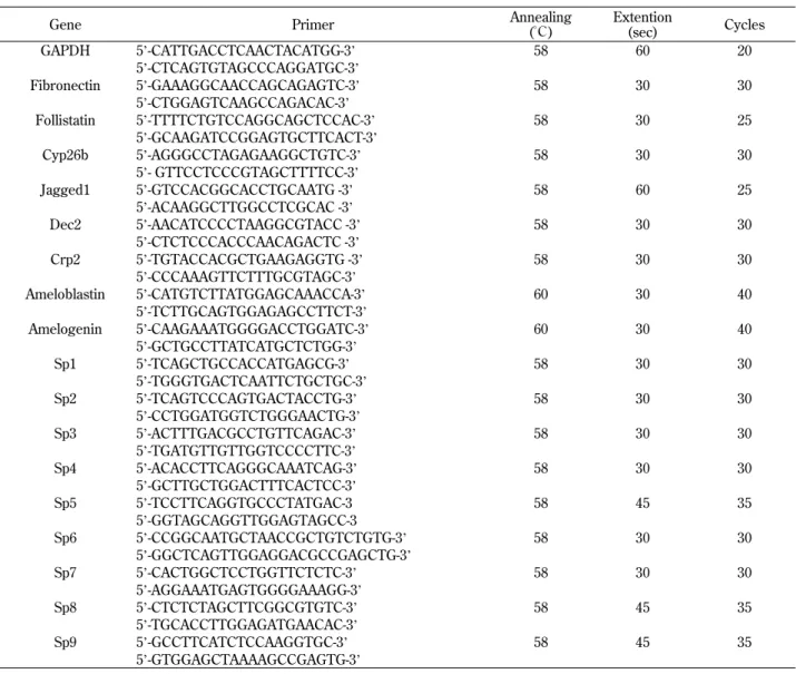
To confirm the microarray results, we selected several up-regulated and down-regulated genes that are related to the differentiation markers or signal-ing molecules includsignal-ing Crp2, a LIM-only protein (13) ; Dec2, a member of the basic helix-loop-helix superfamily (14) ; Cyp26b, a p450 isoform (15) ; follistatin, a BMP antagonist (7) ; and fibronectin, an extracellular matrix (16), and performed RT-PCR analysis with gene-specific primers. Five hundred nanograms of total RNA was used for reversed transcription with an RNA PCR kit (Takara, Shiga, Japan). The synthesized cDNA was used as a tem-plate for PCR. Gene-specific primers and PCR con-ditions are shown in Table 1.
Table 1 Gene-specific primers.
Gene Primer Annealing
("!) Extention (sec) Cycles GAPDH 5’-CATTGACCTCAACTACATGG-3’ 58 60 20 5’-CTCAGTGTAGCCCAGGATGC-3’ Fibronectin 5’-GAAAGGCAACCAGCAGAGTC-3’ 58 30 30 5’-CTGGAGTCAAGCCAGACAC-3’ Follistatin 5’-TTTTCTGTCCAGGCAGCTCCAC-3’ 58 30 25 5’-GCAAGATCCGGAGTGCTTCACT-3’ Cyp26b 5’-AGGGCCTAGAGAAGGCTGTC-3’ 58 30 30 5’- GTTCCTCCCGTAGCTTTTCC-3’ Jagged1 5’-GTCCACGGCACCTGCAATG -3’ 58 60 25 5’-ACAAGGCTTGGCCTCGCAC -3’ Dec2 5’-AACATCCCCTAAGGCGTACC -3’ 58 30 30 5’-CTCTCCCACCCAACAGACTC -3’ Crp2 5’-TGTACCACGCTGAAGAGGTG -3’ 58 30 30 5’-CCCAAAGTTCTTTGCGTAGC-3’ Ameloblastin 5’-CATGTCTTATGGAGCAAACCA-3’ 60 30 40 5’-TCTTGCAGTGGAGAGCCTTCT-3’ Amelogenin 5’-CAAGAAATGGGGACCTGGATC-3’ 60 30 40 5’-GCTGCCTTATCATGCTCTGG-3’ Sp1 5’-TCAGCTGCCACCATGAGCG-3’ 58 30 30 5’-TGGGTGACTCAATTCTGCTGC-3’ Sp2 5’-TCAGTCCCAGTGACTACCTG-3’ 58 30 30 5’-CCTGGATGGTCTGGGAACTG-3’ Sp3 5’-ACTTTGACGCCTGTTCAGAC-3’ 58 30 30 5’-TGATGTTGTTGGTCCCCTTC-3’ Sp4 5’-ACACCTTCAGGGCAAATCAG-3’ 58 30 30 5’-GCTTGCTGGACTTTCACTCC-3’ Sp5 5’-TCCTTCAGGTGCCCTATGAC-3 58 45 35 5’-GGTAGCAGGTTGGAGTAGCC-3 Sp6 5’-CCGGCAATGCTAACCGCTGTCTGTG-3’ 58 30 30 5’-GGCTCAGTTGGAGGACGCCGAGCTG-3’ Sp7 5’-CACTGGCTCCTGGTTCTCTC-3’ 58 30 30 5’-AGGAAATGAGTGGGGAAAGG-3’ Sp8 5’-CTCTCTAGCTTCGGCGTGTC-3’ 58 45 35 5’-TGCACCTTGGAGATGAACAC-3’ Sp9 5’-GCCTTCATCTCCAAGGTGC-3’ 58 45 35 5’-GTGGAGCTAAAAGCCGAGTG-3’
Transient transfection with Sp6-antisense construct
Sp6-antisense expression plasmid was constructed by inversely inserting the rat Sp6 coding DNA frag-ment into pCI neo vector. The Sp6-antisense con-struct was transiently transfected into CHA9 cells in 6-well plates, using Lipofectamine plus reagent (Invitrogen) as described in the section of Immu-nohistochemistry and cytochemistry in MATERI-ALS AND METHODS. After 24 h, at about 80% con-fluence, V1 or CHA9 cells were transfected with 1 μg of Sp6-antisense expression plasmid. Forty-eight hours later, cells were harvested and levels of
fol-listatin gene expression were determined by
RT-PCR.
RESULTS
Characterization of anti-Sp6 antibody
We first developed a rabbit polyclonal antibody against the C-terminal region of rat Sp6 epitope. To check the reactivity and specificity of this antibody, COS7 cells were transiently transfected with FLAG-tagged Sp6 or empty vector as a negative control. Cell lysates were electrophoresed and analyzed by immunoblotting. Both Sp6 and FLAG anti-bodies detected a discrete single band at the same molecular mass, 47 kDa, which was not detectable in the control, indicating that the antibody is spe-cific to Sp6 without background (Fig. 1B).
Localization of Sp6 in vivo
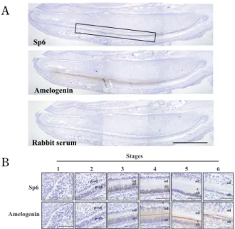
To examine the expression pattern of Sp6 in the tooth, we performed immunohistochemical analy-sis using rat lower incisors at postnatal day 6, be-cause all developmental stages can easily be seen. As shown in Fig. 2A, Sp6 was broadly detected in the ameloblast layer (Fig. 2A, upper). Amelogenin, one of the enamel proteins and a differentiation marker of amelogenesis, was also detected in the same lo-cation as that of Sp6 (Fig. 2A, middle). At high magnification, Sp6 was clearly detectable mainly in the nucleus of differentiating ameloblasts, which are tall, columnar, and polarized cells, and in some odontoblasts (Fig. 2B). Interestingly, Sp6 was not detected in all ameloblasts at the same differentiat-ing stages.
The intracelluar localization of Sp6 was also con-firmed in Sp6-transfected COS7 cells by immuno-cytochemistry with anti-FLAG M2 monoclonal an-tibody (Fig. 3). Fluorescent signals were detected
only in Sp6-transfected samples (Fig. 3A), whereas no fluorescence from the secondary antibody was detected in mock-transfected control with vector alone (Fig. 3B). The signals were detected in both cytoplasm and nucleus. The dotted-like fluores-cence signals were often observed in the nucleus (Fig. 3A).
Establishment of an Sp6-overproducing clone, CHA9
We prepared several HA-tagged Sp6 expression plasmids, and stably transformed an
ameloblast-A A
B B
Fig. 2 Localization of Sp6 protein in rat incisor.
(A) Overview of Sp6 localization in incisor of 6-day-old rat man-dible. Amelogenin and Sp6 were mainly detected in ameloblasts. Scale bar indicates 1.0 mm. The box indicated the area of posi-tion of Sp6-positive cells. (B) The stage-specific localizaposi-tion of Sp6 in amelogenesis. Stage 1, undifferentiated ; stage 2, pre-ameloblast ; stage 3, pre-secretory ; stage 4, secretory ; stage 5, maturation ; stage 6, reduced. From the pre-ameloblast to the maturation stages, Sp6 was detected mainly in the nucleus and weakly in cytosol. Sp6 was also detected in some odontoblasts. enamel ; e, odontoblasts ; od, ameloblasts ; ab, preodontoblasts ; p-od, preameloblasts ; p-ab. Scale bar indicates 100 μm.
Fig. 3 Subcellular localization of Sp6.
COS7 cells were transfected with pCI-neo vector carrying FLAG-tagged SP6 (A), or mock vector as a negative control (B). Sp6 was detected by anti-FLAG antibody and Alexa 488-conjugated secondary antibody. Green color indicates Sp6 localization and blue color indicates nucleus stained with Hoechst 33342.
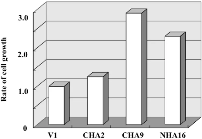
lineage cell line, G5 (11). More than 100 clones were selected by G418 and established. The level of Sp6 production was screened by immunoblot analysis and the highest Sp6 producer, CHA9, was selected (Fig. 4, Lane 4). The effects of Sp6 over-production on cell growth were investigated in some representative clones. CHA9 cells had the most ac-celerated growth rate compared with other clones as shown in Fig. 5. We next examined the effects of Sp6 overproduction on the gene expression of other Sp family members, since they are involved in growth regulation, differentiation, signal transduc-tion, and morphogenesis (3, 17-25). The expression
levels of other Sp family genes were not affected in CHA9 cells, except for Sp8 that was weakly in-duced (Fig. 6A). Sp5, Sp7, and Sp9 were not ex-pressed in CHA9 and V1 cells (Fig. 6A). Levels of mRNA of ameloblastin and amelogenin, two major enamel proteins that are ameloblast differentiation markers, were also assessed by RT-PCR analysis (Fig. 6B). The level of ameloblastin mRNA was sig-nificantly increased in CHA9 cells, although that of amelogenin was not changed.
Fig. 4 Screening for Sp6 high producer clone.
(A) Lane 1, Sp6-CHA in pCIneo transfected into COS7 cells as a positive control ; lane 2, empty vector transfected into G5 cells as a negative control ; lane 3, Sp6-CHA in pCI-neo transfected clone 2 (CHA2) ; lane 4, Sp6-CHA in pCI-neo transfected clone 9 (CHA9) ; lane 5, Sp6-NHA in pCI-neo transfected clone 16 (NHA16). A representative result is shown. Ponceau S staining was performed as a loading control. Asterisk ; non-specific band, arrowhead ; Sp6 protein.
Fig. 5 Effects of Sp6 overproduction on cell growth.
The rate of cell growth was determined by Alamar Blue analy-sis. Vertical axis indicates the relative rate of cell growth com-pared with empty vector-transfected G5 (V1). CHA2, CHA9, and NHA16 corresponded to the transformants that were transfected with different tagged Sp6 expression constructs. The highest Sp6-producer clone, CHA9, had the most accelerated cell growth.
A A
B B
Fig. 6 Gene expression patterns of Sp family members and enamel proteins.
(A) Expression levels of Sp family members determined by RT-PCR. Lane 1, negative control (template ; water) ; lane 2, nega-tive control (template ; no reverse-transcriptase, RT(-)) ; lane 3, control (template ;V1), lane 4, Sp6 overproducer (template ; CHA9) ; lane 5, positive control for PCR (template ; rat day 6 molar).
(B) Expression levels of amelogenin and ameloblastin determined by RT-PCR on the same samples used in (A).
Identification of possible Sp6 target genes by mi-croarray analysis
To identify the possible target genes of Sp6, we screened a total of 20,450 genes by microarray analy-sis. RNA samples were prepared from the Sp6-overproducing clone, CHA9, and the control clone, V1, and hybridized onto the specific oligonucleo-tides to each gene in the microarray. Differentially expressed genes were selected on the basis of a greater than 2-fold or less than 0.5-fold difference in the expression levels between CHA9 and V1 cells. We found that 448 genes were up-regulated and 500 genes were down-regulated by Sp6 overpro-duction in ameloblast-lineage clone, and these are summarized in Table 2. The regulated genes were
also classified by major cellular functions, as shown in Table 3. Both top 30 up-regulated and down-regulated genes are listed in Table 4 and 5, respec-tively.
Table 2 Summary of Sp6 gene regulation by microarray analysis. Known Unknown Total
Up 306 142 448
Down 382 118 500
Total 688 260 948
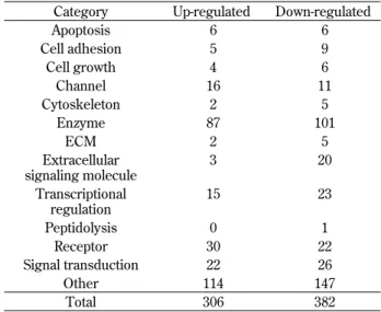
Table 3 Functional classification of Sp6 potential target genes. Category Up-regulated Down-regulated
Apoptosis 6 6 Cell adhesion 5 9 Cell growth 4 6 Channel 16 11 Cytoskeleton 2 5 Enzyme 87 101 ECM 2 5 Extracellular signaling molecule 3 20 Transcriptional regulation 15 23 Peptidolysis 0 1 Receptor 30 22 Signal transduction 22 26 Other 114 147 Total 306 382
Table 4 Up-regulated genes in CHA9.
# up-regulated genes C9/V1
(fold) 1 Q 60501 Hamster p7 preinsertion DNA [TC540005] 25.1 2 Rat alpha-methylacyl-CoA racemase, mRNA [NM_012816] 24.9 3 Rat endothelin receptor type B, mRNA [NM_017333] 19.5 4 Rat gap junction membrane channel protein bet, mRNA [NM_001004099] 18.6 5 Rat glutamate receptor interacting protein 1, mRNA [NM_032069] 17.5 6 Rat collagen, type V, alpha 3, mRNA [NM_021760] 16.8 7 Rat lectin, galactose binding, soluble 7, mRNA [NM_022582] 16.3
8 Rat Dock1 protein, mRNA [XM_219424] 16.3
9 Rat surfactant associated protein C, mRNA [NM_017342] 13.2
10 Q8R4C3 Csr1[TC 556501] 11.3
11 Rat liver glycogen phosphorylase, mRNA [NM_022268] 11.2
12 MOUSE P2Y purinoceptor 5 [TC521815] 10.8
13 Rat serine/threonine kinase 39, STE20/SPS1 homolog, mRNA [NM_019362] 10.5 14 Rat nuclear receptor subfamily 3, group C, member 2, mRNA [NM_013131] 10.4 15 Rat brain derived neurotrophic factor, mRNA [NM_012513] 10.0 16 Rat glutathione-S-transferase, alpha type2, mRNA [NM_017013] 10.0 17 Rat solute carrier family 1, member 3, mRNA [NM_019225] 9.2 18 Rat glutathione S-transferase, alpha 4, mRNA [XM_217195] 9.0
19 MOUSE Asporin [TC557668] 9.0
20 Rat calcium channel, voltage-dependent, alpha2/delta subunit 1, mRNA [NM_012919] 8.6 21 Rat cytosolic cysteine dioxygenase 1, mRNA [NM_052809] 8.6 22 Rat sialyltransferase 10 (alpha-2,3-sialyltransferase VI), mRNA [NM_207602] 8.5 23 Rat basic helix-loop-helix domain containing, class B3 (Dec2), mRNA [NM_133303] 8.4 24 Q70US4 NADH dehydrogenase 6 [TC525243] 8.4 25 Rat transmembrane 7 superfamily member 3, mRNA [NM_001011970] 8.2 26 Rat leukotriene B4 12-hydroxydehydrogenase, mRNA [NM_138863] 7.7
27 Rat desmoglein 5, mRNA [XM_214616] 7.4
28 Rat carbohydrate (keratan sulfate Gal-6) sulfotransferase 1, mRNA [NM_001011955] 7.4 29 Rat glutamate cysteine ligase, modifier subunit, mRNA [NM_017305] 7.0 30 Rat NEDD9 interacting protein with calponin homology and LIM domains, mRNA [XM_215424] 6.8
Validation of Sp6-regulated genes by RT-PCR
RT-PCR analysis showed that expression levels of Jagged 1, Crp2, and Dec2 were enhanced in CHA9 cells compared with V1 (Fig. 7A), whereas those of Cyp 26b, follistatin, and fibronectin were reduced (Fig. 7B). Especially, the expression of follistatin was greatly down-regulated by Sp6 overexpression.
Follistatin gene expression restored by Sp6-antisense construct
In order to establish whether Sp6 directly regu-lates follistatin gene expression, a loss-of-function experiment was performed by transfecting an Sp6-antisense expression plasmid into CHA9 and V1 cells. After transfection, total RNA was isolated and the follistatin gene expression was analyzed by RT-PCR. The results showed that Sp6 knockdown re-stored follistatin gene expression to almost the same level as that in controls (Fig. 8A and B). We also analyzed whether ameloblastin gene expression was down-regulated by Sp6 knockdown technique. The level of ameloblastin mRNA in Sp6-antisense treated CHA9 cells was 76 % reduced compared to that of no treated CHA9 cells (data not shown). The lev-els of Sp6 protein were reciprocally correlated with follistatin gene expression (Fig. 8C). In V1 cells, on the other hand, transfection with Sp6-antisense expression plasmid did not cause any significant changes to follistatin expression levels. Sp6 protein A
A BB
Fig. 7 Confirmation of microarray results by RT-PCR. (A) Representative up-regulated genes in CHA9. RT-PCR was performed on the same samples as in Fig. 6. Lane 1, negative con-trol (RT(-)) ; lane 2, V1 ; lane 3, CHA9.
(B) Representative down-regulated genes in CHA9. Lane 1, nega-tive control (RT(-)) ; lane 2, V1 ; lane 3, CHA9.
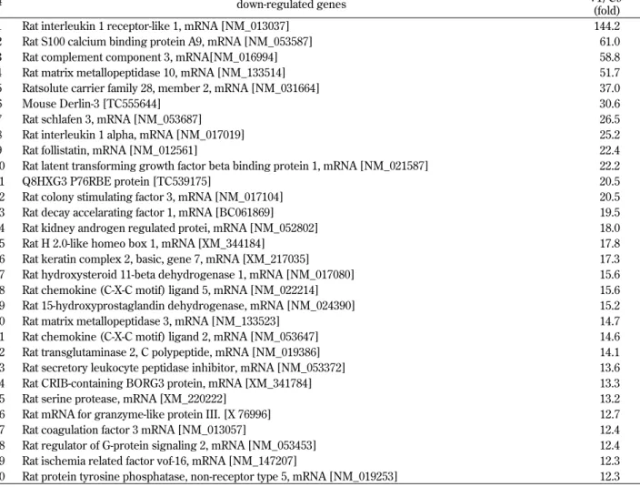
Table 5 Down-regulated genes in CHA9.
# down-regulated genes V1/C9
(fold) 1 Rat interleukin 1 receptor-like 1, mRNA [NM_013037] 144.2 2 Rat S100 calcium binding protein A9, mRNA [NM_053587] 61.0 3 Rat complement component 3, mRNA[NM_016994] 58.8 4 Rat matrix metallopeptidase 10, mRNA [NM_133514] 51.7 5 Ratsolute carrier family 28, member 2, mRNA [NM_031664] 37.0
6 Mouse Derlin-3 [TC555644] 30.6
7 Rat schlafen 3, mRNA [NM_053687] 26.5
8 Rat interleukin 1 alpha, mRNA [NM_017019] 25.2
9 Rat follistatin, mRNA [NM_012561] 22.4
10 Rat latent transforming growth factor beta binding protein 1, mRNA [NM_021587] 22.2
11 Q8HXG3 P76RBE protein [TC539175] 20.5
12 Rat colony stimulating factor 3, mRNA [NM_017104] 20.5 13 Rat decay accelarating factor 1, mRNA [BC061869] 19.5 14 Rat kidney androgen regulated protei, mRNA [NM_052802] 18.0 15 Rat H 2.0-like homeo box 1, mRNA [XM_344184] 17.8 16 Rat keratin complex 2, basic, gene 7, mRNA [XM_217035] 17.3 17 Rat hydroxysteroid 11-beta dehydrogenase 1, mRNA [NM_017080] 15.6 18 Rat chemokine (C-X-C motif) ligand 5, mRNA [NM_022214] 15.6 19 Rat 15-hydroxyprostaglandin dehydrogenase, mRNA [NM_024390] 15.2 20 Rat matrix metallopeptidase 3, mRNA [NM_133523] 14.7 21 Rat chemokine (C-X-C motif) ligand 2, mRNA [NM_053647] 14.6 22 Rat transglutaminase 2, C polypeptide, mRNA [NM_019386] 14.1 23 Rat secretory leukocyte peptidase inhibitor, mRNA [NM_053372] 13.6 24 Rat CRIB-containing BORG3 protein, mRNA [XM_341784] 13.3 25 Rat serine protease, mRNA [XM_220222] 13.2 26 Rat mRNA for granzyme-like protein III. [X 76996] 12.7 27 Rat coagulation factor 3 mRNA [NM_013057] 12.4 28 Rat regulator of G-protein signaling 2, mRNA [NM_053453] 12.4 29 Rat ischemia related factor vof-16, mRNA [NM_147207] 12.3 30 Rat protein tyrosine phosphatase, non-receptor type 5, mRNA [NM_019253] 12.3
was not detected in V1 cells although Sp6 mRNA was weakly detected as shown in Fig. 6A.
DISCUSSION
Sp6 is widely believed to be a DNA-binding tran-scription factor that may have diverse roles in tooth development and morphogenesis (3, 5). Sp6 mRNA was found to be expressed in a tissue-specific man-ner in tooth germ, hair follicle, and limb bud (3). However, there is no evidence that Sp6 is a tran-scription factor.
In this study, we first developed an antibody against Sp6 protein (Fig. 1). By immunohistochemi-cal analysis, we detected Sp6 expression mainly in the nucleus of ameloblasts in the presecretory and secretory stages, and in some odontoblasts (Fig. 2B). Interestingly, we found that amelogenin expression was localized to the same region as that of Sp6 ex-pression (Fig. 2A, middle). Next, we investigated Sp6 target genes with the combined methods of gain of function analysis and microarray analysis. We established an Sp6-overproducing clone, CHA9,
which was transformed by Sp6 expression plasmid using ameloblast-lineage clone, G5 (11). CHA9 cells grow relatively much faster than control cells, which is consistent with previous reports on the cloning of Sp6 (3). Analysis for the effects of Sp6 overproduc-tion on the gene expression of other Sp family mem-bers revealed no gross difference in CHA9 cells compared with control, V1 cells, except for that of Sp8 that was slightly induced in CHA9 cells (Fig. 6A). The physiological role of Sp8 in ameloblasts remains to be determined, although it has been re-ported that Sp8 is expressed during brain and limb bud development and demonstrated to regulate cor-tical patterning and limb outgrowth through FGF8 induction (26, 27). In addition, gene expression of ameloblastin, one of the enamel-producing proteins, was significantly up-regulated in CHA9 cells com-pared with V1 cells (Fig. 6B), although the expres-sion of amelogenin gene was not changed.
These findings suggested that Sp6 may have a regulatory role in tooth development and that Sp6 targets specific genes. We attempted to identify these target genes by pulling out differentially expressed genes by microarray analysis using RNA samples A
A
B
B CC
Fig. 8 Restoration of follistatin gene expression by Sp6 knockdown in CHA9 cells.
Sp6-antisense construct was transiently transfected (+) or not (-) into V1 and CHA9 cells, respectively. (A) RT-PCR results of follista-tin gene expression. (B) Relative ratio of follistafollista-tin gene expression. RT-PCR products in (A) were measured by Quantity One (BIO-RAD). (C) The effect of Sp6-knockdown on Sp6 protein level. Upper panel shows western blot analysis. Lower panel shows Ponceau S staining pattern as a loading control. Sp6 protein was detected by anti-Sp6 antibody (arrowhead). +C ; a positive control (same as lane 1 in Fig.4), M ; molecular size marker.
from CHA9 and V1 cells. A total of 948 genes were found to differ in expression levels by more than 2-fold or less than 0.5-2-fold, and these were considered to be potential Sp6 target genes (Table 2). The genes could be divided into several functional categories, including transcriptional regulation, cell cycle/apop-tosis, metabolism, cell adhesion, enzymatic, ion chan-nel, cytoskeletal, extracellular matrix, extracellular signaling, proteolysis, receptor, and signal trans-duction (Table 3), suggesting many diverse roles for Sp6. Some of the up-regulated and down-regulated genes were related to tooth development. Among these, we focused on follistatin gene since it was greatly suppressed by Sp6 expression and also it has been reported that follistatin may inhibit ameloblast differentiation (7, 8). Follistatin is localized in enamel-free areas of mouse incisors, and follistatin transgenic mouse caused enamel defect in incisor (7, 8). Furthermore, follistatin knockout mouse showed no enamel free area of the incisors (7, 8). In this study, we found that ameloblastin gene ex-pression was enhanced by Sp6 (Fig. 6B), and that Sp6 was co-localized with amelogenin in enamel-forming regions (Fig. 2), suggesting a functional involvement of Sp6 in enamel formation and amelo-genesis. We also found that transfection of an Sp6-antisense construct restored follistatin gene expres-sion in CHA9 cells almost to the levels of V1 cells, supporting a role of Sp6 in the regulation of follis-tatin gene expression.
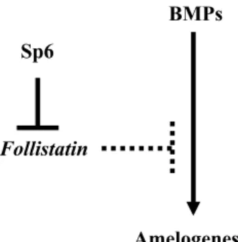
Taken together, our findings suggest that Sp6 may regulate amelogenesis through the downregu-lation of follistatin gene expression. This is consistent with previous reports that demonstrated the involve-ment of follistatin regulation in BMP-mediated amelogenesis (7, 8). Follistatin is also known as an antagonist for activin. There are several reports to suggest a role of activin in tooth development (7, 9, 28-30). However, the expression of activin has been limited at the very early stage in the dental epithelial cells, and at the late stage in mesenchy-mal cells during tooth development (7, 9, 28-30). These findings suggested that follistatin may func-tion in antagonizing BMPs in ameloblasts.
Figure 9 shows the interaction between Sp6, fol-listatin, and BMP in amelogenesis. We propose a role for Sp6 in amelogenesis through the downregu-lation of follistatin gene expression during tooth development and morphogenesis.
ACKNOWLEDGMENTS
This work was partly supported by Grants-in-Aid for scientific research (Nos. 17689051 and 18791368) from the Ministry of Education, Culture, Sports, Sci-ence and Technology, Japan. We thank Ms. Mami Sawatari for technical assistance.
REFERENCES
1. Scohy S, Gabant P, Van reeth T, Hertveldt V, Dreze PL, Van vooren F, Riviere M, Szpirer J, Szpirer C : Identification of KLF 13 and KLF 14 (Sp6), a novel members of Sp/XKLF tran-scription factor family. Genomics 70 : 93-101, 2000
2. Kaczynski J, Cook T, Urrutia R : Sp1-and krup-pel like transcription factors. Genomes Biol 4 : 206, 2003
3. Nakamura T, Unda F, de-Vega S, Vilaxa A, Fukumoto S, Yamada KM, Yamada Y : The Kruppel-like factor epiprofin is expressed by epithelium of developing teeth, hair follicles, and limb buds and promotes cell proliferation. J Biol Chem 279 : 626-634, 2004
4. Hertveldt V, De Mees C, Scohy S, Van Vooren P, Szpirer J, Szpirer C : The Sp6 locus uses sev-eral promoters and generates sense and an-tisense transcripts. Biochimie 89 : 1381-1387, 2007
5. Bouwman P, Philipsen S : Regulation of the ac-tivity of Sp1-related transcription factors. Mol Cell Endocrinol 195 : 27-38, 2002
6. Iemura S, Yamamoto TS, Takagi C, Uchiyama H, Natsume T, Shimasaki S, Sugino H, Ueno N : Direct binding of follistatin to a complex of
Fig. 9 Proposed model for the role of Sp6 in amelogenesis. Based on our gain-of-function and loss-of-function data, we pro-pose a role for Sp6 in amelogenesis through down-regulation of follistatin gene expression. Sp6 downregulates follistatin gene ex-pression, then it may promote amelogenesis.
bone-morphogenetic protein and its receptor inhibits ventral and epidermal cell fates in early Xenopus embryo. Proc Natl Acad Sci USA 95 : 9337-9342, 1998
7. Wang XP, Suomalainen M, Jorgez CJ, Matzuk MM, Werner S, Thesleff I : Follistatin regu-lates enamel patterning in mouse incisors by asymmetrically inhibiting BMP signaling and ameloblast differentiation. Dev Cell 7 : 719-730, 2004
8. Wang XP, Suomalainen M, Jorgez CJ, Matzuk MM, Wankell M, Werner S, Thesleff I : Modu-lation of activin/bone morphogenetic protein signaling by follistatin is required for the mor-phogenesis of mouse molar teeth. Dev Dyn 231 : 98 -108, 2004
9. Wang XP, Suomalainen M, Felszeghy S, Zelarayan LC, Alonso MT, Plikus MV, Maas RL, Chuong CM, Schimmang T, Thesleff I : An integrated gene regulatory network controls stem cell proliferation in teeth. PLoS Biol 6 : 1325-1333, 2007
10. Thesleff I, Wang XP, Suomalainen M : Regu-lation of epithelial stem cells in tooth regen-eration. C R Biol 330 : 561-564, 2007
11. Abe K, Miyoshi K, Muto T, Ruspita I, Horiguchi T, Nagata T, Noma T : Establishment and char-acterization of rat dental epithelial derived amelo-blast-lineage clones. J Biosci Bioeng 103 : 479-485, 2007
12. Ueno A, Miwa Y, Miyoshi K, Horiguchi T, Inoue H, Ruspita I, Abe K, Yamashita K, Hayashi E, Noma T : Constitutive expression of throm-bospondin 1 in MC3T3-E1 osteoblastic cells inhibit mineralization. J Cell Physiol 209 : 322-332, 2006
13. Weiskirchen R, Pino JD, Macalma T, Bister K, Beckerle MC : The cysteine-rich protein fam-ily of highly related LIM domain proteins. J Biol Chem 270 : 28946-28954, 1995
14. Fujimoto K, Shen M, Noshiro M, Matsubara K, Shingu S, Honda K, Yoshida E, Suardita K, Matsuda Y, Kato Y : Molecular cloning and characterization of DEC2, a new member of basic helix-loop-helix proteins. Biochem Bio-phys Res Commun 280 : 164-171, 2001
15. Nelson DR : A second CYP26 P450 in humans and zebrafish : CYP26B1. Arch Biochem Bio-phys 371 : 345-347, 1999
16. Thesleff I, Vainio S, Jalkanen M : Cell-matrix interactions in tooth development. Int J Dev Biol 33 : 91-97, 1989
17. Liu Y, Zhong X, Li W, Brattain MG, Banerji SS : The role of Sp1 in the differential expres-sion of transforming growth factor-beta recep-tor type II in human breast adenocarcinoma MCF-7 cells. J Biol Chem 275 : 12231-12236, 2000
18. Abdelrahim M, Smith R, 3rd, Burghardt R, Safe S : Role of Sp proteins in regulation of vas-cular endothelial growth factor expression and proliferation of pancreatic cancer cells. Cancer Res 64 : 6740-6749, 2004
19. Safe S, Abdelrahim M : Sp transcription factor family and its role in cancer. Eur J Cancer 41 : 2438-2448, 2005
20. Supp DM, Witte DP, Branford WW, Smith EP, Potter SS : Sp4, a member of the Sp1-family of zinc finger transcription factors, is required for normal murine growth, viability, and male fertility. Dev Biol 176 : 284-299, 1996
21. Philipsen S, Suske G : A tale of three fingers : the family of mammalian Sp/XKLF transcrip-tion factors. Nucleic Acids Res 27 : 2991-3000, 1999
22. Zheng L, Iohara K, Ishikawa M, Into T, Takano-Yamamoto T, Matsushita K, Nakashima M : Runx3 negatively regulates Osterix expression in dental pulp cells. Biochem J 405 : 69-75, 2007 23. Bouwman P, Gollner H, Elsasser HP, Eckhoff G, Karis A, Grosveld F, Philipsen S, Suske G : Transcription factor Sp3 is essential for post-natal survival and late tooth development. EMBO J 19 : 655-661, 2000
24. Lv P, Jia HT, Gao XJ : Immunohistochemical localization of transcription factor Sp3 during dental enamel development in rat tooth germ. Eur J Oral Sci 114 : 93-95, 2006
25. Milona MA, Gough JE, Edgar AJ : Expression of alternatively spliced isoforms of human Sp7 in osteoblast-like cells. BMC Genomics 4 : 43, 2003
26. Griesel G, Treichel D, Collombat P, Krull J, Zembrzycki A, van den Akker WM, Gruss P, Simeone A, Mansouri A : Sp8 controls the an-teroposterior patterning at the midbrain-hind-brain border. Development 133 : 1779-1787, 2006 27. Kawakami Y, Esteban CR, Matsui T, Rodríguez-León J, Kato S, Belmonte JC : Sp8 and Sp9, two closely related buttonhead-like transcription factors, regulate Fgf8 expression and limb out-growth in vertebrate embryos. Development 31 : 4763-4774, 2004
pat-terns of bone morphogenetic proteins (Bmps) in the developing mouse tooth suggest roles in morphogenesis and cell differentiation. Dev Dyn 210 : 383-396, 1997
29. Heikinheimo K, Begue-Kirn C,Ritvos O, Tuuri T, Ruch JV : The activin-binding protein follis-tatin is expressed in developing murine molar and induces odontoblast-like cell
differentia-tion in vitro. J Dent Res 76 : 1625-1636, 1997 30. Ferguson CA, Tucker AS, Christensen L, Lau
AL, Matzuk MM, Sharpe PT : Activin is an es-sential early mesenchymal signal in tooth de-velopment that is required for patterning of murine dentition. Genes Dev 12 : 2636-2649, 1998