Portal Venous Gas in Adults:
Clinical Significance, Management, and Outcomes of 25 Consecutive Patients
Masatoku Arai, Shiei Kim, Hiromoto Ishii,
Toru Takiguchi and Hiroyuki Yokota
Department of Emergency & Critical Care Medicine, Nippon Medical School, Tokyo, Japan
Background: Portal venous gas (PVG) is a rare finding and has a grave prognosis. The most common
and critical underlying pathology of PVG is bowel necrosis. However, bowel necrosis is sometimes diffi-cult to accurately diagnose. We retrospectively analyzed data from patients that contributed to the deci-sion to perform emergency surgery and bowel resection.
Methods: Between 2009 and 2019, 25 consecutive adult patients with PVG were identified
retrospec-tively and divided into the Operation and Non-operation groups. The Operation group was further subdivided into the Bowel resection and Non-resection groups. Clinical, laboratory, and radiographic variables were analyzed.
Results: Conservative management was successful for 32% (8/25) of patients (Non-operation group:
mortality 0%); 68% (17/25) were treated surgically (Operation group: mortality 35.3%). In the Operation group, 52.9% (9/17) underwent bowel resection (Bowel resection group: mortality 55.6%); however, bowel resection was unnecessary in 47.1% (8/17) of cases (Non-resection group: mortality 12.5%). Uni-variate analysis revealed significant differences between the Operation and Non-operation groups in GCS, APACHE II, abdominal distention, CRP, lactate, and CT findings of bowel dilatation, pneumatosis intestinalis, and attenuation of contrast effects of the bowel wall. However, with the exception of GCS, there was no significant difference between the Bowel resection and Non-resection groups.
Conclusions: Analysis of clinical, laboratory, and radiographic variables can inform decisions on
conser-vative management. However, 47.1% of the present patients who underwent surgery for suspected bowel necrosis did not require bowel resection, suggesting that this approach alone may not be suffi-cient to avoid non-therapeutic laparotomy. A new approach should be developed to improve this situ-ation. (J Nippon Med Sch 2021; 88: 88―96)
Key words: portal venous gas, pneumatosis intestinalis, mesenteric ischemia, bowel necrosis
Introduction
Portal venous gas (PVG) is a rare finding associated with a grave prognosis1―3
. In 1978, Liebman et al. reported that the mortality rate of patients with PVG was 75%3
. How-ever, recent improvements in imaging modalities, includ-ing CT and ultrasonography, allow detection of even small amounts of PVG, resulting in an increased number of reported cases and a decreased rate of overall mortal-ity1,2,4,5
.
PVG is associated with various pathological conditions,
and clinical significance ranges from benign findings to bowel necrosis1,2,4―8
. The most common underlying pathol-ogy of PVG includes bowel necrosis1―7
. Bowel necrosis is the most critical condition associated with PVG and re-quires emergency laparotomy1―7
. Although decision-making regarding clinical management of PVG is based on a combination of factors, including clinical symptoms, physical examination, laboratory data, and CT2,6,9
, bowel necrosis is sometimes difficult to accurately diagnose, which can affect decisions regarding the performance of
Correspondence to Masatoku Arai, MD, PhD, Department of Emergency & Critical Care Medicine, Nippon Medical School, 1― 1―5 Sendagi, Bunkyo-ku, Tokyo 113―8603, Japan
E-mail: [email protected]
https://doi.org/10.1272/jnms.JNMS.2021_88-201 Journal Website (https://www.nms.ac.jp/sh/jnms/)
Fig. 1 CT scan of a patient with portal venous gas. a: A coronal CT scan showed portal venous gas (PVG) in the liver and pneumatosis intestinalis (PI) throughout the stomach and small intestine. b: An axial CT scan showed PVG in the right and left portal vein (white arrows). c: Air is present in the extrahepatic portal vein and splenic vein (white arrows). This patient underwent emergency laparotomy; how-ever, bowel resection was not needed. He was discharged from our hospital.
emergency surgery and bowel resection4―6,9
. This study retrospectively analyzed clinical, laboratory, and radio-graphic variables contributing to a decision to perform emergency surgery in patients with PVG and assessed the validity of this approach for the clinical management of PVG.
Materials and Methods
This study was approved by the Institutional Review Board of Nippon Medical School Hospital (30-01-1063). The study sample included all patients admitted to the Department of Emergency and Critical Care Medicine of Nippon Medical School during the period from July 2009 through June 2019 with PVG identified on CT scans (Fig.
1). Patients were identified based on comments on CT
findings by staff radiologists, which were included in electronic medical records. Patients younger than 18 years and those who underwent cardiopulmonary resus-citation were excluded. Patients’ medical records were re-viewed to obtain information on demographics, medical history, comorbidities, underlying diseases, early symp-toms, vital signs, CT findings, arterial blood gas analysis data, laboratory data, management, length of hospital
stay, and mortality. Arterial blood gas analysis data and all laboratory data were values measured at the time of PVG diagnosis based on CT findings before the decision to perform emergent surgery (!2 hours before and after performing CT). The severity of illness and the expected risk of hospital mortality were evaluated by calculating the Acute Physiology and Chronic Health Evaluation (APACHE) II score10
.
All patients received PVG diagnoses based on CT find-ings. Decisions regarding clinical management of PVG were made by the attending surgeon and were based on a combination of factors, including clinical symptoms, physical examination, laboratory data, underlying dis-ease, and CT findings. Surgical intervention was per-formed for patients with clinical signs and/or laboratory data suggesting intra-abdominal pathologies that were consistent with radiographic abnormalities, such as bowel obstruction, perforation, and peritonitis. When a combination of clinical, laboratory, and radiographic vari-ables suggested bowel necrosis, surgical exploration was performed without a conclusive diagnosis because of the high mortality rate1―6
. We performed bowel resection only in patients with transmural bowel necrosis when this
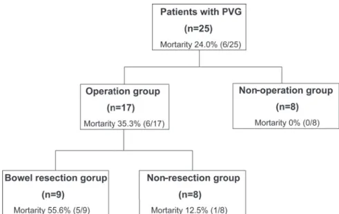
op-Fig. 2 Allocation of patients with portal venous gas, and the mortality rates of each group.
Patients were divided into 2 groups based on whether an emergency opera-tion was performed (Operaopera-tion group, n=17; Non-operaopera-tion group, n=8). The Operation group was further subdivided into 2 groups based on whether bowel resection was performed (Bowel resection group, n=9; Non-resection group, n=8)
-erative finding was observed during exploration.
Patients were classified into 2 groups in relation to whether emergency surgery was performed (Operation group, n=17; Non-operation group, n=8). There were no patients who was not offered operative intervention as a result of futility or refused to give the consent for sur-gery. The Operation group was further subdivided into 2 groups based on whether or not bowel resection was per-formed (Bowel resection group, n=9; Non-resection group, n=8) (Fig. 2). In the Bowel resection group, a di-agnosis of bowel wall necrosis was confirmed by review-ing postoperative histological findreview-ings.
Statistical analyses were performed with the SPSS soft-ware program (SPSS Inc., Chicago, IL, USA). Continuous variables are expressed as medians and ranges; categori-cal data are expressed as proportions and percentages. Intergroup differences were evaluated with the Student’s t-test for continuous variables and the chi-square test for categorical variables. Multivariate analysis was not per-formed because of the insufficient sample size. A P value of <0.05 was considered to indicate statistical signifi-cance.
Results Study Group
On the basis of information obtained from patients’ medical records, the final study sample comprised 25
consecutive patients with PVG diagnosed during the study period (males, n=17; females, n= 8; median age, 75 years; age range, 20-99 years). Seventeen of 25 (68%) pa-tients underwent emergency surgery (Operation group), and 8 of 25 (32%) patients were conservatively managed (Non-operation group) (Fig. 2). In the Non-operation group, no patient crossed over to surgery. The demo-graphics and comorbidities of PVG patients in the Opera-tion and Non-operaOpera-tion groups are shown in Table 1. There were no statistically significant differences in age, sex, or comorbidities between these groups.
Underlying Disease
The underlying diseases associated with PVG were mesenteric ischemia, n=7 (28.0% [non-occlusive mesen-teric ischemia (NOMI), n=5; mesenmesen-teric vascular occlu-sion, n=2]); enterocolitis, n=6 (24.0%); bowel obstruction/ dilatation, n=3 (12.0%); trauma, n=2 (8.0%); unidentified disease, n=3 (12.0%); gastric ulcer, n=1; diverticulitis, n=1; appendicitis, n=1; and intoxication, n=1 (Table 2).
Management
In the Operation group (n=17), free air was not de-tected on CT in any patient. All patients underwent emergency laparotomy for treatment of suspected bowel necrosis. In the Bowel resection group (n=9), bowel wall necrosis was confirmed histologically in all patients (NOMI, n=4; superior mesenteric artery [SMA] thrombo-sis, n=1; enterocolitis, n=1; strangulation ileus, n=1;
para-Table 1 The demographics and comorbidities of PVG patients in the Opera-tion and Non-operaOpera-tion groups
Operation group (n=17)
Non-operation
(n=8) p
Age, median (range), years 75 (51-89) 74.5 (20-99) .810
Sex, male 13/17 (76.5%) 4/8 (50%) .359
Comorbidity
Pulmonary disease 1 0 1.000
Hypertension 6 1 .362
Cardiac vascular disease 7 1 .205
Chronic renal failure 0 1 .320
Liver disease 2 0 1.000 Stroke 5 2 1.000 Diabetes 6 2 1.000 Malignancy 3 2 1.000 Gastric ulcer 1 1 1.000 Abdominal surgery 4 1 1.000 Psychiatry 2 3 .283 Femur fracture 4 1 1.000
PVG: portal venous gas
Table 2 Underlying disease for PVG
Operation group (n=17) Non-operation (n=8)
Total (n=25) Bowel resection (n=9) Non-resection (n=8)
Mesenteric ischemia 5 1 1 7 (28.0%) NOMI 4 1 0 5 (20.0%) SMA thrombosis 1 0 0 1 (4.0%) SMA syndrome 0 0 1 1 (4.0%) Bowel obstruction/dilatation 2 1 0 3 (12.0%) Enterocolitis 1 3 2 6 (24.0%) Appendicitis 1 0 0 1 (4.0%) Gastric ulcer 0 1 0 1 (4.0%) Diverticulitis 0 0 1 1 (4.0%) Trauma 0 1 1 2 (8.0%) Intoxication 0 0 1 1 (4.0%) Unidentified 0 1 2 3 (12.0%)
PVG: portal venous gas, NOMI: non-occlusive mesenteric ischemia, SMA: superior mesenteric artery
lytic ileus, n=1; gangrenous appendicitis, n=1) (Table 2). Resection of the necrotic bowel was performed in the small bowel (n=6), ileocecum (n=1), appendix (n=1) and ascending colon (n=2). One patient required resection of the small bowel and ascending colon. In the surgical pro-cedure, end to end anastomosis of the small intestine was performed in 3 cases, an ileostomy was made in 2 cases, and a colostomy was made in 2 cases. In 2 cases of small bowel necrosis and 1 case of ileocecum necrosis, damage control laparotomy was required, and the patients died before anastomosis was achieved, within 3 days postop-eratively.
In the Non-resection group (NOMI, n=1; enterocolitis, n=3; inguinal hernia with obstruction, n=1; gastric ulcer,
n=1; trauma, n=1; unidentified disease, n=1), despite sus-pected bowel necrosis, the patients did not develop transmural necrosis, and bowel resection could be avoided after a second look, thus resulting in non-therapeutic laparotomy for 7 patients. In one case of in-carceration of inguinal hernia, the ischemic small intes-tine could be salvaged by manipulative reduction (Table
2).
In the Non-operation group, SMA syndrome was de-compressed by nasogastric tube drainage only. Patients with enterocolitis, diverticulitis, and trauma, and those with unidentified disease, were given antibiotics. A pa-tient who had accidentally ingested concentrated hydro-gen peroxide was observed conservatively (Table 2).
Table 3 Early symptoms, physical signs, physical examination and CT findings of the Operation and Non-opera-tion groups
Operation group (n=17) Non-operation group (n=8) p Early symptoms
Emesis 8/17 (47.1%) 3/8 (37.5%) 1.000
Diarrhea 6/17 (54.5%) 1/8 (12.5%) .362
Hematemesis/Melaena 6/17 (35.3%) 2/8 (25.0%) 1.000
Physical signs
Systolic blood pressure (mmHg) 110 (51-146) 123.5 (92-145) .088
Shock (SBP ≦90 mmHg) 7/17 (41.2%) 0/8 (0%) .057
Respiratory rate (/min) 25 (12-38) 22 (18-42) .733
Heart rate (beat/min) 120 (52-155) 94 (54-118) .050
Body temperature (°C) 37.2 (35.2-39.0) 36.9 (35.0-38.1) .571
Glasgow Coma Scale 12 (6-15) 14.5 (9-15) .040
APACHE II 19 (9-25) 9.5 (6-19) .002 Physical examination Abdominal pain 12/17 (70.6%) 5/8 (62.5%) 1.000 Peritoneal sign 3/17 (17.6%) 2/8 (25.0%) 1.000 Distention 13/17 (76.5%) 2/8 (25.0%) .028 CT findings Free air 0/17 (0%) 0/8 (0%) -Ascites 4/17 (44.4%) 5/8 (62.5%) 1.000 Dilatation of bowel 11/17 (64.7%) 1/8 (12.5%) .030 Pneumatosis intestinalis 15/17 (88.2%) 3/8 (37.5%) .017
Attenuation of contrast effect of bowel wall 8/14 (47.1%) 0/8 (0%) .018
Bowel wall thickening 5/17 (29.4%) 2/8 (25.0%) 1.000
Intrahepatic PVG 17/17 (100%) 8/8 (100%)
-Extrahepatic PVG 7/17 (41.2%) 2/8 (25.0%) .661
PVG in bilateral lobes of liver 15/17 (88.2%) 6/8 (75.0%) .570
Superior mesenteric venous gas 11/17 (64.7%) 2/8 (25.0%) .097
Portal vein thrombosis 2/14 (11.8%) 0/8 (0%) .515
PVG: Portal venous gas SBP: Systolic blood pressure
Comparison of the Operation and Non-operation Groups
Table 3 shows the results of the comparison of early
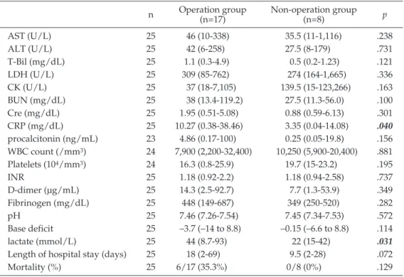
symptoms, physical signs, physical examination and CT findings in the Operation and Non-operation groups. Analysis of physical signs showed significant differences in the Glasgow Coma Scale (GCS) (p=0.04) and APACHE II score (p=0.002). Physical examination findings showed a significant difference in the rate of abdominal disten-tion (p=0.028). Comparison of laboratory values revealed significant differences in C-reactive protein (CRP) (p= 0.04) and lactate (p=0.031) (Table 4). Regarding CT find-ings, bowel dilatation (p=0.03), pneumatosis intestinalis (p=0.017), and attenuation of the contrast effect of the bowel wall (p=0.018) were detected significantly more frequently in the Operation group (Table 3).
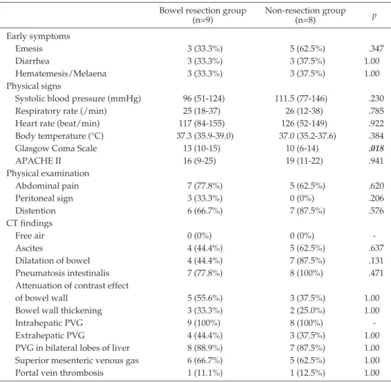
Comparison of the Bowel Resection and Non-resection Groups
We also investigated differences between the Bowel
re-section and Non-rere-section groups. With the exception of the GCS, there was no significant difference between these groups (Table 5, 6).
Outcome
The overall mortality rate for patients with PVG was 24.0% (6/25) (Fig. 2). The mortality rates for the Opera-tion and Non-operaOpera-tion groups were 35.3% (6/17) and 0% (0/8), respectively. The mortality rates for the Bowel resection and Non-resection groups were 55.6% (5/9) and 12.5% (1/8). The mortality rate was significantly higher for the Bowel resection group than for the Non-resection and Non-operation groups (55.6% [5/9] vs. 6.25% [1/16], p=0.012).
Discussion
It has been reported that PVG is not a specific disease entity, rather it represents a diagnostic clue in patients with acute abdominal pathologic conditions1―3
. If treat-ment of the underlying disease is successful, PVG will
Table 4 Laboratory findings and outcomes of the Operation and Non-operation groups
n Operation group (n=17) Non-operation group (n=8) p
AST (U/L) 25 46 (10-338) 35.5 (11-1,116) .238 ALT (U/L) 25 42 (6-258) 27.5 (8-179) .731 T-Bil (mg/dL) 25 1.1 (0.3-4.9) 0.5 (0.2-1.23) .121 LDH (U/L) 25 309 (85-762) 274 (164-1,665) .336 CK (U/L) 25 37 (18-7,105) 139.5 (15-123,266) .163 BUN (mg/dL) 25 38 (13.4-119.2) 27.5 (11.3-56.0) .100 Cre (mg/dL) 25 1.95 (0.51-5.08) 0.88 (0.59-6.13) .301 CRP (mg/dL) 25 10.27 (0.38-38.46) 3.35 (0.04-14.08) .040 procalcitonin (ng/mL) 23 4.86 (0.17-100) 0.25 (0.05-19.8) .156 WBC count (/mm3) 24 7,900 (2,200-32,400) 10,250 (5,900-20,400) .881 Platelets (104/mm3) 24 16.3 (0.8-25.9) 19.7 (15-23.2) .195 INR 25 1.18 (0.92-2.2) 1.18 (0.94-2.58) .737 D-dimer (μg/mL) 25 14.3 (2.5-92.7) 7.7 (1.3-53.9) .349 Fibrinogen (mg/dL) 25 448 (149-687) 349 (250-520) .282 pH 25 7.46 (7.26-7.54) 7.45 (7.34-7.53) .572 Base deficit 25 –3.7 (–14 to 8.8) –0.15 (–6.6 to 8.8) .114 lactate (mmol/L) 25 44 (8.7-93) 22 (15-42) .031
Length of hospital stay (days) 25 18 (2-69) 9.5 (2-28) .072
Mortality (%) 25 6/17 (35.3%) 0/8 (0%) .129
WBC: White blood cell, AST: aspartate aminotransferase, ALT: alanine aminotransferase, T-Bil: Total bilirubin, LDH: lactate dehydrogenase, CK: creatine kinase, BUN: blood urea nitrogen, Cre: creatinine, CRP: C-reactive protein, INR: International Normalized Ratio
disappear naturally and prognosis will be improved11,12
. The most common underlying disease in patients with PVG was shown to be mesenteric ischemia and subse-quent bowel necrosis1―7,9
. In the present study, 28% (7/25) of patients were confirmed to have mesenteric ischemia, which was the main reason for bowel resection (5/9)
(Ta-ble 2). The overall mortality of patients with PVG was
24.0% in the present study; however, the mortality rate was significantly higher in the Bowel resection group than in the Non-resection and Non-operation groups (55.6% vs. 6.25%, p=0.012). Kinoshita et al. previously re-ported1
that, the overall mortality in a review of 182 cases with PVG was 39%; however, the mortality was higher in patients with bowel necrosis (75%). On the ba-sis of those results, they recommended exploratory lapa-rotomy1
.
There have been few reports about the outcomes of conservative management in patients with PVG. Four previous studies of more than 15 cases reported that the mortality rate of conservative management in patients with PVG was 12.5% to 64.7%11,13,15,16
. Although the reports did not describe the decision-making process in detail and did not include the number of cases that crossed over to surgery, the mortality rate of conservative man-agement seemed to be relatively high. It might have in-volved patients not being offered an operation because it
was considered futile or patients who did not receive an operation because they declined surgery. The present study did not include patients for whom surgery was considered futile and those who declined surgery. Thus, we assessed the validity of the decision for clinical man-agement of PVG made by the attending surgeon by evaluating a combination of factors, including the find-ings of clinical symptoms, physical examination, labora-tory data, underlying disease, and CT, as previously re-ported1,2,5,6,9,14,17―19
. These factors were compared between the Operation and Non-operation groups and statistically significant differences were found in GCS14,18
, APACHE II14,18,19
, abdominal distention, CRP levels, lactate levels6
, CT findings of dilatation of bowel9
, pneumatosis intesti-nalis6,9,20
, and attenuation of the contrast effect of the bowel wall8
(Table 3, 4). As a result, 32% (8/25) of the patients with PVG were successfully managed with a conservative approach (mortality rate 0%). However, on comparing the Bowel resection and Non-bowel resection groups, there was no significant difference in any clinical factor except GCS (Table 5, 6). These observations sug-gest that these variables might be useful for making the decision to perform conservative management; however, it might be difficult to differentiate between transmural and partial bowel wall necrosis.
Table 5 Early symptoms, physical signs, physical examination and CT findings of the Bowel resection and Non-resection groups
Bowel resection group (n=9) Non-resection group (n=8) p Early symptoms Emesis 3 (33.3%) 5 (62.5%) .347 Diarrhea 3 (33.3%) 3 (37.5%) 1.00 Hematemesis/Melaena 3 (33.3%) 3 (37.5%) 1.00 Physical signs
Systolic blood pressure (mmHg) 96 (51-124) 111.5 (77-146) .230
Respiratory rate (/min) 25 (18-37) 26 (12-38) .785
Heart rate (beat/min) 117 (84-155) 126 (52-149) .922
Body temperature (°C) 37.3 (35.9-39.0) 37.0 (35.2-37.6) .384
Glasgow Coma Scale 13 (10-15) 10 (6-14) .018
APACHE II 16 (9-25) 19 (11-22) .941 Physical examination Abdominal pain 7 (77.8%) 5 (62.5%) .620 Peritoneal sign 3 (33.3%) 0 (0%) .206 Distention 6 (66.7%) 7 (87.5%) .576 CT findings Free air 0 (0%) 0 (0%) -Ascites 4 (44.4%) 5 (62.5%) .637 Dilatation of bowel 4 (44.4%) 7 (87.5%) .131 Pneumatosis intestinalis 7 (77.8%) 8 (100%) .471
Attenuation of contrast effect
of bowel wall 5 (55.6%) 3 (37.5%) 1.00
Bowel wall thickening 3 (33.3%) 2 (25.0%) 1.00
Intrahepatic PVG 9 (100%) 8 (100%)
-Extrahepatic PVG 4 (44.4%) 3 (37.5%) 1.00
PVG in bilateral lobes of liver 8 (88.9%) 7 (87.5%) 1.00 Superior mesenteric venous gas 6 (66.7%) 5 (62.5%) 1.00
Portal vein thrombosis 1 (11.1%) 1 (12.5%) 1.00
PVG: Portal venous gas
in 47.1% (8/17) of the patients in the Operation group (Table 2). Najafian et al. reported that 4 of 5 patients who underwent an operation resulted in non-therapeutic laparotomy4
. Exploratory laparotomy is the most reliable method for determining the cause of PVG1,2,6
; however, unnecessary laparotomy should be avoided. It is sug-gested that a new approach should be developed to im-prove this situation. Therefore, if a patient’s vital signs are not unstable, diagnostic laparoscopy may be favor-able and less invasive21,22
. In our study, diagnostic la-paroscopy was performed in only one patient in the Non-resection group.
There have been very few reports about laparoscopic approaches in patients with PVG6,7,21―23
. Recently, Koizumi et al7
. reported that, according to an analysis of 1,590 pa-tients with PVG whose data were obtained from a Janese National Inpatient Database, the total number of pa-tients with PVG undergoing surgery for ischemic bowel was 271, and 4.8% (13/271) of those patients received
bowel resection via a laparoscopic approach. However, the number of cases that received laparoscopic explora-tion was not menexplora-tioned. While a laparoscopic approach for exploration may be favorable and less invasive than exploratory laparotomy for patients without unstable vi-tal sings, the evidence-based guideline of the European Association for Endoscopic Surgery on laparoscopy for abdominal emergencies states that laparoscopy does not offer significant advantages in cases of acute mesenteric ischemia24
. The risks and concerns should be considered, including laparoscopy-related complications (e.g., a fur-ther reduction in portal blood flow and intestinal perfu-sion due to the increase in intra-abdominal pressure dur-ing pneumoperitoneum25
), the timing of second-look la-paroscopy, and cost-effectiveness. Further studies will be needed to clarify the risks and benefits of this approach.
Conclusions
Table 6 Laboratory findings and outcomes of the Bowel resection and Non-resection groups
n Bowel resection group (n=9) Non-resection group (n=8) p
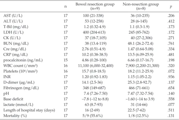
AST (U/L) 17 100 (21-338) 36 (10-235) .206 ALT (U/L) 17 53 (12-258) 28 (6-145) .412 T-Bil (mg/dL) 17 1.2 (0.32-4.9) 1.1 (0.3-1.9) .173 LDH (U/L) 17 400 (204-613) 245 (85-762) .172 CK (U/L) 17 37 (18-7,105) 40 (27-2,306) .271 BUN (mg/dL) 17 38 (13.4-119) 48.1 (26.2-72.4) .761 Cre (mg/dL) 17 2.76 (0.51-4.9) 1.47 (0.64-5.08) .334 CRP (mg/dL) 17 10.2 (0.38-38.5) 13.5 (6.09-25.9) .481 procalcitonin (ng/mL) 15 4.86 (0.28-100) 6.66 (0.17-16.7) .198 WBC count (/mm3) 16 11,100 (6,000-32,400) 7,900 (2,200-21,300) .320 Platelets (104/mm3) 16 15.7 (0.8-18.5) 18.2 (11.2-25.9) .072 INR 17 1.20 (0.92-1.83) 1.15 (1.05-2.2) .936 D-dimer (μg/mL) 17 14.1 (2.5-36) 25.3 (2.8-92.7) .137 Fibrinogen (mg/dL) 17 348 (149-687) 466 (71-661) .654 pH 17 7.4 (7.26-7.50) 7.47 (7.32-7.54) .140 Base deficit 17 –7.8 (–12 to 8.8) –1.60 (–14 to 3.9) .558 lactate (mmol/L) 17 63 (8.7-93) 31 (14-66) .077
Length of hospital stay (days) 17 16 (2-69) 22.5 (7-62) .511
Mortality (%) 17 5/9 (55.6%) 1/8 (12.5%) .131
WBC: White blood cell, AST: aspartate aminotransferase, ALT: alanine aminotransferase, T-Bil: To-tal bilirubin, LDH: lactate dehydrogenase, CK: creatine kinase, BUN: blood urea nitrogen, Cre: cre-atinine, CRP: C-reactive protein, INR: International Normalized Ratio
mortality rate of PVG associated with bowel necrosis, there are some cases of PVG in which conservative man-agement is successful. It is important to carefully con-sider the possibility of conservative management of pa-tients with PVG, based on a combination of factors, in-cluding the clinical symptoms, physical examination findings, laboratory data, underlying disease, and CT findings. This approach appears to be useful for making decisions in relation to conservative management of pa-tients with PVG.
However, in the present study, 47.1% of patients who underwent emergency laparotomy for suspected bowel necrosis did not develop transmural bowel necrosis, and bowel resection could be avoided after a second look procedure, resulting in non-therapeutic laparotomy. It was thus suggested that this approach alone would not be sufficient to accurately predict transmural bowel ne-crosis requiring resection in patients with PVG, and that a new approach should be developed in order to im-prove this situation.
Diagnostic laparoscopy may be a favorable and mini-mally invasive approach; however, there have been very few reports about laparoscopic approaches in patients with PVG. Further studies will be needed in order to clarify the risks and benefits of this approach.
Conflict of Interest: The authors declare no conflict of inter-est.
References
1.Kinoshita H, Shinozaki M, Tanimura H, et al. Clinical fea-tures and management of hepatic portal venous gas four case reports and cumulative review of the literature. Arch Surg. 2001;136(12):1410―4.
2.Nelson AL, Millington TM, Sahani D, et al. Hepatic por-tal venous gas: the ABCs of management. Arch Surg. 2009;144(6):575―81.
3.Liebman PR, Patten MT, Manny J, Benfield JR, Hechtman HB. Hepatic-portal venous gas in adults: etiology, patho-physiology and clinical significance. Ann Surg. 1978;187: 281―7.
4.Najafian H, Habibi M, Reilly T. Hepatic portal vein gas: clinical features and outcomes. Am Surg. 2003;69(6):526―9. 5.Hou SK, Chern CH, How CK, Chen JD, Wang LM, Lee CH. Hepatic portal venous gas: clinical significance of computed tomography findings. Am J Emerg Med. 2004; 22:214―8.
6.Wayne E, Ough M, Wu A, et al. Management algorithm for pneumatosis intestinalis and portal venous gas: treat-ment and outcome of 88 consecutive cases. J Gastrointest Surg. 2010;14(3):437―48.
7.Koizumi C, Michihata N, Matsui H, Fushimi K, Yasunaga H. In-hospital mortality for hepatic portal venous gas: analysis of 1590 patients using a Japanese National Inpa-tient Database. World J Surg. 2018;42(3):816―22.
8.Yamada T, Kan H, Matsumoto S, et al. A case of portal venous gas after rectal surgery without anastomotic leak-age or bowel necrosis. J Nippon Med Sch. 2015;82(4):202― 5.
9.Koami H, Isa T, Ishimine T, et al. Risk factors for bowel necrosis in patients with hepatic portal venous gas. Surg Today. 2015;45(2):156―61.
10.Knaus WA, Draper EA, Wagner DP, Zimmerman JE. APACHE II: a severity of disease classification system. Crit Care Med. 1985;13(10):818―29.
11.Inokuchi R, Fukuda T, Yahagi N, et al. Severe hepatic portal venous gas that spontaneously resolved within a day. Intensive Care Med. 2014;40(9):1369.
12.Huurman VA, Visser LG, Steens SC, et al. Persistent por-tal venous gas. J Gastrointest Surg. 2006;10(5):783―5. 13.Seak CJ, Yen DHT, Ng CJ, et al. Rapid Emergency
Medi-cine Score: A novel prognostic tool for predicting the out-comes of adult patients with hepatic portal venous gas in the emergency department. PLoS One. 2017;12(9):e 0184813.
14.Yoo SK, Park JH, Kwon SH. Clinical outcomes in surgical and non-surgical management of hepatic portal venous gas. Korean J Hepatobiliary Pancreat Surg. 2015;19(4):181― 7.
15.Iannitti DA, Gregg SC, Mayo-Smith WW, et al. Portal ve-nous gas detected by computed tomography: Is surgery imperative? Dig Surg. 2003;20(4):306―15.
16.Heye T, Bernhard M, Mehrabi A, et al. Portomesenteric venous gas: Is gas distribution linked to etiology and out-come? Eur J Radiol. 2012;81(12):3862―9.
17.Seak CJ, Hsu KH, Wong YC, et al. The prognostic factors of adult patients with hepatic portal venous gas in the ED. Am J Emerg Med. 2014;32(9):972―5.
18.Seak CJ, Ng CJ, Yen DH, et al. Performance assessment of the Simplified Acute Physiology Score II, the Acute Physi-ology and Chronic Health Evaluation II score, and the Se-quential Organ Failure Assessment score in predicting the outcomes of adult patients with hepatic portal venous gas in the ED. Am J Emerg Med. 2014;32:1481―4.
19.Wu JM, Tsai MS, Lin MT, Tien YW, Lin TH. High APACHE II score and long length of bowel resection im-pair the outcomes in patients with necrotic bowel in-duced hepatic portal venous gas. BMC gastroenterol. 2011;11:18.
20.Bani Hani M, Kamangar F, Goldberg S, et al. Pneumatosis and portal venous gas: do CT findings reassure? J Surg
Res. 2013;185(2):581―6.
21.Taniguchi K, Asakuma M, Nagayabu K, et al. Exploring the use of single-port surgery in the conservative man-agement of hepatic portal vein gas: A case report. Medi-cine (Baltimore). 2018;97(48):e13446.
22.Shah NR, Dossick DS, Madura JA, Heppell JP. Use of di-agnostic laparoscopy in a patient with gastric pneumato-sis and portal venous gas. Case Rep Gastroenterol. 2013;7 (2):261―5.
23.Napolitano L, Waku M, Costantini R, Mazahreh T, Inno-centi P. Portal vein gas due to gangrenous cholecystitis treated by a laparoscopic procedure: report of a case. Surg Today. 2009;39(10):909―12.
24.Agresta F, Ansaloni L, Baiocchi GL, et al. Laparoscopic approach to acute abdomen from the Consensus Develop-ment Conference of the Società Italiana di Chirurgia En-doscopica e nuove tecnologie (SICE), Associazione rurghi Ospedalieri Italiani (ACOI), Società Italiana di Chi-rurgia (SIC), Società Italiana di ChiChi-rurgia d’Urgenza e del Trauma ( SICUT ) , Società Italiana di Chirurgia nell’Ospedalità Privata (SICOP), and the European Asso-ciation for Endoscopic Surgery (EAES). 2012;26(8):2134― 64.
25.Schilling MK, Redaelli C, Krähenbühl L, et al. Splanchnic microcirculatory changes during CO2 laparoscopy. J Am Coll Surg. 1997;184(4):378―82.
(Received, (Accepted, (J-STAGE Advance Publication,
November February March 21, 2019) 26, 2020) 31, 2020)
Journal of Nippon Medical School has adopted the Creative Com-mons Attribution-NonCommercial-NoDerivatives 4.0 International License (https://creativecommons.org/licenses/by-nc-nd/4.0/) for this article. The Medical Association of Nippon Medical School re-mains the copyright holder of all articles. Anyone may download, reuse, copy, reprint, or distribute articles for non-profit purposes under this license, on condition that the authors of the articles are properly credited.