Studies on the Proteases of Marine Bacteria II
: Effect of Cations on Protease Formation
著者
SAKATA Taizo, UEDA Kazuaki, KAKIMOTO Daiichi
journal or
publication title
鹿児島大学水産学部紀要=Memoirs of Faculty of
Fisheries Kagoshima University
volume
26
page range
63-69
別言語のタイトル
海洋細菌のプロテアーゼに関する研究 II : プロテ
アーゼ産生に及ぼすカチオンの影響について
Vol. 26 pp. 63-69 (1977)
Studies on the Proteases of Marine Bacteria—II Effect of Cations on Protease Formation
Taizo Sakata, Kazuaki Ueda and Daiichi Kakimoto*
Abstract
Extracellular ptotease(s) of a marine Pseudomonas sp. 1-6 was produced dependent of polypep-tone concentration of the suspending medium but independent of yeast extract in the presence of polypeptone and artificial sea water (ASW).
In a basal suspension medium containing polypeptone (0.5%), yeast extract (0.1%) and NaCl (3.0%), protease formation of marine isolates did not occur during 6 hr incubation. The addition of Ca++ ion to the basal medium stimulated protease formation but Mg++ ion did not replace Ca++ ion.
In the experiment of washed cell suspensions, K+ ion was necessary for protease formation of both marine isolates and Bacillus subtilis. The effect of K+ ion was almost identical with
that of yeast extract on protease formation. On the other hand, the supplement of Ca++ ion was indispensable to marine isolates but not to B. subtilis except as a trace ion contaminant.
Marine bacteria have been known to require the salts of sea water for both growth and the production of extracellular enzymes. Kato et al1) reported that in a marine
psychrophilic bacterium, Pseudomonas sp. No. 548, both the growth and the protease activity were at maximum in a medium containing sea water at full strength. Studies by Tojo et al2> indicated that CaCl2 activated the production of protease in marine isolates. However, very few detailed studies on the factors affecting the ex
tracellular enzyme production of marine bacteria have been done.
We3^ have examined protease formation by marine bacteria and showed that pro tease production of many marine bacteria was enhanced in media containing Ca++ ion. This paper describes experiments on the formation of extracellular protease by cells washed and resuspended in various media. The purpose of this investigation is
to determine the factors which control extracellular enzyme production.
Materials and Methods
Organisms. Marine bacteria, strains 1-6 and C-18 were isolated from sea water collected in the offshore sea of the Sata peninsula, Japan. A marine Pseudomonas sp.
1-6 was principally used throughout this work. A marine pseudomonad 1055-1 and
a terrestrial bacterium, Bacillus subtilis 6051 were obtained from Hokkaido Univer-Laboratory of Microbiology, Faculty of Fisheries, Kagoshima University, Japan.
64 Mem. Fac. Fish., Kagoshima Univ. Vol. 26 (1977)
sity and Kyoto University, respectively.
Washed cell experiment. Bacterial cells were grown in ZoBell 2216E liquid
medium or nutrient broth. After growth for 12 hr at 25 C, the cells were harvested and washed with ASW or salts solution. The washed cells were resuspended in the appropriate suspending medium and optical density of cell suspension was adjusted to approximately 1.0 at 540 nm. The cell suspensions (100 ml) were shaken in 300 ml flasks at 25 C for 6 hrs and samples were withdrawn to determine cell growth (change in optical density at 540 nm). Immediately after the cell suspensions were centrifuged at 7,000 rpm for 15 min and the supernatant fluids were assayed for pro tease activity.
The suspending media. Each basal medium is composed as follows. ZoBell 2216E medium: 0.5% polypeptone (Daigo Eiyo) 0.1% yeast extract (Daigo Eiyo) and Herbst's artificial sea water (ASW). Medium PSW: 0.5% polypeptone and ASW. Medium PS: 0.5% polypeptone and 3.0% NaCl. Medium PYS: 0.5% polypeptone, 0.1% yeast extract and 3.0% NaCl.
Enzyme assay. Protease activity was assayed by a modification of the casein
digestion method of Kunitz4). The assay mixture contained 2.0 ml of 0.6% casein
in 0.05 M sodium carbonate buffer (pH 10.0) and 0.5 ml culture fluid. After 2 hr incubation at 30 C, the reaction was stopped with 2.5 ml of 0.44 M TCA solution. The precipitated protein was filtered by Toyo filter paper No. 5c and the absorbance of the filtrate was measured at 275 nm in a 1.0 cm cuvette by use of a Hitachi Model 101 spectrophotometer.
Results
Time course of extracellular protease production and growth
Fig. 1 shows the time course of protease formation and growth in growing cultures
Fig. 1. Q d i? •H > 0.8 0.6
^ ^ \
0.4 0.2 n L-# I I 1 1 I 1 g 0 12 24 36 48 60 72 Cultural period (hr)Protease formation by Pseudomonas sp. 1-6 during a cultivation in ZoBell 2216E
modified medium. 1.5 1.010 +> 10 a 0.52 o u 0 »
of strain 1-6. The maximum growth and protease activity of the cultures were ob served after 24 hr under the experimental conditions tested. The production rate of extracellular protease was found to be very high in logarithmic phase from 6 to
24 hr and then to slow down. In the following suspension experiments active grow
ing cells in the logarithmic phase at 21 hr were taken as samples.
When the washed cells obtained from a culture growing in ZoBell 2216E at 12 hr
were resuspended in fresh ZoBell 2216E medium, extracellular protease continued to be produced in the suspension fluid after 1 hr of lag period as shown in Fig. 2. Al though the cell density of the suspension was very high (O. D. 1.0 at 540 nm), con siderable cell growth occured during the incubation period. In this suspension ex periment, extracellular protease was considered to be produced by active growing cells.
~ 0.4
1 1 2 3 4 5 6
Incubation time (hr)
Fig. 2. Time course of protease formation by a washed cell suspension in ZoBell 2216E
modified medium.
Effect of organic components on protease formation
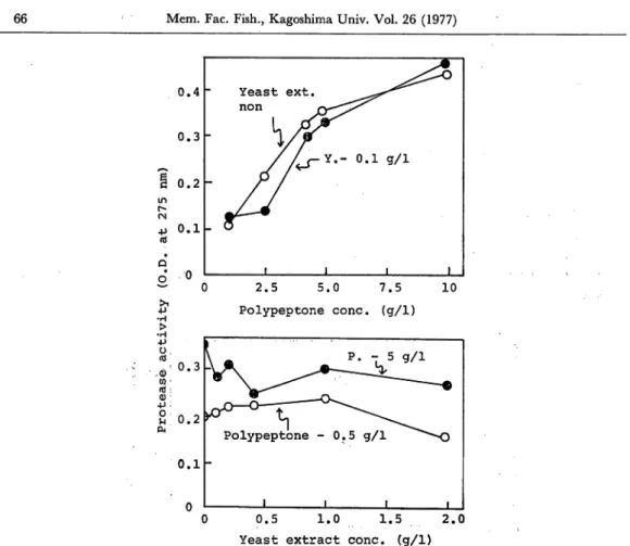
Marine isolate 1-6 produced extracellular protease activity both in growing culture and in washed cell suspension in ZoBell 2216E medium. An investigation into the components of the medium which favor the formation of protease was undertaken. Compared with several organic components of the media, polypeptone was found to be most suitable for enzyme formation rather than phytone, beef extract, casein and casamino acids. Fig. 3 shows the effects of polypeptone and yeast extract concentra tion on protease formation during 6 hr incubation in suspending medium. Protease activity of culture filtrates increased rapidly with an increase of polypeptone concen tration up to 5 g/1 and then gradually increased at concentrations over 5 g/1. In the presence of polypeptone, a wide range of yeast extract concentrations from 0 to 2 g/1 did not stimulate protease formation substantially. Protease formation was found to be independent of yeast extract concentration in the presence of appropriate levels of polypeptone and ASW.
66 Mem. Fac. Fish., Kagoshima Univ. Vol. 26 (1977) 0.4 Yeast ext. ^^^^ n o n 0.3 0.2 0.1 n 1 1 1 1 ~ 0 J? •H > •H •P O, rd (d: Q) •M O i H CM 2.5 5.0 7.5 Polypeptone cone, (g/1) 10 0.5 1.0 1.5 2.0
Yeast extract cone, (g/1)
Fig. 3. Effect of the concentration of organic compounds on protease formation.
Effect of cations on protease formation
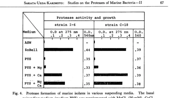
Protease formation of the marine isolate 1-6 requires at least polypeptone and in organic components of ASW. Among the inorganic components of ASW, the effect
of mono- and divalent cations on protease formation was examined and the results are shown in Fig. 4. When the washed cells were resuspended in a basal medium
containing polypeptone (0.5%), yeast extract (0.1%) and NaCl (3.0%), protease for
mation did not occure during 6 hr incubation. The addition of Ca++ ion to the basal medium stimulated protease formation but Mg++ ion did not replace Ca++ ion as a factor affecting protease formation. Fig. 5 shows effect of Ca++ concentration on protease formation; The highest activity was observed at 5 mM of Ca++ and at higher concentration than 5 mM protease formation was reduced somewhat.
It is well known that marine bacteria require inorganic ions at relatively high lev
els for growth in comparison with terrestrial bacteria. A comparison of protease
formation among marine isolates 1-6, 1055-1 and terrestrial Bacillus subtilis in differ ent media is given in Fig. 6. In this experiment K+ ion was used instead of yeast
Medium \
Protease activity and growth
strain 1-6 strain C-18 O.D at 275 nm .1 .2 .3 .4 O.D. 540nn O.D. at 275 nm .1 .2 .3 .4 O.D. 540 ASW ZoBell PYS PYS + Mg PYS .+ Ca PYS + Mg Ca i i i 1 1 1 1 -.44 .35 .33 o37 .35 l 1 1 I 1 1 .39 o37 .36 039 .38 Fig. 4. Protease formation of marine isolates in various suspending media. The basal
suspending medium (medium PYS) was supplemented with MgCl2 (20 mM), CaCl2 (20 mM) or MgCl2 (10 mM) and CaCl2 (10 mM). Bacterial growth was expressed as the difference in optical density at 540 nm (pre and post incubation).
Medium N^
Protease activity and growth
O.D. at 275 nm .1 .2 .3 O.D. 540 PYS R.M.+Ca 1 i i 1 .40 PYS+ Ca 1 PYS+ Ca 3 PYS+ Ca 5 PYS+ CalO PYS+ Ca20 .37 .38 .39 .39 .39
Fig. 5. Effect of Ca++ ion concentration on protease formation by washed cells suspended in media PYS. In the case of medium PYS, CaCl2 (5 mM)
was added to the enzyme reaction mixture (R.M. + Ca).
extract since the effect of K+ ion, ash components of yeast extract and yeast extract were found to be almost identical on protease formation. Marine isolates produced protease activity in a medium containing polypeptone, Na+, K+ and Ca++. A sub stantial decrease in protease production was observed if any one of the four compo nents was absent. On the other hand, protease formation by B. subtilis occured in the combination of polypeptone, Na+ and K+. It is apparent that marine isolates
68 Mem. Fac. Fish., Kagoshima Univ. Vol. 26 (1977)
Medium \
Protease activity and growth
s t r a i n 1-6 s t r a i n 1055-1 O.D. a t 275 nm .1 .2 O.D. 540 O.D, a t 275 nm . 1 o2 O.Do 540 PS (3%) • i t .20 1 i i -PS + K .40 1 .35 PS+K+Ca ••HBHHHH .45 .35 PS+K+Mg • .43 .35 PS + Ca olO • .05 Medium\ Bacillus s u b t i l i s O.D. a t 275 nm .1 .2 03 .4 05 06 O.D. 540 PS(0.5%) PS + K PS+K+Ca • • i i i i I .10 .25 .20 PS + Ca 1 .05
Fig. 6. Protease formation of marine isolates and B. subtilis in various suspending media.
The basal suspending medium containing 3% and 0.5% of NaCl was used for marine isolates and B. subtilis, respectively and supplemented with MgCl2 (10 mM), CaCl2 (10mM)orKCl(2mM).
require Ca++ ion but that B. subtilis does not for protease formation except as a con taminant in media components.
Discussion
A specific function for the inorganic ions of sea water in the physiological proper ties of marine bacteria has been reported by many workers. MacLeod5* summa
rized that inorganic ions of sea water act on marine bacteria in activation and sta bilization of enzymes, transportation of substrates and prevention of cell lysis. With
respect to the possibility that inorganic ions were required for the production of ex tracellular protease, Kato et al1* reported that the ratio of proteolytic activity to growth of marine Pseudomonas sp. No. 548 increased with a higher content of sea water in the medium. In the case of marine Pseudomonas 1-6, protease formation was observed in a suspension medium composed of polypeptone and ASW or in a
medium containing polypeptone, Na+, K+ and Ca++. Na+ and K+ are expected to
required for protease formation by active growing cells.
On the other hand, Mg++
ion was more effective than monovalent cations in preventing disruption of the cellsand stimulating cell growth but was not an indispensable factor for protease forma
tion.Protease activity was observed to be very high in a medium containing Ca++ ion.
On the other hand, after 6 hr incubation in a medium without Ca++, no more pro
tease activity was detected even with the addition of Ca++ ion to culture filtrate.
Ca++ ion is thought to stimulate the production of or release of protease from the cells.
However, Ca++ ion has been found to activate and stabilize enzyme proteins. It is
very difficult to determine whether Ca++ ion stimulates enzyme protein synthesis or
stabilizes enzyme protein in the culture filtrate.
References
1) Kato N., T. Nagasawa, Y. Tani and K. Ogata (1972): Protease formation by a marine psy chrophilic bacterium. Agr. Biol. Chem., 36, 1177-1184.
2) Tojo T., T. Tokuyama and K. Asano (1974): Studies on the marine bacteria producing
pro-tease-II. Bull Coll. Agr. & Vet. Med. Nihon Univ., 32, 140-152.
3) Sashihara N., T. Sakata and D. Kakimoto (1975): Study on the protease of marine bacteria.
Mem. Fac. Fish., Kagoshima Univ., 24, 149-160.
4) KunttzM. (1946): Crystalline soybean trypsin inhibitor-II. /. Gen. Physiol, 30, 291-310. 5) MacLeod R. A. (1968): On the role of inorganic ions in the physiology of marine bacteria.