IRUCAA@TDC : Identification of unknown body using DNA analysis and dental characteristics in chest X-ray photograph
10
0
0
全文
(2) 145. Bull Tokyo Dent Coll (2005) 46 (4): 145–153. Case Report. Identification of Unknown Body Using DNA Analysis and Dental Characteristics in Chest X-ray Photograph Kiyoshi Minaguchi, Sayaka Maruyama, Iku Kasahara, Chizuru Nohira, Yoichi Hanaoka, Tohkai Tsai, Hitoshi Kiriyama and Nobuyuki Takahashi* Department of Forensic Odontology, Tokyo Dental College, 1-2-2 Masago, Mihama-ku, Chiba 261-8502, Japan * Scientific Crime Laboratory, Chiba Prefectural Police Headquarters, 1-71-1 Chuominato, Chuo-ku, Chiba 260-0024, Japan. Received 5 December, 2005/Accepted for Publication 18 January, 2006. Abstract An unknown skeletonized body was identified by DNA analysis and dental information. The body had already been cremated when a candidate for the unknown body was proposed. Therefore, for DNA analysis we used teeth that had been kept for a long time after use for serological examination. We also used a chest X-ray photograph of the candidate and photographs of dentition, as well as dental X-ray photographs taken when the unknown body was found. Because DNA obtained from teeth was highly degraded, we amplified three PCR fragments to determine the 766 bp mitochondrial DNA (mtDNA) sequence including HV1 and HV2. Polymorphism of the ABO locus was also analyzed using small PCR fragments. Although the isolated DNA was contaminated, probably with DNA from a different individual, DNA polymorphisms of mtDNA and the ABO locus could be analyzed. We discuss the reliability of our conclusions from the point of view of the necessity of constructing an accurate mtDNA database. Although a dentist who had treated the teeth of the unknown body could not be found, a chest X-ray photograph for medical diagnosis was very useful in comparing dental characteristics, as it included an image of the frontal part of the lower jaw and upper teeth. Key words:. Personal identification —Skeletonized body — DNA analysis— Mitochondrial DNA—Dental identification. Introduction It is generally accepted that dental identification is a very reliable method for the personal identification of an unknown body, when the appropriate antemortem record of the deceased person has been obtained. However, it is not easy to locate a candidate of a certain unknown body using dental. characteristics alone. In addition, even if a possible candidate has been located by some other means, sometimes, it is still difficult to find the dentist who treated the teeth of that candidate. In such cases, we should remember that antemortem records other than those obtained from dental clinics are also available for dental identification. Teeth offer another advantage as an aid in personal. 145.
(3) 146. Minaguchi K et al.. A. C. B. Fig. 1 Dental characteristics of skeletonized body A: Frontal view of skull. B: Upper and lower dentition. Although 13 teeth were lost, a lot of dental fillings were found in remaining teeth. C: Left view of upper dentition. Left central incisor was protruded.. identification. They are very good sources for DNA analysis, even in cases where the specimens are highly decomposed11,12,16). In order to perform personal identification by DNA analysis, we have to find a source of DNA derived from the candidate, or a blood relative of the candidate. In the latter case, it would be better to consider which kind of DNA polymorphisms were most suitable, which would depend on the blood relationship between the possible blood relative and the candidate, rather than simply using autosomal microsatellite polymorphism detection kits. In this paper, we present a case of identifying an unknown body by DNA analysis using teeth and dental information obtained from a chest X-ray photograph.. Case A skeletonized female body was found in a forest. Because the body was wearing a pair of. pajamas but no shoes, the police decided that the woman was involved in a criminal case. She had had a lot of dental treatment. This information was open to the public through the Internet. However, the body could not be identified for a long time. A year and eight months later, a candidate for the unknown person was proposed. We were asked to determine whether the body corresponded to the candidate.. Process of Searching for a Candidate and Results of Examination We examined the dental characteristics of the skeletonized body (Fig. 1). The skull had 10 inlay fillings and a crown. Two teeth had root canal fillings (Figs. 1, 2, and 3). She had protruded central incisors (Fig. 1). Because no other useful information was available, we tried to find a candidate using dental information alone. We prepared photographs.
(4) 147. DNA and Dental Identification. 16. 14 13. 14 13 14 13. 47 47 47. 46. 21. 21. 24 25. 45. 46. 45 44. 44 43 42. 35. 36. 36. 37. Fig. 2 Dental X-ray photographs of skull Kinds of teeth are shown on top and at bottom of each tooth.. Fig. 3 Dental chart of skull. of dental treatment, X-rays, and a photograph showing the characteristic features of her incisors. These photographs were uploaded at an Internet site. However, this method proved unsuccessful. A candidate for the unknown body was proposed from different sources. Although a dentist who treated her teeth could not be found and the body had already. been cremated, three teeth from the unknown body, which had been used for serological examination, had been kept as pieces of evidence. Therefore, we performed DNA analysis using these teeth to determine the relationship between the body and the candidate’s mother and sister. In addition, because the police found a chest X-ray photograph of.
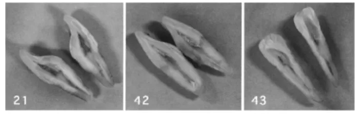
(5) 148. Minaguchi K et al.. Fig. 4 Teeth used for serological test Position of each tooth is shown. Inner surface of pulp chamber had been scraped off.. the candidate, we used that also as an aid for personal identification. 1. DNA analysis The materials offered for DNA analysis consisted of blood samples obtained from a woman and her daughter, and three pieces of teeth, 21, 42, and 43 (Fig. 4). We were asked to examine (1) DNA polymorphisms in each sample, (2) whether the woman was the mother of the unknown body, and (3) whether the daughter of the woman was sister of the unknown body. Isolation of DNA from the blood samples was performed as described elsewhere11). The tooth samples had been used for serological tests on the ABO blood group system. They had been cut into two pieces to expose the pulp chambers, and most of the inner surfaces of those chambers had been scraped off for that examination. To isolate DNA from the unknown body, we chose two pieces of tooth 42 and washed them using physiological saline solution in a sterile test tube twice for 1.5 hours each in order to remove DNA contamination. The tooth was crushed into powder using a Multi-beads Shocker (Yasui Co., Ltd.), and then decalcified in 3 ml of 0.5 M EDTA for 2 days. Although not all of the tooth powder dissolved in the EDTA, it was centrifuged, washed in 3 ml of Tris-EDTA (TE) buffer twice, and re-suspended in TE, after which it was digested in proteinase K overnight. DNA was then extracted with phenol/chloroform. The EDTA solution used for decalcification and the TE buffer used for washing the tooth powder were concentrated by Centricon-100 (Millipore), and DNA was. further extracted from these solutions using the same protocol as described above. Finally, three kinds of DNA sample, derived from the tooth powder, the TE-washing solution, and the EDTA solution, were prepared. Using these samples, mitochondrial DNA (mtDNA) polymorphisms and polymorphisms of the ABO locus were investigated. In the analysis of the mtDNA polymorphisms for the woman and her daughter, we determined nucleotide sequences of 1,137 bp from position 15999 to 567, which included two hypervariable (HV) regions, HV1 (from position 16025–16400) and HV2 (from position 28–369). Because it was difficult to amplify large DNA fragments from the tooth sample, we amplified three fragments of 16072–16420 (349 bp), 16356–67 (281 bp), and 8–389 (382 bp), and determined nucleotide sequences of 766 bp from 16123 to 319. The method for mtDNA sequencing and their primers are described elsewhere10). When we compared sequences obtained from the woman, her daughter, and the tooth sample with the reference sequence2,3), each showed the same haplotype with mutational positions at 16136–16172–16174–16183–16189–16217– 16284–16309–16519 in the HV1 region and 73–199–202–207–263–309.1–309.2–315.1 in the HV2 region. When intergenic COII/ tRNALys deletion7,10) was examined in the three samples, all of them bore the 9-bp deletion. These results showed that this haplotype belonged to the B4b1a1 haplogroup10). The same haplotype was not found in the 1,201 Japanese database composed of data provided by Seo et al.14), Imaizumi et al.8), Maruyama et al.10), Tanaka et al.15) and Nohira et al.13). To.
(6) DNA and Dental Identification. 149. Fig. 5 SSCP gel electrophoresis of ABO locus Migration positions of allelic products are indicated on both sides of gels. A: Electrophoresis of 138 bp PCR products. Lanes 1–5 show control samples with known genotypes, and lanes 6 and 7 show samples from the woman and her daughter, respectively. Lanes 8–10 show samples obtained from unknown body. Three allelic products, Oa, Og and B, were detected in samples from tooth powder and TE-washing solution. However, two allelic products, Oa and Og, were detected in sample from EDTA solution. Lane 1: AOa, lane 2: AB, lane 3: OaOa, lane 4: BOa, lane 5: OaOg, lane 6: DNA from woman, BOa, lane 7: DNA from daughter, BOg, lane 8: DNA from tooth powder, lane 9: DNA from TE-washing solution, lane 10: DNA from EDTA solution. B: Electrophoresis of 89 bp PCR products. Lanes 1–5 and 10 show control samples with known genotypes, and lanes 6 and 7 show samples from woman and her daughter, respectively. Lanes 8, 9, and 11 contain samples obtained from unknown body. Three allelic products, Oa, Og and B, were detected in the samples from tooth powder and TE-washing solution. However, two allelic products, Oa and Og, were detected in sample from EDTA solution. Lane 1: AB, lane 2: AA, lane 3: BOa, lane 4: OaOa, lane 5: OaOg, lane 6: DNA from woman, BOa, lane 7: DNA from daughter, BOg, lane 8: DNA from tooth powder, lane 9: DNA from TE-washing solution, lane 10: OaOg, lane 11: DNA from EDTA solution.. construct a database, we classified all of the data into phylogenetic haplogroups, compared mutational position in each sample, and discarded questionable sequences. The most similar haplotype in the database was 16136– 16183–16189–16217–16284–16519–73–199– 202–207–263–309.1–309.2–315.1, which is one of the most frequent haplotypes seen in 1.4% of the Japanese population. However, the present haplotype differed at three positions, 16172–16174–16309, from this common haplotype. Therefore, it was concluded that an extremely rare haplotype was shared between the sequence of the tooth sample and those of the woman and her daughter. In analysis of the ABO gene, a part of exon 6 of the locus was amplified1). This amplicon includes nucleotides at positions 261 and 297 in exon 6, the mutations of which essentially coincide with the result of the serological. ABO blood grouping. An additional nucleotide sequence polymorphism is present in this region, resulting in the identification of four kinds of alleles, A, B, Oa, and Og types1). In this case, we examined the same mutations using two kinds of primer pairs which amplified different-sized PCR products. The method for PCR amplification and detection of the mutation was described previously16). To amplify an 89 bp small segment of the ABO locus, we used oligonucleotide 5⬘-CTGCTCGTTGAGGATGTCGA-3⬘ as the reverse primer. The PCR products were analyzed by SSCP gel electrophoresis. Figure 5A shows the electrophoresis of the 138 bp PCR products. The woman and her daughter showed two kinds of paired-bands: the mother shared the B and Oa alleles, and the daughter shared the B and Og alleles. In contrast, three kinds of paired-bands derived from the Oa,.
(7) 150. Minaguchi K et al.. Og and B alleles were detected in the DNA samples isolated from the tooth powder and TE-washing solution, and only two kinds of paired-bands derived from the Oa and Og alleles were detected in the DNA sample isolated from the EDTA solution. Although the bands for the B allele from the tooth powder and the TE-washing solution were fainter than those for the Oa and Og alleles, these results showed that the DNA samples isolated from the tooth powder and the TEwashing solution contained DNA from more than two individuals. We further amplified the 89-bp PCR products from the same samples (Fig. 5B). The same results as those using 138 bp fragments were obtained. The bands for the B allele from the tooth powder and the TE-washing solution were fainter than those for the Oa and Og alleles. These results suggested that the ABO genotype of the tooth sample was probably OaOg, and that the bands for the B allele might derive from contaminated DNA. From the results of this DNA analysis, we concluded that there was no contradiction in assuming that the woman was the mother of the unknown body, and that the daughter of the woman was the sister. 2. Comparison of dental characteristics The police found out that the candidate for the unknown body had consulted a physician, and that her chest X-ray photograph had been taken. Because an image of the X-ray photo included frontal parts of the upper and lower jaws (Fig. 6), we compared them with the photographs and dental X-ray photographs we took when the unknown body was found (Figs. 1 and 2). The shape of the radioopaque images for the tooth restorations was compared with the tooth fillings found in the skull. The patterns of the fillings for 35 and 36 were similar to those found in the dental X-ray photographs of the skull. In addition, we believe that similar patterns for the fillings for 14, 44, 45, and 46 in the chest X-ray photograph might be obtained if a dental X-ray photograph of the skull was taken from the same direction. The. Fig. 6 Part of chest X-ray photograph Dental filling restored lower molars and upper premolar can be observed.. characteristic feature of protrusion of the left upper central incisor was also recognized in the chest X-ray photograph of the candidate (Figs. 1, 2, and 6). These results suggested that the chest X-ray photograph was probably taken from the candidate.. Discussion 1. Reliability of DNA analysis Human mtDNA consists of a doublestranded, closed, circular molecule present in 1,000–10,000 copies per cell5). The complete nucleotide sequence of the 16,569 bp human mitochondrial DNA was determined in 19812), and this sequence is used as a reference sequence to show mutational positions in mtDNA genome. It carries a highly variable, single, non-coding region (1,122 bp), the so-called “control region”. The high copy number and high mutation rate of mtDNA, as well as maternal inheritance and lack of recombination, offer considerable advantages to the use of mtDNA sequencing in forensic science. Because we found that the tooth samples were highly degraded, and that the subjects for comparison by DNA analysis were female relatives of a candidate, we decided to analyze maternally inherited mtDNA polymorphisms. In addition, because the ABO blood groups of the unknown body had been examined by serological tests when the body was found, we also analyzed polymorphism of the ABO locus. In the present case, we performed DNA.
(8) DNA and Dental Identification. analysis using a tooth which had been used for blood typing. Therefore, in order to eliminate contamination from the tooth samples, we washed them twice in a sterile physiological saline solution. However, DNA isolated from the teeth still contained contaminated DNA. Sequence variations determined using DNA extracted from the tooth powder and that extracted from the EDTA solution were identical and did not show background peaks in the electrophoretogram, suggesting DNA contamination. In this case, we amplified three fragments, 16072–16420, 16356–67, and 8– 389, to construct a target sequence of 766 bp. When a DNA sample is highly degraded, we cannot amplify large fragments. Therefore, we must amplify more than two small fragments to construct one target sequence. In such cases, if DNA from two individuals is included in the sample, there is the possibility that a different source sequence will be amplified in each fragment. In order to confirm whether a certain sequence obtained from a DNA sample derived from a single individual, it is important to confirm whether the mtDNA haplotype bears characteristics found in the established database. If a similar haplotype is found in an accurate database, the results will support the assumption that the sequence derives from a single individual, and is not a combination of more than two individuals. In the present case, the sequence obtained from the tooth samples possessed common mutational positions, 16136–16183–16189– 16217–16284–16519–199–202–207, to those found in the B4b1a1 haplogroup. Recent studies on mtDNA polymorphisms including coding and control region information have revealed highly reliable phylogenetic relationships among mtDNA haplotypes. The preset haplogroup B4b1a1 is widely distributed in East Asia, but is fairly restricted to the Japanese and Korean populations, and the presence of those mutational positions in this haplogroup have been fully established in other studies9,10,15). These results supported our belief that the three fragments amplified from the tooth sample were derived from a single individual. Some warn against various types of artifactural. 151. errors in mitochondrial DNA polymorphic databases4,6). Quotation of such databases might make examiners overlook artifactural recombination of compiled mtDNA haplotypes constructed from more than two sequences. We are confident that making efforts to minimize errors in the database of mitochondrial DNA polymorphisms will increase reliability in the use of mtDNA sequencing in forensic casework. In the analysis of the ABO locus, it was confirmed that the isolated DNAs from the tooth powder and TE-washing solution were contaminated with a different source of DNA. However, because the same mtDNA sequences were obtained from the DNA samples of the tooth powder, the TE-washing solution, and the EDTA solution, we believe it was reasonable to conclude that the DNA from a single individual predominated in these particular samples. The band for the B allele was fainter than those for the Oa and Og alleles (Fig. 5A). The same PCR profile was reproduced in a different PCR with different amplification sizes (Fig. 5B). These results suggested that two kinds of well-amplified products, Oa and Og, probably derived from a single individual belonging to the haplogroup B4b1a1 and that the allelic product of the B allele could be considered to derive from contaminated DNA. The reason why the mtDNA sequence from the contaminated DNA was not detected may be due to the difference in the degree of degradation, or difference in amount between the target DNA and the contaminated DNA. We were informed that the individual who performed the ABO blood group serological test was B type, so it is possible that the contaminated DNA derived from him. 2. Dental characteristics Because the unknown body had had a lot of dental treatment, it would have been possible to identify the body using dental information alone if the dentist who treated her had been found. Although the chest X-ray photograph found in this case was not taken for dental treatment, it was very useful for personal.
(9) 152. Minaguchi K et al.. identification, since it included a part of dental arch. If the skull had not been cremated, the X-ray photograph could have been used as sufficient evidence to conclude that the unknown body derived from the candidate. Tooth hard tissues remain undegraded for a long time, even in a skeletonized body. When an X-ray photograph of the teeth is found as an antemortem record of an unknown skeletonized body, essentially, it can be used as an extremely useful evidence for personal identification. We should bear in mind that X-ray CTs of the skull taken for medical inspection are also useful for dental identification, because traces of teeth can be examined in its urgent view. 3. Concluding remarks We concluded that the unknown body corresponded to the candidate, since analysis of mtDNA haplotype and ABO genotype supported a mutual blood relationship, and dental characteristics found in the chest X-ray photograph did not exclude the possibility. Because of its high copy number, mtDNA is suitable for DNA analysis from highly degraded specimens. When a certain mtDNA sequence is obtained, we compare the haplotype with an established database. Therefore, the accuracy of the database is a major premise in increasing reliability of mtDNA sequencing in forensic casework. Although teeth are one of the most suitable sources for DNA analysis, especially from highly degraded and skeletonized bodies, we should always bear in mind that dental characteristics are not at all inferior to DNA analysis, and that a chest X-ray photograph is sometimes available for dental identification.. Acknowledgements This study was supported by a Grant-inAid for Scientific Research (17390567 and 16659171) from the Ministry of Education, Culture, Sports, Science and Technology of Japan.. References 1) Akane A, Yoshimura S, Yoshida M, Okii Y, Watabiki T, Matsubara K, Kimura K (1996) ABO genotyping following a single PCR amplification. J Forensic Sci 41:272–274. 2) Anderson S, Bankier AT, Barrell BG, de-Bruijn MH, Coulson AR, Drouin J, Eperon IC, Nierlich DP, Roe BA, Sanger F, Schreier PH, Smith AJ, Staden R, Young IG (1981) Sequence and organization of the human mitochondrial genome. Nature 290:457–465. 3) Andrews RM, Kubacka I, Chinnery PF, Lightowlers RN, Turnbull DM, Howell N (1999) Reanalysis and revision of the Cambridge reference sequence for human mitochondrial DNA. Nat Genet 23:147. 4) Bandelt HJ, Lahermo P, Richards M, Macaulay V (2001) Detecting errors in mtDNA data by phylogenetic analysis. Int J Legal Med 115:64– 69. 5) Bogenhagen D, Clayton DA (1974) The number of mitochondrial deoxyribonucleic acid genomes in mouse L and human HeLa cells. Quantitative isolation of mitochondrial deoxyribonucleic acid. J Biol Chem 249:7991– 7995. 6) Forster P (2003) To err is human. Ann Hum Genet 67:2–4. 7) Horai S, Murayama K, Hayasaka K, Matsubayashi S, Hattori Y, Fucharoen G, Harihara S, Park KS, Omoto K, Pan IH (1996) mtDNA polymorphism in East Asian populations, with special reference to the peopling of Japan. Am J Hum Genet 59:579–590. 8) Imaizumi K, Parsons TJ, Yoshino M, Holland MM (2002) A new database of mitochondrial DNA hypervariable regions I and II sequences from 162 Japanese individuals. Int J Legal Med 116:68–73. 9) Lee SD, Shin CH, Kim KB, Lee YS, Lee JB (1997) Sequence variation of mitochondrial DNA control region in Koreans. Forensic Sci Int 87:99–116. 10) Maruyama S, Minaguchi K, Saitou N (2003) Sequence polymorphisms of the mitochondrial DNA control region and phylogenetic analysis of mtDNA lineages in the Japanese population. Int J Legal Med 117:218–225. 11) Minaguchi K (1999) DYS19 system in the Japanese population and its detection using teeth as a source of DNA. Bull Tokyo Dent Coll 40:21–26. 12) Minaguchi K, Haga T (2000) Polymorphism of the D12S66 system in the Japanese population and its detection using degraded DNA. Bull Tokyo Dent Coll 41:15–20. 13) Nohira C, Maruyama S, Minaguchi K, Nambiar.
(10) DNA and Dental Identification. P (2005) Phylogenetic analysis of mtDNA lineages in the Japanese and Malay populations. DNA Polymorphism 13:251–252. (in Japanese) 14) Seo Y, Stradmann-Bellinghausen B, Rittner C, Takahama K, Schneider PM (1998) Sequence polymorphism of mitochondrial DNA control region in Japanese. Forensic Sci Int 97:155– 164. 15) Tanaka M, Cabrera VM, Gonzalez AM, Larruga JM, Takeyasu T, Fuku N, Guo LJ, Hirose R, Fujita Y, Kurata M, Shinoda K, Umetsu K, Yamada Y, Oshida Y, Sato Y, Hattori N, Mizuno Y, Arai Y, Hirose N, Ohta S, Ogawa O, Tanaka Y, Kawamori R, Shamoto-Nagai M, Maruyama W, Shimokata H, Suzuki R, Shimodaira H (2004) Mitochondrial genome variation in eastern Asia and the peopling of Japan. Genome Res. 153. 14:1832–1850. 16) Utsuno H, Minaguchi K (2004) Influence of template DNA degradation on the genotyping of SNPs and STR polymorphisms from forensic materials by PCR. Bull Tokyo Dent Coll 45: 33–46. Reprint requests to: Dr. Kiyoshi Minaguchi Department of Forensic Odontology, Tokyo Dental College, 1-2-2 Masago, Mihama-ku, Chiba 261-8502, Japan Tel: 81-43-270-3786 Fax: 81-43-270-3788 E-mail: [email protected].
(11)
図


+2

関連したドキュメント
The approach based on the strangeness index includes un- determined solution components but requires a number of constant rank conditions, whereas the approach based on
In solving equations in which the unknown was represented by a letter, students explicitly explored the concept of equation and used two solving methods.. The analysis of
In particular, we consider a reverse Lee decomposition for the deformation gra- dient and we choose an appropriate state space in which one of the variables, characterizing the
Interesting results were obtained in Lie group invariance of generalized functions [8, 31, 46, 48], nonlinear hyperbolic equations with generalized function data [7, 39, 40, 42, 45,
Inside this class, we identify a new subclass of Liouvillian integrable systems, under suitable conditions such Liouvillian integrable systems can have at most one limit cycle, and
In order to be able to apply the Cartan–K¨ ahler theorem to prove existence of solutions in the real-analytic category, one needs a stronger result than Proposition 2.3; one needs
This paper presents an investigation into the mechanics of this specific problem and develops an analytical approach that accounts for the effects of geometrical and material data on
In this paper, we have proposed a modified Tikhonov regularization method to identify an unknown source term and unknown initial condition in a class of inverse boundary value
