1. Introduction
Genetically modifi ed (GM) organisms are increasingly generated for use as agricultural, bio-industrial, and food products. Their release into the environment has raised public concern about the potential risk of horizontal gene transfer, including the integrated gene transfer from GM organisms to bacteria in natural environments.
Gene transfer among bacteria occurs in natural environ-ments10,12). Recombinant DNA, such as antibiotic resistance
marker genes, is also transferred from GM bacteria to other bacteria in natural environments8,18,20). Since several kinds of
GM plants useful for agriculture have been developed and commercialized, gene transfer from GM plants to bacteria in the fi eld has been examined extensively as is seen else-where17). Occurrence of such events in the fi eld has not been
demonstrated so far14), however, problems and diffi culties
have been pointed out in detecting such events with ex-tremely low frequencies7,14). Occurrence of gene transfer
from GM plants to bacteria in gut microbiota of animals has not been demonstrated, either3,6,11,19).
Various kinds of GM fi sh have been developed, mainly as tools for biological and environmental research. GM fi sh have also been developed for food, although no commer-cially valuable GM food fi sh have been obtained yet9). To
our knowledge, however, there are no reports dealing with
gene transfer from GM fi sh to bacteria in fi sh fl ora and/or surrounding aquatic environments.
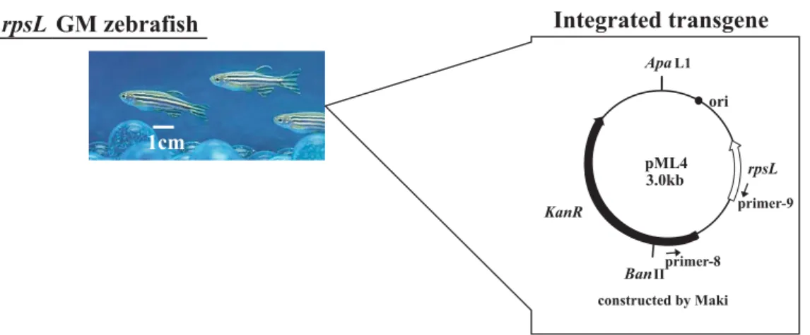
We previously developed GM rpsL zebrafi sh containing a
shuttle vector plasmid (pML4) for detecting mutagens in water1). pML4 contains the rpsL gene as a target gene for
detecting mutations and a kanamycin (Km)-resistance gene as a marker for transformation. Before we can use this rpsL
GM zebrafi sh to monitor environmental mutagens outside the laboratory (i.e., in the river), we have to clarify in
ad-vance the level of integrated DNA transfer from the GM fi sh to environmental bacteria. Therefore, we tried to deter-mine the frequency of DNA transfer from GM fi sh to bacteria existing in or near the fi sh. Here, we showed that the amounts of pML4 DNA in environmental bacteria cultured from feces, gut, and putrescent tissues of GM fi sh were lower than the detection limit in all samples.
2. Materials and Methods
2.1. Fish and transgene
A GM zebrafi sh line carrying approximately 350 copies of the pML4 plasmid per haploid genome was established from the zebrafi sh AB line for a mutagenicity assay1). The
GM fi sh have been maintained as hemizygous GM fi sh in rearing water (0.1% Instant Ocean salts, Tomita Pharma-ceutical Co., Ltd. Tokushima, Japan) at 26°C over 10
gen-Examination of Integrated pML4 DNA Transfer from Genetically
Modifi ed Zebrafi sh to Bacteria
A
KIKOH. H
ASHIMOTO, K
IMIKOA
MANUMA*, K
AZUHIROI
WASAKIand Y
ASUNOBUA
OKINational Institute for Environmental Studies, 16-2 Onogawa, Tsukuba, Ibaraki 305–8506, Japan
* TEL: +81–(0)29–850–2390 FAX: +81–(0)29–850–2588 * E-mail: [email protected]
(Received; 29 September, 2008/Accepted; 11 November, 2008)
The increased use of genetically modifi ed (GM) organisms has raised concerns that the transgenes could be transferred to organisms in the wild. We examined the transfer of a foreign gene integrated into the chromosomal DNA of rpsL transgenic
zebrafi sh to coexisting bacteria. This GM fi sh carries pML4 plasmid DNA containing a kanamycin (Km)-resistance gene as the foreign DNA. We isolated Km-resistant bacteria from feces, gut, and putrescent tissues and examined pML4 DNA trans-fer to them by colony direct PCR to amplify specifi c pML4 sequences. Km-resistant bacteria were grown from feces, but pML4 sequences were not detected in a stored sample of the colonies. We isolated 751 Km-resistant anaerobic bacteria from the gut, but none of them contained the pML4 sequence. To isolate Km-resistant bacteria from putrescent tissues, a GM fi sh euthanized on ice was incubated in rearing water at 28°C for 7 days. The number of Km-resistant bacteria increased with putrefaction, to about 10 times more at 7 days than that at day 0, but the pML4 sequence was not detected in approximately 900 colonies isolated from the putrescent tissues at day 7. These fi ndings suggest that the Km-resistant bacteria isolated in this study were not produced by pML4 gene transfer from the GM fi sh.
erations. The pML4 construct, which contains KanR, a Km-resistance gene5), is shown in Fig. 1. The zebrafi sh AB
line was used as non-GM control fi sh.
2.2. Isolation of bacteria and preparation of PCR samples
We used three kinds of media, Brain Heart Infusion (BHI; BD, NJ, USA), Sabouraud Dextrose Broth (SDB; BD), and Nutrient Broth (NB; BD), of which constituents and/or pH are diff erent among them, for the cultivation of a wide variety of microorganisms according to the methods of Rawls et al.15)
Feces collected from several GM fi sh and non-GM fi sh (17.8 mg and 4.7 mg wet weight, respectively) were suspend-ed in BHI msuspend-edium and spread onto BHI, SDB, or NB agar plates with or without Km (50 mg/l). Colonies were counted after the plates were aerobically cultured at 28°C for 3 days. Fifty colonies obtained from GM fi sh on Km-containing plates were randomly and individually picked for PCR. All the colonies from GM fi sh remaining on each Km plate type (BHI-Km, SDB-Km, or NB-Km) were scraped and combined into a pooled bacterial cell suspension in 10 mM Tris-HCl-1mM EDTA (TE) buff er (pH 8.0) for PCR; each pooled sample consisted of thousands of colonies grown on each type of plate.
A gut collected from a GM fi sh (19.4 mg wet weight) was immediately put into BHI medium, cut into pieces, and homogenized. The gut suspension was spread onto BHI, SDB, or NB plates with or without Km (50 mg/l) in an anaerobic globe box (Anaerorator model FEB-350, Sanki Kagaku Kougei Co., Ltd, Tokyo). These plates were cultured anaerobically in a BBL GasPak (BD) at 28°C for 3 days. A total of 751 colonies of Km-resistant bacteria were isolated individually. The 751 colonies were pooled into 76 bacterial cell suspensions containing 9 or 10 colonies each in TE buff er for PCR.
To obtain putrescent fi sh, a GM fi sh was killed by anes-thesia on ice and placed in 50 ml rearing water and incu-bated at 28°C for 7 days. Just before the incubation, part of the abdomen (skin and intestine, 23.6 mg wet weight, Fig. 2a) was collected as control tissues (0 day), cut into pieces
Fig. 1. Zebrafi sh and pML4 plasmid.
Fig. 2. Putrescence of euthanized GM zebrafi sh.
A GM fi sh euthanized on ice was placed in rearing water and incubated at 28°C for 7 days.
a, GM fi sh after death. Red circle indicates the part that was removed immediately as control tissues; b, GM fi sh at 0 days after removing control tissues; c, GM fi sh after 7 days incuba-tion. Red circle indicates the part removed as putrescent tissue at 7 days.
ed. Each tissue (before and after putrescence) was suspend-ed in BHI msuspend-edium and spread onto BHI, SDB, or NB plates with or without Km (50 mg/l). After the plates were aerobically cultured at 28°C for 3 days, approximately 300 Km-resistant colonies were picked from each plate (6 plates each of BHI-Km, SDB-Km, or NB-Km) cultured from con-trol (0 day) or putrescent (7 days) tissues. The 300 colonies were pooled into samples containing 10 colonies each, to obtain approximately 30 bacterial cell suspensions in TE buff er for each kind of plate and PCR was performed.
2.3. Detection of pML4 sequence by PCR
Colony direct PCR was performed using primers specifi c for pML4; primer-8 (5’-ATCGCGAGCCCATTTATACC-3’) and primer-9 (5’-GGCACGTTGCTTTTCGCAAC-3’) (Fig. 1). We performed PCR in a fi nal volume of 20 μl of Mg2+
plus Ex Taq buff er (Takara Bio Inc., Shiga, Japan)
contain-ing 0.2 mM dNTPs, 10 pmole of each primer, 0.5 unit of
Ex Taq polymerase (Takara Bio Inc.) and 0.05 OD660 units
of the bacterial cell suspension. The PCR conditions were 94°C for 2 min; 35 cycles of 94°C for 30 sec, 63°C for 30 sec, and 72°C for 1 min; and 72°C for 5 min. PCR
prod-scraped and pooled samples from feces was estimated from an experiment employing, as a standard, E. coli RR1
trans-formed with pML4, and it was 2.6 bacterial cells per 20 μl of the PCR mixture (Fig. 3). A similar detection limit was also obtained by using another primer set specific for pML4. For colonies isolated from the gut and putrescent abdomen, 10 colonies were combined into one bacterial cell suspension for PCR, and therefore, the bacterial cell number from each isolated colony in the PCR mixture was much higher than the 2.6 cells/20 μl detection limit described above.
2.4. Calculation of pML4 DNA transfer frequency
The pML4 DNA transfer frequency in Km-resistant bacteria is calculated by the following formula:
(pML4 DNA transfer frequency in Km-resistant bacteria)=(Number of bacteria with transferred pML4 DNA)/(Number of Km-resistant bacteria checked by PCR)
The pML4 DNA transfer frequency in total bacteria cul-tured without Km was calculated by the following formula:
(pML4 DNA transfer frequency in total bacteria) ={pML4 DNA transfer frequency in Km-resistant bacteria ×Number of total bacteria (CFU/g dry sample)}/{Number of Km-resistant bacteria (CFU/g dry sample)}
3. Results
3.1. pML4 DNA transfer from GM fish feces to bacteria
Table 1 shows the number of bacteria cultured from feces collected from GM fi sh or non-GM fi sh using 3 kinds of medium. After 3 days of culture, approximately 107 to 109
CFU/g were grown on each plate without Km from both GM and non-GM fi sh. Considerable numbers of Km-resis-tant colonies grew from the feces of GM fi sh on BHI-Km and NB-Km plates (8.5×10⁸ and 2.0×10⁵ CFU/g dry feces,
respectively), and these numbers were very similar to those cultured from feces of non-GM fi sh (8.5×10⁸ and 2.1×10⁵
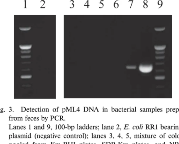
CFU/g dry feces on BHI-Km and NB-Km plates, respec-tively). To examine whether the Km-resistant colonies origi-Fig. 3. Detection of pML4 DNA in bacterial samples prepared
from feces by PCR.
Lanes 1 and 9, 100-bp ladders; lane 2, E. coli RR1 bearing no
plasmid (negative control); lanes 3, 4, 5, mixture of colonies pooled from Km-BHI plates, SDB-Km plates, and NB-Km plates, respectively; lanes 6, 7, and 8, E. coli RR1 bearing the
pML4 plasmid at 0.26, 2.6, and 2.6×10⁶ cells/20 μL reaction
mixture, respectively (positive control).
Table 1. Km resistance and pML4 DNA transfer to bacteria from GM fish feces. Medium (CFU/g dry feces)Total bacteria Km-resistant bacteria
(CFU/g dry feces) Km resistance rate
Number of bacteria used for PCR Number of pML4 DNA transferred bacteria pML4 DNA transfer frequency in total bacteria BHI GM 2.3×10⁹ 8.5×10⁸ 3.7×10⁻¹ 59645 <2.6 <1.6×10⁻⁵ Non-GM 1.8×10⁹ 8.5×10⁸ 4.7×10⁻¹ n.d. n.d. n.d. SDB GM 2.2×10⁸ 1.5×10⁷ 6.8×10⁻² 7677 <2.6 <2.3×10⁻⁵ Non-GM 4.4×10⁷ 6.4×10⁵ 1.5×10⁻² n.d. n.d. n.d. NB GM 2.5×10⁹ 2.0×10⁵ 8.0×10⁻⁵ 2879 <2.6 <7.2×10⁻⁸ Non-GM 1.6×10⁹ 2.1×10⁵ 1.3×10⁻⁴ n.d. n.d. n.d. n.d.: not determined.
The detection limit was estimated to be 2.6 bacterial cell per 20 μL for PCR of scraped and pooled samples. Therefore, the pML4 trans-fer frequency in Km-resistant bacteria used for PCR was calculated to be below 2.6 cells/number of Km-resistant bacteria used for PCR, and the pML4 transfer frequency in total bacteria was calculated to be below 2.6 cells/number of Km-resistant bacteria for PCR×Km-resistance rate. (For example, for the BHI plate, the value was calculated to be below 2.6/59645×3.7×10–1=1.6×10–5.)
nating from GM fi sh contained pML4 DNA, we randomly isolated 50 colonies each grown from GM fi sh feces on the BHI, SDB, and NB plates. All the remaining colonies on each plate were scraped and combined with colonies from other plates containing the same medium into 3 pooled samples and subjected to PCR along with individually iso-lated colonies. After gel electrophoresis analysis, no PCR products were detected in the 50 individual colonies or in the pooled samples (Fig. 3). The pML4 DNA transfer fre-quency in the 3 pooled samples was calculated to be below 10–5–10–8 (Table 1).
3.2. pML4 DNA transfer from GM fi sh gut to anaerobic bacteria
Anaerobic intestinal bacteria from GM fi sh gut were cultured on BHI, SDB, or NB plates with or without Km (Table 2). Substantial numbers of Km-resistant bacteria were detected only on BHI-Km plates. The Km-resistance rate of bacteria cultured on BHI plates was 8.7×10⁻³ as
calculated from the number of Km-resistant colonies (9.6×10⁴ CFU/g dry gut) divided by the total number of
colonies (1.1×10⁷ CFU/g dry gut). We did not scrape
to-gether all the colonies on the plates, but isolated 751 colonies individually to avoid contamination of GM fi sh tissues in the PCR samples. We looked for pML4 DNA in the 751 isolated Km-resistant intestinal bacteria by PCR but did not detect any PCR products corresponding to pML4 DNA, indicating that pML4 was transferred to less than one out of 751 isolated bacteria. From these results, we estimated the pML4 transfer frequency to be under 1.2×10⁻⁵ (1.2×
10–5=1/751×8.7×10–3).
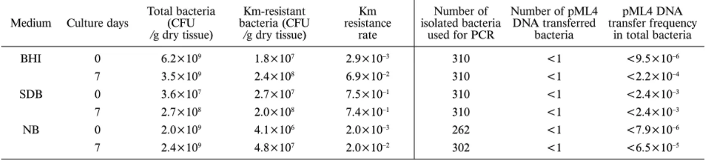
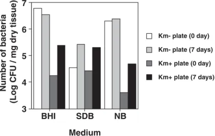
3.3. pML4 DNA transfer from putrescent GM fi sh tissue to bacteria
We next examined whether the gene could be transferred
from decomposing fi sh tissue. After the fi sh was killed by exposure to cold, we removed a part of abdomen contain-ing skin and gut as day 0 control tissues (Fig. 2a, b). The remainder of the fi sh was incubated in rearing water at 28°C. After 5 days of incubation, the outlines of the dead fi sh became unclear, likely due to the putrescence of the tissues. On day 7, we collected some of this tissue from around the abdomen (Fig. 2c). Both control and putrescent tissues were cultured aerobically on BHI, SDB, and NB plates. Considerable numbers of Km-resistant bacteria grew from the control tissue harvested on day 0, but after 7 days incubation, the numbers of Km-resistant bacteria increased to 7.4–13.3 times that at day 0 (Table 3, Fig. 4). We isolat-ed approximately 300 colonies from each plate, and PCR was performed as described in the Materials and Methods. No amplifi ed products were detected in any of the bacterial samples from BHI-Km, SDB-Km, or NB-Km plates at either 0 and 7 days, indicating that pML4 DNA transfer frequencies were below 10–3.
3.4. DNA sequence homology to Km-resistance genes deposited in GenBank
Considerable numbers of Km-resistant bacteria were cultured even from feces of non-GM fi sh (Table 1). Km is an aminoglycoside antibiotic and resistance to Km is often due to enzymatic inactivation of Km. The DNA sequences of many bacterial genes encoding aminoglycoside-modifying enzymes that confer Km-resistance have been already deter-mined (Table 4).
We performed a homology search between KanR in pML4 and all of the Km-resistance genes we found in the DNA Data Bank of Japan and in the review of Shaw et al.16) (Table 4), and found only very low homologies
be-tween KanR and other bacterial genes, except for the uncul-tured eubacterium plasmid pIE1130 (99.1% homology) and Table 2. Km resistance and pML4 transfer to bacteria from GM fish gut.
Medium (CFU/g dry gut)Total bacteria bacteria (CFU Km-resistant
/g dry gut) Km resistance rate
Number of isolated bacteria used for PCR Number pML4 DNA transferred bacteria pML4 DNA transfer frequency in total bacteria BHI 1.1×10⁷ 9.6×10⁴ 8.7×10⁻³ 751 <1 <1.2×10⁻⁵ SDB 1.9×10⁵ 0 n.d. n.d. n.d. ND NB 5.4×10⁷ 0 n.d. n.d. n.d. ND
ND: Not detected, n.d.: not determined.
Table 3. Km-resistant bacteria and pML4 transfer to bacteria from dead GM fish. Medium Culture days Total bacteria (CFU
/g dry tissue) Km-resistant bacteria (CFU /g dry tissue) Km resistance rate Number of isolated bacteria used for PCR Number of pML4 DNA transferred bacteria pML4 DNA transfer frequency in total bacteria BHI 0 6.2×10⁹ 1.8×10⁷ 2.9×10⁻³ 310 <1 <9.5×10⁻⁶ 7 3.5×10⁹ 2.4×10⁸ 6.9×10⁻² 310 <1 <2.2×10⁻⁴ SDB 0 3.6×10⁷ 2.7×10⁷ 7.5×10⁻¹ 310 <1 <2.4×10⁻³ 7 2.7×10⁸ 2.0×10⁸ 7.4×10⁻¹ 310 <1 <2.4×10⁻³ NB 0 2.0×10⁹ 4.1×10⁶ 2.0×10⁻³ 262 <1 <7.9×10⁻⁶ 7 2.4×10⁹ 4.8×10⁷ 2.0×10⁻² 302 <1 <6.5×10⁻⁵
4. Discussion
As a fi rst trial for investigating gene transfer from GM fi sh to environmental bacteria, we cultured Km-resistant bacteria from feces, gut, and putrescent tissues of GM fi sh and examined by PCR whether they contained pML4 DNA sequences. Appreciable numbers of Km-resistant bacteria were cultured in all samples, but the pML4 sequences in
transfer from the GM fi sh. The pML4 DNA transfer fre-quencies were calculated to be below 10–3 to 10–8 (Tables
1–3). Notably, similar numbers of total and Km-resistant bacteria were grown from GM and non-GM feces and similar frequencies of Km resistance were observed (Table 1), suggesting that these Km-resistant bacteria are probably indigenous to natural environments. These fi ndings are con-sistent with the existence of many Km-resistant bacteria
Fig. 4. CFU of total and Km-resistant bacteria isolated from euthanized GM zebrafi sh before (day 0) and after (day 7) putrescence. Table 4. Km resistance genes in bacteria.
Km-resistant bacteria Enzymea (gene) DDBJ/EMBL/GenBank accession no.
Escherichia coli (Kanamycin resistance transposon Tn903; KanR)b V00359 Campylobacter coli
plasmid pIP1433 Kanamycin resistance protein (aphA-3) M26832
Pseudomonas aeruginosa Aminoglycoside acetyltransferase (aac(6')-Iae) AB104852
Staphylococcus aureus Kanamycin nucleotidyltransferase (aadD) AF181950
Streptomyces albulus Kanamycin acetyltransferase (kat) AB116646
Streptomyces kanamyceticus 16S rRNA methylase (kmr) AB164229
Streptomyces kanamyceticus Aminoglycoside 6'-N-acetyltransferase (kac) AB164230 Uncultured eubacterium
plasmid pIE1130 Aminoglycoside phosphotransferase (aph(3')-I) AJ271879
Vibrio cholerae Aminoglycoside adenylyltransferase (aadB) AF221902
Pseudomonas aeruginosa PST-1c Aminoglycoside-(3)-N-acetyltransferase (aac(3)-IIIa) X55652
Pseudomonas aeruginosac n.d.d (aac(3)-IIIb) L06160
Pseudomonas aeruginosac Aminoglycoside 3’-N-acetyltransferase (aac(3)-IIIc) L06161
Unidentifi ed bacteriumc Aminoglycoside adenylyltransferase (ant(2’’)-Ia) X04555 Escherichia coli plasmid RP4c Aminoglycoside-3’-phosphotransferase (aph(3’)-Ib) M20305 Klebsiella pneumoniaec Aminoglycoside-3’,5’’-phosphotransferase-I (
aph(3’)-Ic) M37910
Escherichia coli c Neomycin phosphotransferase (aph(3’)-IIa) V00618 Streptococcus faecalisc 3’,5’’-Aminoglycoside phosphotransferase type III
(aph(3’)-IIIa) V01547
Acinetobacter baumanniic 3’-Aminoglycoside phosphotransferase (aph(3’)-VIa) X07753 Campylobacter jejunic Kanamycin phosphotransferase (aph(3’)-VIIa) M29953
a, Registered name in DDBJ/GenBank.
b, The transgene integrated in the GM fish used in this study carries this gene, KanR. c, Reported in the review of Shaw et al.16)
confi rmed by the survey of the previously identifi ed bacteria; almost all Km-resistant bacterial genes listed (Table 4) have no homology with KanR in pML4. Boon reported that a considerable percentage of bacteria isolated from river water in rural, semi-rural, and urban sites show resistance to several antibiotics, including Km2).
The variation in the DNA transfer frequency (below 10-3
to 10-8) depended on the percentage of Km-resistant bacteria
among the total cultured bacteria and the number of colo-nies isolated for PCR. To obtain more precise data, we may need to isolate much higher numbers of Km-resistant colo-nies for PCR, for example, from putrescent tissues cultured in SDB medium (Table 3). The transformation frequencies of naturally transformable bacteria were reported to be 10–2
to 10–9 12), and the transformation frequencies of a bacterial
strain specialized to receive a GM plant transgene were at the 10–8 level13). Considering these reports and the
non-con-jugative nature of the pML4 plasmid, the frequency of transformation of the bacteria surrounding GM fi sh by pML4 might be lower than natural bacterial transformation and GM plant DNA transfer. To detect such rare events us-ing the strategy employed here, we might need to isolate much higher numbers of Km-resistant colonies and to cul-ture them to create much more bacterial colonies. This strategy, however, contains serious problems; bacteria re-ceiving pML4 may not always become Km-resistant because the Km-resistance gene in pML4 may not always be ex-pressed in a heterologous bacterial host. Moreover, only a small proportion of the bacteria present in the feces, gut, and fi sh environment are culturable. To estimate more precisely the possibility of DNA transfer from GM fi sh to surrounding bacteria, new strategies might be required, such as the recently developed gnotobiotic zebrafi sh technique15),
and/or the establishment of a new transgenic fi sh line bearing a marker gene that is effi ciently selectable and func-tional in a broad range of bacteria.
Despite the limitations to the experiments described above, we showed at least that an integrated transgene of GM fi sh, pML4, did not cause transfer of culturable Km resistance to bacteria existing with the GM fi sh under the experimental conditions, suggesting that using rpsL GM
fi sh1) for monitoring aquatic environments is unlikely to
cause a serious problem with pML4 transfer.
Acknowledgments
We thank Yukari Sakashita for her excellent technical contributions.
This study was supported by the Global Environment Re-search Fund from the Ministry of the Environment of Japan.
References
1) Amanuma, K., H. Takeda, H. Amanuma, and Y. Aoki. 2000. Transgenic zebrafi sh for detecting mutations caused by com-pounds in aquatic environments. Nat. Biotechnol. 18: 62–65. 2) Boon, P.I., and M. Cattanach. 1999. Antibiotic resistance of
native and faecal bacteria isolated from rivers, reservoirs and sewage treatment facilities in Victoria, south-eastern Australia. Lett. Appl. Microbiol. 28: 164–168.
3) Chambers, P.A., P.S. Duggan, J. Heritage, and J.M. Forbes. 2002. The fate of antibiotic resistance marker genes in trans-genic plant feed material fed to chickens. J. Antimicrob. Chemother. 49: 161–164.
4) de Vries, J., M. Heine, K. Harms, and W. Wackernagel. 2003. Spread of recombinant DNA by roots and pollen of transgenic potato plants, identifi ed by highly specifi c biomonitoring using natural transformation of an Acinetobacter sp. Appl. Environ.
Microbiol. 69: 4455–4462.
5) Gondo, Y., Y. Shioyama, K. Nakao, and M. Katsuki. 1996. A novel positive detection system of in vivo mutations in rpsL
(strA) transgenic mice. Mutat. Res. 360: 1–14.
6) Gruzza, M., M. Fons, M.F. Ouriet, Y. Duval-Ifl ah, and R. Ducluzeau. 1994. Study of gene transfer in vitro and in the digestive tract of gnotobiotic mice from Lactococcus lactis
strains to various strains belonging to human intestinal fl ora. Microb. Releases. 2: 183–189.
7) Heinemann, J.A., and T. Traavik. 2004. Problems in monitor-ing horizontal gene transfer in fi eld trials of transgenic plants. Nat. Biotechnol. 22: 1105–1109.
8) Jiang, S.C., and J.H. Paul. 1998. Gene transfer by transduc-tion in the marine environment. Appl. Environ. Microbiol. 64: 2780–2787.
9) Kapuscinski, A.R. 2005. Current scientifi c understanding of the environmental biosafety of transgenic fi sh and shellfi sh. Rev. Sci. Tech. 24: 309–322.
10) Kellenberger, E. 1994. Genetic ecology: a new interdisciplinary science, fundamental for evolution, biodiversity and biosafety evaluations. Experientia. 50: 429–437.
11) Kharazmi, M., S. Sczesny, M. Blaut, W.P. Hammes, and C. Hertel. 2003. Marker rescue studies of the transfer of recombi-nant DNA to Streptococcus gordonii in vitro, in foods and
gnotobiotic rats. Appl. Environ. Microbiol. 69: 6121–6127. 12) Lorenz, M.G., and W. Wackernagel. 1994. Bacterial gene
transfer by natural genetic transformation in the environment. Microbiol. Rev. 58: 563–602.
13) Nielsen, K.M., J.D. van Elsas, and K. Smalla. 2000. Transfor-mation of Acinetobacter sp. strain BD413 (pFG4ΔnptII) with
transgenic plant DNA in soil microcosms and eff ects of kana-mycin on selection of transformants. Appl. Environ. Microbiol. 66: 1237–1242.
14) Pontiroli, A., P. Simonet, A. Frostegard, T.M. Vogel, and J.M. Monier. 2007. Fate of transgenic plant DNA in the environ-ment. Environ. Biosafety Res. 6: 15–35.
15) Rawls, J.F., B.S. Samuel, and J.I. Gordon. 2004. Gnotobiotic zebrafi sh reveal evolutionarily conserved responses to the gut microbiota. Proc. Natl. Acad. Sci. USA. 101: 4596–4601. 16) Shaw, K.J., P.N. Rather, R.S. Hare, and G.H. Miller. 1993.
Molecular genetics of aminoglycoside resistance genes and familial relationships of the aminoglycoside-modifying enzymes. Microbiol. Rev. 57: 138–163.
17) Smalla, K., and T.M. Vogel eds. 2007. Thematic issue on hori-zontal gene transfer. Environ. Biosafety Res. 6(1-2).
18) Stewart, G.J., and C.D. Sinigalliano. 1990. Detection of hori-zontal gene transfer by natural transformation in native and introduced species of bacteria in marine and synthetic sedi-ments. Appl. Environ. Microbiol. 56: 1818–1824.
19) Tuohy, K., M. Davies, P. Rumsby, C. Rumney, M.R. Adams, and I.R. Rowland. 2002. Monitoring transfer of recombinant and nonrecombinant plasmids between Lactococcus lactis
strains and members of the human gastrointestinal microbiota
in vivo-impact of donor cell number and diet. J. Appl.
Micro-biol. 93: 954–964.
20) Williams, H.G., M.J. Day, J.C. Fry, and G.J. Stewart. 1996. Natural transformation in river epilithon. Appl. Environ. Mi-crobiol. 62: 2994–2998.