6 ―104― 報 告
汎血球減少を呈した骨髄原発悪性リンパ腫の 1 例
東京女子医科大学八千代医療センター血液内科 ア サ ノ チ ヒ ロ コ ダ マ ショウコ マ ス ダ ミチヒコ 浅野 千尋・兒玉 聖子・増田 道彦 (受理 平成 30 年 6 月 22 日)A Case of Bone Marrow Lymphoma with Pancytopenia Chihiro ASANO, Shoko KODAMA and Michihiko MASUDA
Department of Hematology, Tokyo Women s Medical University Yachiyo Medical Center
Bone marrow lymphoma is a rare disease. Herein, we report a case of a 77-year old woman who was diag-nosed with primary bone marrow lymphoma. She presented with fatigue and dyspnea 1 month before hospitali-zation. Two weeks before hospitalization, she underwent blood tests, and her complete blood count revealed pan-cytopenia. A bone marrow biopsy was performed and showed abnormal and diffuse lymphoid cells. On immuno-histochemistry, these cells were positive for leukocyto common antigen (LCA), CD20, and Bcl-2. Upon further ex-amination, there was no palpable lymphadenopathy. A positron emission/computed tomography (PET/CT) scan revealed fluorodeoxyglucose (FDG) uptake only by the vertebra and pelvic bone. The patient received R-T-COP chemotherapy (rituximab, cyclophosphamide, pirarubicin, vincristine, and prednisolone). Despite chemotherapy, the disease became refractory, and the patient died 3 months after chemotherapy. Based on this case, we recom-mend for pancytopenia to be considered a symptom of bone marrow lymphoma.
Key Words: bone marrow lymphoma, pancytopenia
緒 言 悪性リンパ腫は全身のどこの臓器にも発生し得る 疾患である.骨髄内に二次性に浸潤する症例は散見 されるが骨髄原発の悪性リンパ腫は比較的まれで, 症例数が少ないため好発年齢や性差など統計的な結 果がなく報告も少数にとどまっている.今回汎血球 減少で発症した骨髄原発悪性リンパ腫を経験したの で報告する. 症 例 患者:77 歳,女性. 主訴: 怠感,労作時の息切れ. 既往歴:69 歳時,骨粗鬆症. 現病歴:東京女子医科大学八千代医療センター (以下,当院)入院の 1 か月前より 怠感,労作時の 息切れを認めていた.入院の 2 週間前に検診を受け, 血液検査で白血球 1,300/μl,血色素 9.5 g/dl,血小板 4.7×10⁴/μl と汎血球減少を認めた.入院前日近医を 受診し,再検査を行ったがさらに汎血球減少の進行 を認めたため当科紹介となった. 入院時現症:血圧,体温等バイタルに異常なし. 眼球結膜に黄染なく,眼瞼結膜に貧血あり.表在リ ンパ節腫脹なく,胸腹部に異常所見はなかった. 入院時検査所見:白血球 1,410/μl(好中球 64.5 %, リンパ球 25.5 %,単球 4.0 %,好酸球 5.5 %,好塩基 球 0.5 %),Hb 6.8 g/dl,血小板 3.5×10⁴/μl,網赤血球 1.9×10⁴/μl と汎血球減少を認めたが,末梢血中に異 :浅野千尋 〒276―8524 千葉県八千代市大和田新田 477―96 東京女子医科大学八千代医療センター血液内科 Email: [email protected] doi: doi:10.24488/jtwmu.88.4_104
Copyright Ⓒ 2018 Society of Tokyo Women s Medical University
! # $ 東女医大誌 第 88 巻 第 4 号 頁 104∼107 平成 30 年 8 月 " # %
7
―105―
Fig. 1 Histological bone marrow findings
Large abnormal lymphocytes were observed with diffuse spread, under hematoxylin and eosin staining (A, B) and were positive for CD20 (C) and bcl-2 (D).
A
B
D
C
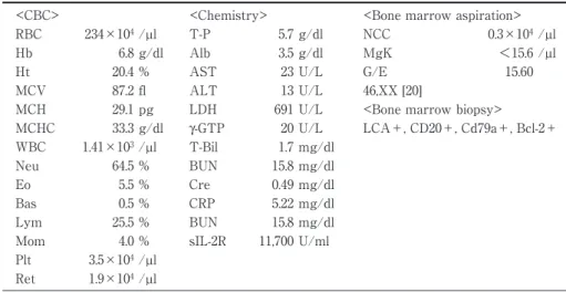
Table 1 Laboratory findings at the time of diagnosis
<CBC> <Chemistry> <Bone marrow aspiration> RBC 234×104/µl T-P 5.7 g/dl NCC 0.3×10⁴/µl Hb 6.8 g/dl Alb 3.5 g/dl MgK <15.6 /µl Ht 20.4 % AST 23 U/L G/E 15.60 MCV 87.2 fl ALT 13 U/L 46,XX [20]
MCH 29.1 pg LDH 691 U/L <Bone marrow biopsy> MCHC 33.3 g/dl γ-GTP 20 U/L LCA+, CD20+, Cd79a+, Bcl-2+ WBC 1.41×10³/µl T-Bil 1.7 mg/dl
Neu 64.5 % BUN 15.8 mg/dl Eo 5.5 % Cre 0.49 mg/dl Bas 0.5 % CRP 5.22 mg/dl Lym 25.5 % BUN 15.8 mg/dl Mom 4.0 % sIL-2R 11,700 U/ml Plt 3.5×10⁴/µl
Ret 1.9×10⁴/µl
型リンパ球は認めなかった.生化学検査では LDH 691 U/l,sIL-2R 11,700 U/ml と上昇していた(Table 1). 臨床経過:入院後に施行した骨髄検査の骨髄塗抹 標本では低形成でほとんど造血細胞はなく再生不良 性貧血を疑ったが,骨髄生検で骨梁間にびまん性に 異型細胞の増生を認めた.免疫染色にて leukocyto common antigen(LCA),CD20,CD79a,Bcl-2 陽性, CD5,CD10,cyclinD1,TdT 陰 性,Ki-67 陽 性 率 は 40∼50 %で最終的に悪性リンパ腫(びまん性大細胞 B 型)と診断された(Fig. 1).CT 検査では明らかな リンパ節腫脹は認められず,肝脾腫もなかったが, PET 検査では全身骨の骨髄のみに異常集積を認め, 骨髄原発と考えられた(Fig. 2).入院 20 日目に R-T-COP 療法(rituximab,cyclophosphamide,piraru-bicin,vincristine,prednisolone)を施行した.治療 後 LDH は一旦は正常化し,sIL-2R 6,640 U/ml と低 下し改善傾向であったが,骨髄回復期に LDH 値は 上昇を認めた(Fig. 3).救援療法を検討したがご本人 はこれ以上の積極的治療は希望されなかったため緩 和医療を行い,入院 69 日目永眠となった. 考 察 骨髄原発悪性リンパ腫は悪性リンパ腫の中でもま れである.特徴としてはリンパ節腫脹に乏しく,末 梢血中に異型リンパ球を認めないため診断に苦慮す る例が多い.骨髄からの発生のため,検査結果では
8
―106―
Fig. 2 Positron emission tomography showing uptake in the bone marrow
Fig. 3 Clinical course and treatment
R-T-COP: rituximab, cyclophosphamide, pirarubicin, vincristine, and prednisolone.
0 100 200 300 400 500 600 700 800 0 500 1000 1500 2000 2500 3000 3500 4000 4500 1 2 3 4 5 6 7 8 9 10 11 12 13 14 15 16 17 18 19 20 21 22 23 LDH WBC /μl U/L Day
R-T-COP
ほとんどの症例に血球減少を認める1)∼3) .発熱などの B 症状を認める場合があるが血球貪食症候群を示す こともある.血球貪食症候群はマクロファージなど の抗原提示細胞と CD8 陽性 T 細胞が活性化し高サ イトカイン血症となり,組織障害や血球減少,凝固 異常などを起こす疾患で,成人ではウイルス感染や 悪性リンパ腫など造血器腫瘍によって二次性に発症 することが多い.梶浦らは 9 例中 6 例に血球貪食症 候群を併発していたと報告している4) .本症例では骨 髄生検で明らかな血球貪食像は認められなかった. 診断基準は明確になっているわけではないが,①骨 髄内のみに腫瘍細胞の浸潤を認めるが,末梢血には 腫瘍細胞を認めないこと,②CT などの検査でリン パ節,肝臓,脾臓や他の節外に病変を認めないこと, ③他の骨腫瘍がないこと,などが提唱されている5) . またchronic lymphocytic leukemia / small lympho-cytic lymphona ( CLL / SLL ) , plasmablastic lym-phoma,mantle cell lymphoma や splenic marginal zone lymphoma など骨髄以外に病変が発見されに くい疾患も除外される.診断には骨髄生検は必須で あるが,最近では PET/CT 検査の有用性が示唆さ れている6)9 ―107― 組織型としては B 細胞性が多く,T 細胞性はまれ である.Martinez ら5) は 21 例の骨髄原発悪性リンパ 腫の症例を検討しているが T 細胞性は 2 例のみで あった.また 19 例の B 細胞性の中でもびまん性大 細胞 B 型が 15 例と一番多く,続いてろ胞性が 4 例 であった. 治療方針も確定していないため CHOP 療法(R-CHOP 療法)に基づき治療されているが,予後は不 良で,びまん性大細胞 B 型では全生存率の中央値は 1.8 年であった5)7) .本症例でも一旦は治療効果が認め られたが次回治療前に病勢が悪化し,治療不応と考 えられている. 骨髄原発悪性リンパ腫の症例は身体所見では診断 が難しく,血液検査で汎血球減少を認める例が多い. 汎血球減少を認める症例では骨髄原発悪性リンパ腫 の可能性を考慮すべきである. 結 論 骨髄原発の悪性リンパ腫は非常にまれであるが, 検査所見として汎血球減少を示すことがあるため原 因不明の汎血球減少の場合は同疾患を考慮すること も必要と考える. 開示すべき利益相反はない. 文 献
1)Niscola P, Palombi M, Fratoni S et al: Primary diffuse large B-cell lymphoma of the bone marrow in a frail and elderly patient successfully treated with rituximab, cyclophosphamide, doxorubicin, vincristine, and prednisone. Blood Res 48: 296―297, 2013
2)Yacoub A, Aljitawi OS, Cui W : Primary Bone Marrow DLBCL, unraveling the unique biology of this very rare presentation. Blood 122: 5065, 2013 3)Hishizawa M, Okamoto K, Chonabayashi K et al:
Primary large B-cell lymphoma of the bone mar-row. Br J Hematol 136: 351, 2007
4)Kajiura D, Yamashita Y, Mori N: Diffuse large B-cell lymphoma initially manifesting in the bone marrow. Am J Clin Pathol 127: 762―769, 2007 5)Martinez A, Ponzoni M, Agostinelli C et al:
Pri-mary bone marrow lymphoma: an uncommon ex-tranodal presentation of aggressibe non-Hodgkin lymphomas. Am J Surg Pathol 36: 296―304, 2012 6)Moritani K, Nakano N, Yonezawa S et al:
Useful-ness of positron emission tomography-CT for diag-nosis of primary bone marrow lymphoma in chil-dren. Pediatr Hematol Oncol 12: 1―6, 2018
7)Chang H, Hung YS, Lin TL et al: Primary bone marrow diffuse large B cell lymphoma: a case se-ries and review. Ann Hematol 90: 791―796, 2011