Activated Protein Kinase C Attenuates Ca
2+Overloading and
Re-oxygenation Hypercontracture in Isolated Rat Cardiomyocytes following
Chemical Hypoxia
Shigeto Miyasaka, Takashi Ichiba, Hiroaki Kuroda and Shigetsugu Ohgi
Second Department of Surgery, Faculty of Medicine, Tottori University, Yonago 683-0826, Japan
The aims of this study were i) to test the effects of protein kinase C (PKC) activation on the intracellular Ca2+ concentration of rat cardiomyocytes during chemical hypoxia-reoxygenation, ii) to determine the contribution of the sarcoplasmic reticulum (SR) and the Na+-Ca2+ exchange on the regulation of intracellular Ca2+ following PKC activation and iii) to test the role of PKC-dependent intracellular pH changes in intracellular Ca2+ regulation. We used the isolated adult rat cardiomyocyte perfusion model. Cardiomyocytes were loaded with the Ca2+-fluorescent probe Fluo-3 and the pH-fluorescent probe SNARF1. Cells were subjected to 50 min of glucose-free and NaCN chemical hypoxia followed by 30 min of simulated reoxygenation. The activation of PKC significantly inhibited the hypoxia-induced increase of Fluo-3 fluorescent intensity (control; 692 ± 100% and activated PKC; 322 ± 43%: P < 0.05). This inhibitory effect was not affected by the inhibition of the SR Ca2+ uptake induced by thapsigargin, but was cancelled by the inhibition of the Na+-Ca2+ exchange with dichlorobenzamil (thapsigargin 337 ± 47%; dichlorobenzamil 609 ± 100%). PKC activation also attenuated the decrease in intracellular pH during chemical hypoxia, even in the presence of the Na+-H+ exchange inhibitor amiloride (control; 6.54 ± 0.02, PKC; 6.72 ± 0.03, PKC + amiloride; 6.73 ± 0.03). We concluded that the PKC attenuation of Ca2+ overloading to rat cardiomyocytes during chemical hypoxia-reoxygenation does not depend on the Ca2+ uptake by the SR, but does require a Na+-Ca2+ exchange. Since PKC attenuated the increasing intracellular H+ during chemical hypoxia, a low H+ concentration may be important for the maintenance of Ca2+ extrusion via the Na+-Ca2+ exchange.
Key words: Ca2+ overload; intracellular pH; isolated rat cardiomyocytes; Na+-Ca2+ exchanger; protein kinase C
Ischemic preconditioning reduces myocardial injury induced by sustained ischemia-reperfu-sion, with preservation of myocardial function and infarct size reduction. Previous findings suggest that the activation of protein kinase C (PKC) plays a role in the cardioprotective effect of ischemic preconditioning (Liu et al., 1994; Li and Kloner, 1995; Kitakaze et al., 1996). PKC activation by Ca2+ or pharmacologic agents mimics ischemic preconditioning (Miyawaki
and Ashraf, 1977; Armstrong and Ganote, 1994; Tsuchida et al., 1994; Miyawaki et al., 1996). It has been proposed that PKC attenu-ates an increase in intracellular Ca2+ concentra-tion during ischemia/reperfusion via i) intra-cellular alkalization (MacLeod and Harding, 1991), ii) activation of the sarcoplasmic reticu-lum (SR) calcium pump (Movesesian et al., 1984), iii) modification of the Na+-Ca2+ ex-change (Steenbergen et al., 1993) and iv)
inhib-Abbreviations: amil, amiloride; [Ca2+]
i, intracellular Ca2+ concentration; %[Ca2+]i,percentage of the pre-ischemic fluorescene intensity; DCB, dichlorobenzamil; Fluo-3, Fluo-3 acetoxymethylester; %L, percentage of the prehypoxic length; PKC, protein kinase C; pHin, intracellular pH; PMA, phorbol 12-myristate 13-acetate; SNARF1, SNARF1 acetoxymethylester; SR, sarcoplasmic reticulum;thaps, thapsigargin
ition of the L-type Ca2+ channel (Leatherman et al., 1987; Zheng et al., 1992). However, the precise mechanism underlying the cardiopro-tection afforded by PKC activation is unknown. The present study i) examined how PKC affects the intracellular Ca2+ concentration ([Ca2+]
i) and cell length of isolated rat cardio-myocytes during chemical hypoxia-reoxygena-tion, ii) tested whether the SR and Na+-Ca2+ exchanger on the sarcolemma contribute to the attenuation of Ca2+ overloading during ische-mia under the activation of PKC and iii) exa-mined how PKC affects intracellular pH (pHin) and the regulation of the intracellular Ca2+ during chemical hypoxia-reoxygenation.
Materials and Methods Isolation of rat cardiomyocytes
Ventricular myocytes were isolated from adult rat hearts by enzymatic dissociation (Satoh et al., 1994; Siegmund et al., 1994). In brief, 8-week-old male Wistar rats weighing 200 to 250 g were anesthetized by inhalation of diethyl ether. After an injection of 50 U of heparin into the inferior vena cava, the hearts were quickly excised and attached to a Langendorff appara-tus. Each heart was perfused through the coro-nary artery with i) Ca2+-free Tyrode solution (136.5 mmol/L NaCl, 5.4 mmol/L KCl, 0.53 mmol/L MgCl2, 5.5 mmol/L HEPES, 5.5 mmol/ L glucose, adjusted to pH 7.4 with NaOH) at 37˚C for 3 min, ii) Tyrode solution containing collagenase type I (0.04 g/100 mL, Sigma Chemical Co, St. Louis, MO) for 20 min and iii) modified Kraftbruhe (KB) solution (70 mmol/L KOH, 40 mmol/L KCL, 20 mmol/L KH2PO4, 3 mmol/L MgCl2, 50 mmol/L glutamic acid, 10 mmol/L glucose, 10 mmol/L HEPES, 0.5 mmol/L EGTA, titrated to pH 7.4 with KOH). The ventricles were excised and minced in modified KB solution. The cells were filtered through a stainless steel mesh (150 µm) and incubated for 2 h.
The Guidelines for Animal Experimentation at the Faculty of Medicine, Tottori University, were followed.
Loading of Fluo-3 and SNARF1
The culture medium was changed from KB solution to Medium 199 (GIBCO, Grand Island, NY) containing 1% bovine serum albumin (wt/ vol), the Ca2+-selective fluorescence probe 10 µmol/L Fluo-3 acetoxymethylester (Fluo-3) (Dojindo, Kumamoto, Japan). Pluronic F127 [0.075% (wt/vol)] (Molecular Probes, Inc., Evgene, OR) was used as a detergent for cell permeation of the dye (Satoh et al., 1994; Lopez et al., 1996). Cells were loaded for 30 min at 37˚C, followed by incubation for 30 min with Tyrode solution to allow hydrolysis of the Fluo-3. The fluorescent pH indicator SNARF1 ace-toxymethylester (SNARF1) (Molecular Probes) at 10 µmol/L was loaded in the same way as Fluo-3 (Seksek et al., 1991; Zaguilan et al., 1991).
Apparatus and measurement of [Ca2+]i ,
cell length and pHin
Cardiomyocytes attached to a glass-bottomed dish (content volume 1 mL) on the stage of an inverted conforcal laser microscope (InSIGHT, Meridian Instruments Far East, Tokyo, Japan) were perfused (0.6 mL/min) with Tyrode solution. The temperature of the dish was maintained at 37˚C by a temperature controller (Medical Systems Corp, NY).
The ion-selective fluorescent probes were excited by an argon laser, at wave lengths of 488 nm for Fluo-3 and 514 nm for SNARF1. The light emitted from the cells was monitored by a CCD camera (Meridian Instruments) using interference filters (530 nm for Fluo-3 and 580 nm and 644 nm for SNARF1). Images were recorded by a video cassette recorder (SONY, Tokyo) at 30 frames per second.
The fluorescence intensity of Fluo-3 was analyzed by the InSIGHT PLUS-IQ analysis system (Meridian Instruments). Because Fluo-3 has no spectral shift on binding Ca2+, the abso-lute value of [Ca2+]
i can not be measured (Minta et al., 1989; Hayashi et al., 1994). Changes in [Ca2+]
i are expressed as the percentage of the preischemic fluorescence intensity (%[Ca2+]
The fluorescence intensity of SNARF1 was analyzed by the InSIGHT PLUS-IQ analysis system with a ratio of 580/644 nm. The in situ calibration of SNARF1 was conducted by the nigericin-K+ method (Seksek et al., 1991; Zaguilan et al., 1991). The fluorescence inten-sity ratio of 580/644 nm was converted to pHin using a published calibration curve.
Cell length was measured from the Fluo-3 fluorescence image by the InSIGHT PLUS-IQ analysis system, and changes in cell length are expressed as the percentage of the prehypoxic cell length (%L).
The %[Ca2+]
i, pHin and %L were calculated from the mean of 30 frames (1 s) at each experi-mental point.
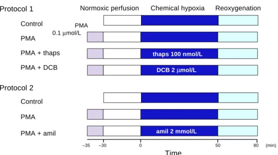
Experimental protocol 1
Effects of PKC activation on Ca2+ over-loading and cell length during chemical hypoxia-reoxygenation (Fig. 1).
Control: Cardiomyocytes loaded with
Fluo-3 were perfused with Tyrode solution equil-ibrated with 100% O2 for 30 min. The cells
were then subjected to chemical hypoxia for 50 min and reoxygenation for 30 min. The chem-ical hypoxic solution was based on Tyrode so-lution but contained 2 mmol/L NaCN, no glucose and a pH of 6.7.
PMA: Cells were exposed to 0.1 µmol/L phorbol 12-myristate 13-acetate (PMA) (Wako, Osaka, Japan) for 5 min before normoxic perfu-sion. The cells were subsequently perfused with oxygenated Tyrode solution and subjected to chemical hypoxia-reoxygenation in the same way as the controls.
PMA + thaps: The procedure was similar
to the PKC group except that 100 nmol/L thapsigargin (thaps) (Funakoshi, Tokyo) was included in the perfusion solution administered during chemical hypoxia (Wu and Feher, 1995; Meldrum et al., 1996).
PMA + DCB: The procedure was similar to
the PKC group except that 20 µmol/L 2-4-dichlorobenzamil (DCB) (Molecular Probes) was included in the perfusion solution admin-istered during chemical hypoxia (Schiffman et al., 1990; Kawada et al., 1992).
Fluo-3 signals were obtained 1 min before
Protocol 1 Control PMA PMA + thaps PMA + DCB Protocol 2 Control PMA PMA + amil
Normoxic perfusion Chemical hypoxia Reoxygenation PMA 0.1 µmol/L thaps 100 nmol/L DCB 2 µmol/L amil 2 mmol/L –35 –30 0 50 80 (min) Time
Fig. 1. Schematic representation of the 2 cardiomyocyte treatment protocols. Cells were perfused with 100%
oxygenated Tyrode solution during normoxic perfusion and reoxygenation periods. Chemical hypoxia was achieved by the perfusion of Tyrode solution with 2 mmol/L NaCN without glucose at pH 6.7. In the activated PKC group, cells were perfused with Tyrode solution with 0.1 µmol/L PMA for 5 min from –35 to –30 min. amil, amiloride; DCB, dichlorobenzamil; PKC, protein kinase C; PMA, phorbol 12-myristate 13-acetate; thaps, thapsigargin.
pre 5 10 15 20 25 30 35 40 45 50 5 10 15 20 25 30 pre 5 10 15 20 25 30 35 40 45 50 5 10 15 20 25 30
chemical hypoxia as prehypoxic data and for every 5-min period during chemical hypoxia-reoxygenation.
Experimental protocol 2
Effects of PKC activation on intracellular acido-sis during chemical hypoxia-reoxygenation (Fig. 1).
Control: Cells loaded with SNARF1
under-went the same procedure as the control group in protocol 1.
PMA: Cells loaded with SNARF1 underwent
the same procedure as the PMA group in protocol 1.
PMA + amil: This procedure was similar to
that used for PMA, except that 2 mmol/L
amil-Fig. 2. The time course of
changes in %[Ca2+]
i during chemical hypoxia-reoxygena-tion in untreated (open circles, n = 11) and activated PKC-treated (closed circles, n = 11) rat car-diomyocytes. Data are mean ± SEM of control and PMA-treated cells. PKC, protein kinase C; PMA, phorbol 12-myristate 13-acetate.
Fig. 3. The time course of %
changes in initial cell length during chemical hypoxia-reoxy-genation in untreated (open circles, n = 11) and activated PKC (closed circles, n = 11) rat cardiomyocytes. PKC, protein kinase C; PMA, phorbol 12-myristate 13-acetate.
Chemical hypoxia Reoxygenation
Control PMA Time (min) 100 90 80 70 60 50 40 30 20 %L (%) (%)
Chemical hypoxia Reoxygenation
Control PMA Time (min) 900 800 700 600 500 400 300 200 100 0 %[Ca 2+ ]i
oride (amil) (an inhibitor of the Na+-H+ exchang-er) was administered during chemical hypoxia. The data were collected at the same point as those for protocol 1.
Statistical analysis
All data are expressed as mean ± SEM. The sig-nificance of differences among groups was deter-mined using an analysis of variance with Fisher’s PLSD post hoc test. Values of P < 0.05 were considered significant. All statistical ana-lysis was performed using the software program StatView version J-4.5 on a Macintosh com-puter.
Results Isolated cardiomyocytes
We selected the following cells, each loaded with fluorescent dye to use as material in the experiment: i) cells that were rod-shaped, ii) those with clear cross-striations and iii) quiescent cells. The mean initial cell length was 106 ± 3 µm (n = 42), and there were no significant differences in cell length among these 4 groups. The cells which had entirely balled up into blebbed forms with an absent sarcomere pattern were defined as hyper-contractured cells.
Effects of PKC activation on [Ca2+]i and
cell length
In the control and PMA groups, [Ca2+]i in-creased significantly 25 min after the induction of chemical hypoxia (Fig. 2). The %[Ca2+]i rose continuously to the end of chemical hypoxia in both groups. However, the increase
of [Ca2+]i in the PMA group was considerably attenuated from 35 to 50 min of chemical hypoxia compared with controls. Finally, the %[Ca2+]i of the control and PMA groups reached 692 ± 100% (n = 11) and 322 ± 43% (n = 11) respec-tively in chemical hypoxia (P < 0.05) (Fig. 4). After reoxygenation, %[Ca2+]i decreased in both groups and reestablished preischemic levels within 15 min.
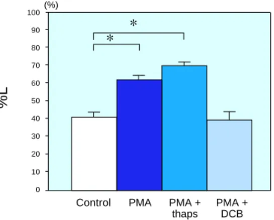
Ischemic cell shortening (rigor shortening) was observed 15 min after the initiation of chemical hypoxia in both groups (Fig. 3). The cell shapes changed rapidly from rod to square with a length reduction to about 62% of the initial length. At the end of chemical hypoxia, the %L values of the control and PMA groups were reduced to 62.9 ± 1.7% and 62.5 ± 1.5%, respectively. In the control cells, hypercontra-ction occurred at the initiation of reoxygena-tion. In contrast, hypercontraction was not ob-served in the PMA group, and some cells slight-ly recovered their length. At 30 min after re-oxygenation, the %L values of the control and PMA groups were 40.8 ± 2.6% and 61.8 ± 2.1% (P < 0.05), respectively (Fig. 5).
Fig. 5. Cell length changes in isolated rat
cardiomyocytes treated with PMA, thaps or DCB. In the PMA group and the PMA + thaps group, cell lengths were maintained compared with the controls, but in the PMA + DCB group, the cell lengths were not significantly different from those of the controls at the end of reoxygenation. *P < 0.05. DCB, dichlor-obenzamil; PMA, phorbol 12-myristate 13-acetate; thaps, thapsigargin.
(%)
Control PMA PMA + PMA + thaps DCB 1000 900 800 700 600 500 400 300 200 100 0 %[Ca 2+ ]i (%)
Control PMA PMA + PMA + thaps DCB 100 90 80 70 60 50 40 30 20 10 0 %L
Fig. 4. Intracellular Ca2+ concentration changes in isolated rat cardiomyocytes treated with PMA, thaps or DCB. The PMA group and the PMA + thaps group showed lesser increases of %[Ca2+]i than the controls, but the PMA + DCB group showed no sig-nificant difference from the controls at the end of chemical hypoxia. *P < 0.05. DCB, dichloro-benzamil; PMA, phorbol 12-myristate 13-acetate; thaps, thapsigargin.
**
pre 2 5 10 15 20 25 30 35 40 45 50 5 10 15 20 25 30
Contribution of SR and the Na+-Ca2+ ex-changer
In the PMA + thaps group, the increase of %[Ca2+]i during chemical hypoxia was attenu-ated. The PMA + thaps value of %[Ca2+]i at 50 min of hypoxia was 337 ± 47% (n = 10), with no significant difference between the PMA and PMA + thaps group (Fig. 4).
The alteration of %L in the PMA + thaps group was similar to that of the PMA group; hypercontraction was not observed after reoxy-genation, and the final %L was 69.4 ± 1.78%, not significantly different from the PMA group value (Fig. 5). These results indicate that the effects of PKC activation were not abolished despite the inhibition of SR Ca2+ uptake.
In the PMA + DCB group, the %[Ca2+]i at 50 min of hypoxia and final %L were 609 ± 100% and 39.4 ± 4.6% (n = 10), respectively. These values were not significantly different from those of the controls (Figs. 4 and 5). These results suggest that the effects of PKC activation were abolished by inhibition of the Na+-Ca2+ exchanger.
Effects of PKC activation on pHin during
chemical hypoxia
The pHin values before chemical hypoxia in the control, PMA and PMA + amil groups were 7.30 ± 0.03 (n = 10), 7.33 ± 0.02 (n = 9) and
7.25 ± 0.03 (n = 9), respectively. In all groups, a decrease of pHin was observed during chem-ical hypoxia (Fig. 6).
In the PMA and PMA + amil groups, intra-cellular acidification was significantly reduced compared with the control group at 25 min be-fore reoxygenation. The final pHin values dur-ing chemical hypoxia of the control, PMA and PMA + amil groups were 6.54 ± 0.02, 6.72 ± 0.03 and 6.73 ± 0.03, respectively (Fig. 6).
In the PMA + amil group, the effect of PKC activation was observed despite inhibition of the Na+-H+ exchanger.
Discussion
This study demonstrated that PKC activation by PMA affects the [Ca2+]i, L and pHin of isolated rat cardiomyocytes during chemical hypoxia. In-tracellular Ca2+ is a very important factor in cell damage during ischemia-reperfusion (Smith and Allen, 1988). A Ca2+ overloading of car-diomyocytes accelerates ATP depletion and activates Ca2+-dependent enzymes, leading to irreversible changes including hypercontraction at reperfusion (Nicotera et al., 1989; Ladilov et al., 1997). The reduction of a Ca2+ overload may thus reduce reperfusion injury, preserve cardiac function and limit infarction size.
The results of the present study indicate that the activation of PKC reduced the increase of [Ca2+]i during chemical hypoxia. This suggests
Chemical hypoxia Reoxygenation
Control PMA PMA + amil Time (min) 7.6 7.4 7.2 7 6.8 6.6 6.4 6.2 pH in
Fig. 6. The time course of
changes in pHin during chem-ical hypoxia-reoxygenation in untreated rat cardiomyocytes (open circles, n = 10 ), those with activated PKC (closed cir-cles, n = 9 ), and those with acti-vated PKC and inhibited Na+ -H– exchanger (squares, n = 9 ). amil, amiloride; PKC, protein kinase C; PMA, phorbol 12-myristate 13-acetate.
that activating PKC might protect the myocar-dium from ischemia and reperfusion injury, as previously reported (Miyawaki and Ashraf, 1977; Armstrong and Ganote, 1994; Tsuchida et al., 1994; Miyawaki et al., 1996).
Cell-length changes of isolated cardio-myocytes may be useful as an index for systolic and diastolic function of the myocardium. We observed that the rat cardiomyocytes shortened to 65% of their initial length during chemical hypoxia. This hypoxic shortening is reversible, and myocytes have the ability to undergo twitch contractions if the cells have suffered no irre-versible ischemic damage. When the ischemic injury was more severe, reoxygenation caused a sudden further contraction of the cells at reperfusion, to 36% of their initial length. Hyper-contracted cells never recover their length and have no contraction ability (Stern et al., 1985). Pathologically, hypercontraction may prevent recovery of cardiac function. The lengths of the present PKC-activated cells were maintained after reoxygenation compared with the controls, which suggests that activated PKC may protect myocardial function. Meldrum and colleagues (1996) demonstrated the effects of activated PKC on cardiac function and the leakage of creatinine kinase during ischemia-reperfusion, using the Langendorff model. Our results support their study; we examined the same phenomena at the cellular level.
Several mechanisms for the attenuation of Ca2+ overload during ischemia by activated PKC have been proposed. Movsesian and co-workers (1984) reported that PKC stimulates Ca2+ uptake from cytosol by the SR. Steenbergen and coworkers (1993) suggested that PKC activation modifies the Na+-Ca2+ exchanger. Lacarda and colleagues (1988) proposed that activated PKC inhibits the L-type Ca2+ channel. However, the precise mechanism of how PKC attenuates a Ca2+ overload remains uncertain. In the present study, we tested the contribution of Ca2+ uptake by SR and Ca2+ extrusion by the Na+-Ca2+ exchanger under the activation of PKC. The inhibition of Ca2+ uptake from the cyto-sol into the SR by thapsigargin did not block the effects of activated PKC during chemical hypo-xia. This result suggests that the SR did not
play an important role in the effect of PKC. Since the SR Ca2+ pump requires ATP to func-tion, its activation by PKC might increase ATP consumption, and the pump would thus not function during severe ischemic ATP depletion. However, when the Na+-Ca2+ exchanger was inhibited, the effects of PKC activation were abolished. This indicates that the attenu-ation of a Ca2+ overload by PKC depends on the Na+-Ca2+ exchanger. The Na+-Ca2+ exchanger is a passive Ca2+ transport system that is depen-dent on the electrical membrane potential and the intra- and/or extra-cellular concentrations of Ca2+ and Na+. The Na+-Ca2+ exchanger is thus a potentially important mechanism in the regulation of Ca2+ under the condition of severe ATP depletion.
The activation of PKC also reduced intra-cellular acidification during chemical hypoxia. Meldrum and colleages (1997) and Rehring and colleages (1996) reported that the activation of PKC by PMA or α1 adrenoreceptor stimulation limits ischemic acidosis in rat hearts. This ef-fect was independent of the Na+-H+ exchanger, because it occurred despite Na+-H+ exchanger inhibition. Macleod and Harding (1991) pro-posed that PKC does not activate the Na+-H+ exchanger directly but rather the Na+-H+ ex-changer function was modified by the change in pHin. The mechanisms underlying the reduc-tion of intracellular acidificareduc-tion following PKC activation are unknown, although Murry and coworkers (1990) showed that precondition-ing reduced the production of lactate, resultprecondition-ing in a drop in cytosolic pH during ischemia.
Kohmoto and colleagues (1990) and Coravoeuf and coworkers (1981) suggested that intracellular acidosis impairs Ca2+ extrusion via the Na+-Ca2+ exchanger. Intracellular H+ may directly inhibit the Na+- Ca2+ exchanger (Philipson et al., 1982), and may also indirectly inhibit it by increasing the Na+ influx via the Na+-H+ exchanger (Baker and McNaughton, 1976). If activated PKC attenuates intracellular acidosis by stimulation of the Na+-H+ exchang-er, the intracellular Na+ would increase and in-hibit the Na+-Ca2+ exchanger. Thus, PKC may not directly stimulate the Na+-Ca2+ exchange but rather may inhibit it via a reduction of H+
production. Our present results suggest that a reduced pHin leads to a reduced Na+ influx via the Na+-H+ exchanger and limits increases in [Ca2+]
i by maintaining the function of the Na+ -Ca2+ exchanger. This hypothesis is the same as that of Steenbergen (1993), who reported similar ionic changes using a nuclear magnetic resonance (NMR) analysis of ischemically preconditioned guinea pig hearts.
Since the signal transduction pathways following PKC activation are complex, there are other possible mechanisms for cardio-protection from Ca2+ overload and reoxyge-nation hypercontracture. Ladilov and co-workers (1997) suggested that the prevention of the ischemic dephosphorylation of contractile proteins reduces the susceptibility of cells to reoxygenation hypercontracture. The reduction of myofilament sensitivity to calcium may be mediated by PKC-dependent pathways and contribute to the reduction of the susceptibility of isolated cardiomyocytes to reoxygenation hypercontracture.
In summary, the activation of PKC attenu-ates the Ca2+ overload of rat cardiomyocytes and facilitates the maintenance of cell length during chemical hypoxia-reoxygenation. These effects do not depend on the Na+-H+ exchanger or SR, but require the Na+-Ca2+ exchanger func-tion and a reducfunc-tion of intracellular acidosis.
Acknowledgments: We would like to thank Prof. J.
Hasegawa, Dept. of Clinical Pharmacology and Prof. Y. Hiji, the First Dept. of Physiology, Faculty of Medicine, Tottori University for their appropriate advice and valuable suggestions to this study.
References
1 Armstrong S, Ganote CE. Preconditioning of isolated rabbit cardiomyocytes: effects of glyco-lytic blockade, phorbol esters, and ischaemia. Cardiovasc Res 1994;28:1700–1706.
2 Baker PF, McNaughton PA. Kinetics and ener-getics of carcium efflux from intact squid giant axons. J Physiol 1976;259:103–114.
3 Coraboeuf E, Gautier P, Guiraudou P. Potential and tension changes induced by sodium removal in dog Purkinje fibers: role of an electrogenic
sodium-calcium exchange. J Physiol 1981;311: 605–622.
4 Hayashi H, Satoh H, Noda N, Terada H, Hirano M, Yamashita Y, et al. Simultaneous measure-ment of intracellular Na+ and Ca2+ during K+-free perfusion in isolated myocytes. Am J Physiol 1994;266:C416–C422.
5 Kawada T, Yoshida Y, Sakurai H, Imai S. Myocardial Na+ during ischemia and accumu-lation of Ca2+ after reperfusion: a study with monesin and dichlorobenzamil. Jpn J Pharmacol 1992;59:191–200.
6 Kitakaze M, Noda K, Minamino T, Komamura K, Funaya H, Shinozaki Y, et al. Role of acti-vation of protein kinase C in the infarct size-limiting effect of ischemic preconditioning through activation of ecto-5'-nucleotidase. Cir-culation 1996;93:781–791.
7 Kohmoto O, Spitzer KW, Movsesian MA, Barry WH. Effects of intracellular acidosis on [Ca2+]
i transients, transsarcolemmal Ca2+ fluxes, and contraction in ventricular myocytes. Circ Res 1990;66:622–632.
8 Lacarda AE, Rampe D, Brown AM. Effects of protein kinase C activators on cardiac Ca2+ channels. Nature 1988;335:249–251.
9 Ladilov YV, Siegmund B, Balser C, Piper HM. Simulated ischemia increases the susceptibility of rat cardiomyocytes to hypercontracture. Circ Res 1997;80:69–75.
10 Leatherman GF, Kim D, Smith TW. Effect of phorbol esters on contractile state and calcium flux in cultured chick heart cells. Am J Physiol 1987; 253:H205–H209.
11 Li Y, Kloner RA. Does protein kinase C play a role in ischemic preconditioning in rat hearts? Am J Physiol 1995;268:H426–H431.
12 Liu Y, Ytrehus K, Downey JM. Evidence that translocation of protein kinase C is a key event during ischemic preconditioning of rabbit myo-cardium. J Mol Cell Cardiol 1994;26:661–668. 13 Lopez JR, Jahangir R, Jahangir A, Shenk WK,
Terzic A. Potassium channel openers prevent potassium-induced calcium loading of cardiac cells. Possible implications in cardioplegia. J Thorac Cardiovasc Surg 1996;112:820–831. 14 Macleod KT, Harding SE. Effects of phorbol
ester on contraction, intracellular pH and intra-cellular Ca2+ in isolated mammalian ventricular myocytes. J Physiol 1991;444; 481–498. 15 Meldrum DR, Cleveland JC Jr, Meng X,
Sharidan BC, Gamboni F, Cain BS, et al. Protein kinase C isoform diversity in preconditioning. J Surg Res 1997;69:183–137.
16 Meldrum DR, Cleveland JC, Mitchell MB, Rowland RT, Banarjee A, Harken AH. Const-ructive priming of myocardium against ischemia-reperfusion injury. Shock 1996;6:238–234. 17 Meldrum DR, Cleveland JC, Sheridan BC,
Rowland RT, Banerjee A, Harken AH. Cardiac preconditioning with calcium: clinically acces-sible myocardial protection. J Thorac Cardiovasc Surg 1996;112:778–786.
18 Minta A, Kao JPY, Tsien RY. Fluorescent indi-cators for cytosolic calcium based on rhodamine and fluorescein choromophores. J Biol Chem 1989;264:8171–8178.
19 Miyawaki H, Ashraf M. Isoproterenol mimics calcium preconditioning-induced protection against ischemia. Am J Physiol 1977;272:H927– H936.
20 Miyawaki H, Zhou X, Ashraf M. Calcium pre-conditioning elicits strong protection against ischemic injury via protein kinase C signaling pathway. Circ Res 1996;79:137–146.
21 Movsesian M, Nishikawa M, Adelstein RS. Phosphorylation of phospholamban by calcium-activated, phospholipid-dependent protein kinase. J Biol Chem 1984;259:8029–8032. 22 Murry CE, Richard VJ, Reimer KA, Jennings
RB. Ischemic preconditioning slows energy metabolism and delays ultrastructural damage during a sustained ischemic episode. Circ Res 1990;66:913–931.
23 Nicotera P, McConkey DJ, Dypbukt JM, Jones DP, Orrenius S. Ca2+-activated mechanisms in cell killing. Drug Metab Rev 1989;20:193–201. 24 Philipson KD, Bersohn MM, Nishimoto AY.
Effects of pH on Na+-Ca2+ exchange in canine cardiac sarcolemmal vesicles. Circ Res 1982;50: 287–293.
25 Rehring TF, Friese RS, Cleveland JC Jr, Meng X, Robertson FG, Harken AH, et al. Alpha-adorenergic preservation of myocardial pH dur-ing ischemia is PKC isoform dependent. J Surg Res 1996;63:324–327.
26 Satoh H, Hayashi H, Noda N, Terada H, Kobayashi A, Hirano M, et al. Regulation of [Na+]i and [Ca2+]
i in guinea pig myocytes: dual loading of fluorescent indicators SBFI and fluo-3. Am J
Physiol 1994; 266:H568–H576.
27 Schiffman SS, Frey AE, Suggs MS, Cragoe EJ, Erickson RP. The effect of amiloride analogs on teste responses in gerbil. Physiol Behav 1990;47: 435–441.
28 Seksek O, Toulme NH, Sureau F, Bolard J. SNARF-1 as an intracellular pH indicator in laser microspectrofluorometory: a critical assessment. Analyl Biochem 1991;193:49–54.
29 Siegmund B, Ladilov YV, Piper HM. Import-ance of sodium for recovery of calcium control in reoxygenated cardiomyocytes. Am J Phisiol 1994;267:H506–H513.
30 Smith GL, Allen DG. Effects of metabolic block-ade on intracellular calcium concentration in isolated Ferret ventricular muscle. Circ Res 1988;62:1223–1236.
31 Steenbergen C, Perlman ME, London RE, Murphy E. Mechanism of preconditioning: ionic altera-tions. Circ Res 1993;72:112–125.
32 Stern MD, Chien AM, Capogrossi MC, Pelto DJ, Lakatta EG. Direct observation of the “oxygen paradox” in single rat ventricular myocytes. Circ Res 1985;56:899–903.
33 Tsuchida A, Liu Y, Liu GS, Cohen MV, Downey JM. α1-Adrenergic agonists precondition rabbit ischemic myocardium independent of adenosine by direct activation of protein kinase C. Circ Res 1994;75:576–585.
34 Wu QY, Feher JJ. Effect of ischemia and ischemia-reperfusion on ryanodine binding and Ca2+ up take of cardiac sarcoplasmic reticulum. J Mol Cell Cardiol 1995;27:1965-1975.
35 Zaguilan RM, Martinez GM, Lattanzio F, Gillies RJ. Simultaneous measurement of intracellular pH and Ca2+ using the fluorescence of SNARF-1 and fura-2. Am J Pysiol 1991;260:C297–C307. 36 Zheng JS, Christie A, Levy MN, Scarpa A. Ca2+
mobilization by extracellular ATP in rat cardiac myocytes: regulation by protein kinase C and A. Am J Physiol 1992;263:C933–C940.
![Fig. 2. The time course of changes in %[Ca 2+ ] i during chemical hypoxia-reoxygena-tion in untreated (open circles, n](https://thumb-ap.123doks.com/thumbv2/123deta/5784049.1027936/4.892.148.546.158.423/fig-course-changes-chemical-hypoxia-reoxygena-untreated-circles.webp)