• 検索結果がありません。

RUNX2 expression during early healing of tooth-extraction wounds in rats

N/A
N/A
Protected

Academic year: 2021

シェア "RUNX2 expression during early healing of tooth-extraction wounds in rats"

Copied!
7
0
0

読み込み中.... (全文を見る)

全文

(1)

Abstract: Determining the molecular mechanisms involved in the healing of wounds created by tooth extraction will likely increase understanding of jawbone healing after dental surgery. Runt-related transcription factor 2 (RUNX2) is required for mesenchymal stem cells to differentiate to osteopro- genitor cells. Therefore, we used a rat model to analyze RUNX2 expression during wound-socket healing after tooth extraction. Immunohistochemical analyses of wound tissue immediately after tooth extraction revealed RUNX2 expression in monocytic cells in the coagulum and, to a lesser extent, in remnants of the periodontal ligament. Shortly thereafter, fibroblastic cells proliferated in the coagulum and large poly- morphic cells were enclosed within the newly formed bone matrix. Western blot analysis showed that RUNX2 expression peaked from 12 h to 1 day after extraction and then rapidly declined. These findings indicate that the osteogenic commitment of cells derived from hematopoietic tissue in the extraction wound was greater than that of cells in remnants of the periodontal ligament. Thus, cells derived mainly from hematopoietic tissue and RUNX2 expression are essential in the differentiation of mesenchymal stem cells to osteoprogenitor cells immediately after tooth extraction. (J Oral Sci 57, 319-325, 2015)

Key words: RUNX2; transcription factor; osteogenesis;

tooth-extraction socket; wound healing.

Introduction

Determining the cellular and molecular mechanisms involved in the healing of wounds created by tooth extraction may increase understanding of jawbone healing after dental surgery. Healing of tooth-extraction wounds normally involves blood coagulation, interaction of blood coagulum with proliferative fibrous tissue, callus formation, and bone maturation through remodeling (1).

Fibrous tissue, which organizes the blood coagulum and has a role in osteogenesis in the socket, mainly consists of fibroblast-like cells, endothelial-like cells, and puta- tive mesenchymal stem cells (2). The differentiation pathway for mesenchymal stem cells is regulated by tissue-specific transcription factors. For example, in mesenchymal stem cells the chondroblastic lineage is induced by sex-determining region Y (SRY)-box 9, the myoblastic lineage by myogenic differentiation 1 family proteins, the adipoblastic lineage by peroxisome prolifer- ator-activated receptor γ, and the osteoblastic lineage by runt-related transcription factor 2 (RUNX2) (3).

RUNX2 is a member of the runt domain family of transcription factors and is crucial in the differentiation and proliferation of mesenchymal stem cells in their transition to osteoprogenitor cells (4). Binding of ECM proteins to integrins, mechanical loading, fibroblast growth factor-2 (FGF2), parathyroid hormone (PTH), and bone morphogenetic protein (BMP) all regulate the onset of RUNX2 expression (4). RUNX2 is also known as core-binding factor α1 (CBFα1) and β (PEBP2αA) Journal of Oral Science, Vol. 57, No. 4, 319-325, 2015

Original

RUNX2 expression during early healing of tooth-extraction wounds in rats

Hirotaka Sato 1) and Yutaka Takaoka 2)

1)

Department of Pathology, Division of Anatomical and Cellular Pathology, Iwate Medical University, Iwate, Japan

2)

Division of Medical Informatics and Bioinformatics, Kobe University Graduate School of Medicine, Kobe, Japan

(Received June 4, 2015; Accepted August 10, 2015)

Correspondence to Dr. Hirotaka Sato, Department of Pathology, Division of Anatomical and Cellular Pathology, Iwate Medical University, 2-1-1 Nishitokuta, Yahaba-cho, Shiwa-gun, Iwate 028-3694, Japan

Fax: +81-19-908-8018 E-mail: [email protected] doi.org/10.2334/josnusd.57.319

DN/JST.JSTAGE/josnusd/57.319

(2)

320

and AML3. Osteoprogenitor cells, preosteoblasts, osteo- blasts, and osteocytes express RUNX2 (2,5,6).

Information is limited on the contribution of RUNX2 to the healing of wounds created by tooth extraction (2,6).

In the present study we comprehensively evaluated the sources of and temporal changes in RUNX2 expression, using immunodetection techniques to analyze tooth- extraction wounds during the period from immediately after extraction until formation of new bone.

Materials and Methods

Animals and wound models

Forty-eight 10-week-old male Wistar rats underwent right maxillary incisor tooth extraction under general anesthesia using intraperitoneal administration of pento- barbital sodium (40 mg/kg). After surgery, eight rats were killed with an overdose of pentobarbital sodium deliv- ered intraperitoneally at 12 h, and 1, 3, 5, 7, and 10 days.

Right craniomaxillary tissues were dissected. The Ethics Committee on Animal Experiments of Iwate Medical University approved the experimental protocol (20-018).

The study was conducted in accordance with local laws and regulations and with guidelines established by the United States National Institutes of Health regarding the care and use of animals for experimental procedures.

Tissue preparation

One half of the dissected right craniomaxillary tissues was fixed in 4% paraformaldehyde (PFA), decalcified with 10% Na

2

EDTA (pH 7.5) for 2 weeks, dehydrated using an ascending series of alcohol concentrations, and embedded in paraffin. Serial sections (thickness, 6 μm) were prepared on the sagittal plane for histopathological examination, stained routinely with hematoxylin and eosin, and analyzed using the immunohistochemical techniques described below. The lateral socket walls of the other half of the dissected tissues were removed, and the entire contents were harvested for Western blot analysis.

Immunohistochemistry

Sections were deparaffinized using Hemo-Clear (Falma Co., Ltd., Tokyo, Japan) and subjected to antigen retrieval in 10 mM sodium citrate buffer, pH 6.0, at 95°C for 5 min. After endogenous peroxidase activity was blocked with 0.3% hydrogen peroxidase in methanol, sections were incubated overnight with a 1:100 dilu- tion of an anti-RUNX2 antibody (rabbit anti-PEBP2αA [M70] polyclonal antibody, Santa Cruz Biotechnology, Inc., Santa Cruz, CA, USA) and then with an anti-rabbit peroxidase-conjugated secondary antibody (EnVision

+ Dual Link System Peroxidase, DakoCytomation, Denmark A/S, Glostrup, Denmark) at room temperature in humidified chambers. Antigen-antibody complexes were visualized using 3’3-diaminobenzidine tetrahydro- chloride (Wako, Osaka, Japan), and the sections were counterstained with methyl green. Normal rabbit immu- noglobulin G (Santa Cruz Biotechnology, Inc.) diluted to an equivalent protein concentration served as a negative control in place of the primary antibody.

Western blotting

Wound tissues were homogenized in cold RIPA buffer (50 mM Tris-HCl, pH 8.0, 150 mM NaCl, 0.5% sodium deoxycholate, 0.1% SDS) containing a mixture of protease inhibitors (Complete Protease Inhibitor Cock- tail, Roche Diagnostics GmbH, Penzberg, Germany) using a sonicator (Branson Sonifier Cell Disruptor, model S-150D; Danbury, CT, USA) for 30 s at an output frequency of 22.5 kHz and with the probe-intensity gradation set to 2-3. The homogenate was centrifuged for 10 min at 4°C and 16,060 × g, and the supernatant was used for Western blotting analysis. Protein concentra- tions were determined using the Bradford assay (7), with bovine serum albumin as the standard. Electrophoresis of the extracted samples (20 µg, 10 µL) was performed using a 10 % SDS-polyacrylamide gel according to the method of Laemmli (8). After electrophoresis, proteins were electroblotted onto a polyvinylidene fluoride (PVDF) membrane (Hybond-P, GE Healthcare UK Ltd, Buckinghamshire, UK). The PVDF membranes were incubated overnight at 4°C in 5 % nonfat dry milk in 0.2% Tween-PBS to reduce nonspecific binding of the antibodies, incubated for 2 h with the primary antibody, and then incubated for 1 h with the secondary antibody.

Antibody-antigen complexes were visualized using the ECL Plus detection Kit (Amersham BioScience UK Ltd., Buckinghamshire, UK) and X-ray film (Fuji Photo Film, Tokyo, Japan).

Results

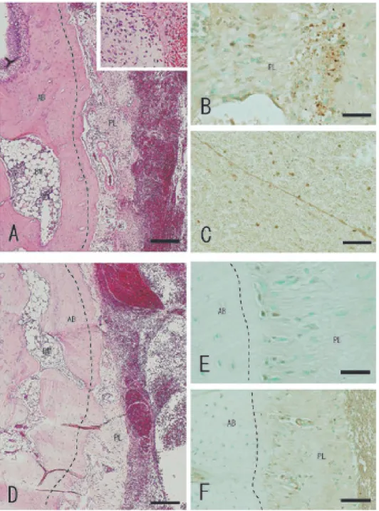
Histopathological findings

Bleeding occurred from the ruptured periodontal liga-

ment and the tissues around the socket at 12 h after tooth

extraction. The socket was filled with blood coagulum,

which was composed of densely aggregated erythrocytes,

leukocytes, and a fibrin network. Polymorphonuclear

leukocytes and cell fragments regarded as apoptotic

bodies were present in the remnants of the periodontal

ligament attached to the socket periphery (Fig. 1A and

inset). RUNX2 expression was detected in the nucleus

of some periodontal ligament cells, in the cell fragments

(3)

in the periodontal ligament (Fig. 1B), and on the nuclear membrane in some monocytic cells in the coagulum (Fig.

1C). On day 1, the blood coagulum was compartmental- ized by the fibrinous cord, and the irregular stump of the periodontal ligament was smoother than its surface at 12 h (Fig. 1D). There was slight thickening of the

endosteal lining of the bone marrow space adjacent to the socket. RUNX2 expression was detected on the nuclear membrane in some monocytic cells in the coagulum, in the nucleus of some osteoblasts or preosteoblasts on the alveolar bone (Fig. 1E), and in the cytoplasm of vascular cells in the periodontal ligament (Fig. 1F).

Fig. 1 Histopathological analysis of extraction sockets 12 h and 1 day after tooth extraction. (A, D) Histology; (B, C, E, F)

RUNX2 expression. (A) The blood coagulum comprised erythrocytes, leukocytes, and the fibrin network. Polymorphonuclear leukocytes and cell fragments in remnants of the periodontal ligament (inset). (B) RUNX2 expression in nuclei of fibroblastic cells and cell fragments in the periodontal ligament. (C) RUNX2 on the nuclear membrane of monocytic cells in the coagulum.

(D) Blood coagulum compartmentalized by the fibrinous cord and the smoothed stump of the periodontal ligament. (E) RUNX2

expression in nuclei of osteoblasts or preosteoblasts on the alveolar bone. (F) RUNX2 expression in the cytoplasm of vascular cells

in the periodontal ligament. AB: alveolar bone, PL: periodontal ligament, BM: bone marrow. Dashed lines represent the alveolar

wall. Scale bars: (A) 200 µm (inset, 70 µm), (B) 34 µm, (C) 67 µm, (D) 200 µm, (E) 34 µm, (F) 67 µm.

(4)

322

Three days after extraction, osteoblasts with basophilic cytoplasm produced bone matrix on the alveolar bone.

Thickening of the endosteum was prominent, particularly on the socket side. Spindle-shaped fibroblastic cells with round nuclei proliferated in the coagulum (Fig. 2A and

inset). RUNX2 expression was detected in the nucleus

of some periodontal ligament cells (Fig. 2B) and in the

nuclei of proliferating fibroblastic cells in the coagulum

(Fig. 2C). On day 5, bone matrix with many large

polymorphic cells was present in the coagulum, and the

Fig. 2 Histopathological analysis of the extraction sockets on days 3 and 5. (A, D) Histology; (B, C, E, F) RUNX2 expression. (A)

Osteoblasts produced bone matrix on the alveolar bone. Thickening of the endosteum became prominent, particularly on the socket

side (arrows). Spindle-shaped fibroblastic cells proliferated in the coagulum (inset). (B) RUNX2 expression in nuclei of periodontal

ligament cells. (C) RUNX2 expression in nuclei of proliferating fibroblastic cells in the coagulum. (D) Proliferating fibroblastic

cells associated with the remnant of the periodontal ligament. Thickening of the endosteum with bone matrix production on the

socket side (arrows). Bone matrix including large polymorphic cells in the coagulum (inset). (E) RUNX2 expression in the nuclei

of periodontal ligament cells. (F) RUNX2 expression in cells enclosed within the bone matrix and fibroblastic cells among the bone

matrices. AB, alveolar bone; PL, periodontal ligament; BM, bone marrow, BMT; bone matrix. Dashed lines represent the alveolar

wall. Scale bars: (A) 200 µm (inset, 138 µm), (B) 29 µm, (C) 44 µm, (D) 250 µm (inset, 138 µm), (E) 43 µm, (F) 67 µm.

(5)

proliferating fibroblastic cells were associated with the remnant of the periodontal ligament. Thickening of the endosteum and bone matrix production were apparent on the socket side (Fig. 2D and inset). RUNX2 expression was detected in the nuclei of some periodontal ligament cells (Fig. 2E), cells enclosed within the bone matrix, and fibroblastic cells among the bone matrices (Fig. 2F).

Trabeculae of woven bone in the socket on day 7

coalesced at the fundus to the side of the socket. The remnants of the periodontal ligament lapsed into hyaline degeneration, and the proliferative response of the endos- teum next to the alveolar wall was attenuated (Fig. 3A).

RUNX2 expression was detected in osteoblasts on the surface of the alveolar bone (Fig. 3B) and on the inner surface of the bone marrow space next to the socket, in osteoblasts on the surface of the newly formed bone, and

Fig. 3 Histopathological analysis of extraction sockets on days 7 and 10. (A, D) Histology; (B, C, E) RUNX2 expression. (A)

Coalescent trabeculae of woven bone at the fundus to the side of the socket. (B) RUNX2 in osteoblasts on the surface of alveolar bone. (C) RUNX2 expression in osteoblasts on the surfaces of new bone and osteocytes within. (D) Fused trabeculae of new bone extensively filled the socket. Remnants of the periodontal ligament replaced by bone tissue. (E) RUNX2 expression in osteoblasts on the surface of the bone tissue and osteocytes enclosed within. AB, alveolar bone; PL, periodontal ligament; BM, bone marrow;

NB, newly formed bone; BV, blood vessel. Dashed lines represent the alveolar wall. Scale bars: (A) 200 µm, (B) 37 µm, (C) 86

µm, (D) 200 µm, (E) 58 µm.

(6)

324

in osteocytes enclosed within the new bone (Fig. 3C).

The intensity of RUNX2 expression was lower than at day 5. Trabeculae of new bone, which fused together to increase their width, extensively filled the socket on day 10 (Fig. 3D). The area occupied by remnants of the periodontal ligament was replaced by bone tissue.

RUNX2 expression was detected in some osteoblasts on the surface of the new bone and in some osteocytes enclosed within (Fig. 3E). In addition, signal intensity diminished. There were few RUNX2

+

cells in the fibrous tissue lying among trabeculae of new bone (Fig. 3E) and a few RUNX2

+

osteoblasts lining the bone marrow space next to the socket. Observation of periodontal tissue around maxillary molars in the immunostained sections revealed weak RUNX2 expression only in the nuclei of the cells on the root-side half of the periodontal ligament (data not shown). Tooth extraction resulted in loss of the root-side half of the periodontal ligament, including RUNX2

+

cells.

Western blot analysis of RUNX2 in the tooth- extraction wound

Western blot analysis showed that RUNX2 (approxi- mately 50 kDa) levels were highest in the wound at 12 h and 1 day after extraction and rapidly decreased from day 3 to day 10 (Fig. 4).

Discussion

Osteogenic cells derived from hematopoietic tissue—

which are present in the coagulum—and cells in the remnants of the periodontal ligament participate in osteogenesis in the wound socket after teeth are extracted from rats. In the present study, we detected RUNX2 expression immediately after tooth extraction in mono-

cytic cells in the coagulum and in the remnants of the periodontal ligament. Shortly thereafter, we observed that fibroblastic cells proliferated in the coagulum and that large polymorphic cells were enclosed within the newly formed bone matrix in the socket. Although studies of human tooth-extraction wounds (2,6) did not determine RUNX2 levels immediately after extraction, RUNX2 was detected in proliferating spindle-shaped cells (regarded as osteoprogenitor cells) and in periodontal ligament cells. Because RUNX2 expression suggests osteogenic commitment (3,9-11), these findings indicate that cells in the coagulum and in the remnant of the periodontal ligament participate in osteogenesis.

Osteogenic cells derived from hematopoietic tissue are crucial in osteogenesis in tooth-extraction wounds in rats.

In the present study, Western blot analysis demonstrated that RUNX2 expression peaked from 12 h to 1 day after extraction, which corresponds to the coagulation stage.

Immunohistochemical analyses revealed that monocytic cells in the coagulum were the primary source of RUNX2 expression at this stage. These findings indicate that the osteogenic commitment of cells derived from hemato- poietic tissue in the extraction wound was greater than that of cells in the remnants of the periodontal ligament.

RUNX2 plays a crucial role in the differentiation of mesenchymal stem cells to osteoprogenitor cells in the coagulum immediately after extraction. In the present study, immunohistochemical and Western blot analyses revealed that RUNX2 expression peaked when monocytic cells were differentiating into fibroblastic cells in the coagulum immediately after extraction and dramatically declined thereafter. RUNX2 is essential in differentiation of mesenchymal stem cells to osteoprogenitor cells during early osteogenesis but not during the maturation stages of bone (4-6,12). This indicates that RUNX2 functions mainly in the differentiation of mesenchymal stem cells present in the subset of monocytic cells that differentiate into fibroblastic osteoprogenitor cells immediately after tooth extraction.

In the remnants of the periodontal ligament immedi- ately after extraction, RUNX2 expression is associated with apoptosis caused by load-induced microdamage. We show here that RUNX2 was present in cell fragments that represent apoptotic bodies in the remnants of the peri- odontal ligament, at 12 h after tooth extraction. RUNX2 expression (4,12) and apoptosis (13,14) are induced by mechanical forces on local tissue.

Taken together, the present findings indicate that cells derived mainly from hematopoietic tissue and RUNX2 expression are crucial in the differentiation of mesen- chymal stem cells to osteoprogenitor cells immediately

Fig. 4 Western blot analysis of RUNX2 expression in the extrac-

tion wound. RUNX2 (approximately 50 kDa) expression peaked

between 12 h and 1 day, then decreased rapidly from day 3 to day

10 postextraction. M, molecular weight markers.

(7)

after tooth extraction. Further studies of factors regu- lating onset of RUNX2 expression in extraction wound healing are required.

Acknowledgments

This study was supported, in part, by a Grant-in-Aid for Strategic Medical Science Research Center from the Ministry of Education, Culture, Sports, Science, and Technology of Japan, 2010-2014.

Conflicts of interest

No competing financial interests to declare.

References

1. Lin WL, McCulloch CA, Cho MI (1994) Differentiation of periodontal ligament fibroblasts into osteoblasts during socket healing after tooth extraction in the rat. Anat Rec 240, 492-506.

2. Devlin H, Sloan P (2002) Early bone healing events in the human extraction socket. Int J Oral Maxillofac Surg 31, 641-645.

3. Jimi E, Hirata S, Shin M, Yamazaki M, Fukushima H (2010) Molecular mechanisms of BMP-induced bone formation:

cross-talk between BMP and NF-κB signaling pathways in osteoblastogenesis. Jpn Dent Sci Rev 46, 33-42.

4. Camilleri S, McDonald F (2006) Runx2 and dental develop- ment. Eur J Oral Sci 114, 361-373.

5. Tu Q, Zhang J, James L, Dickson J, Tang J, Yang P et al.

(2007) Cbfa1/Runx2-deficiency delays bone wound healing and locally delivered Cbfa1/Runx2 promotes bone repair in animal models. Wound Repair Regen 15, 404-412.

6. Nahles S, Nack C, Gratecap K, Lage H, Nelson JJ, Nelson K (2013) Bone physiology in human grafted and non-grafted

extraction sockets--an immunohistochemical study. Clin Oral Implants Res 24, 812-819.

7. Bradford MM (1976) A rapid and sensitive method for the quantitation of microgram quantities of protein utilizing the principle of protein-dye binding. Anal Biochem 72, 248-254.

8. Laemmli UK (1970) Cleavage of structural proteins during the assembly of the head of bacteriophage T4. Nature 227, 680-685.

9. Nakashima K, Zhou X, Kunkel G, Zhang Z, Deng JM, Behringer RR et al. (2002) The novel zinc finger-containing transcription factor osterix is required for osteoblast differen- tiation and bone formation. Cell 108, 17-29.

10. Kusafuka K, Sasaguri K, Sato S, Takemura T, Kameya T (2006) Runx2 expression is associated with pathologic new bone formation around radicular cysts: an immunohisto- chemical demonstration. J Oral Pathol Med 35, 492-499.

11. Ohta S, Yamada S, Matuzaka K, Inoue T (2008) The behavior of stem cells and progenitor cells in the periodontal ligament during wound healing as observed using immunohisto- chemical methods. J Periodont Res 43, 595-603.

12. Kanno T, Takahashi T, Ariyoshi W, Tsujisawa T, Haga M, Nishihara T (2005) Tensile mechanical strain up-regulates Runx2 and osteogenic factor expression in human periosteal cells: implications for distraction osteogenesis. J Oral Maxil- lofac Surg 63, 499-504.

13. Bochaton-Piallat ML, Gabbiani F, Redard M, Desmoulière A, Gabbiani G (1995) Apoptosis participates in cellularity regulation during rat aortic intimal thickening. Am J Pathol 146, 1059-1064.

14. Clark WD, Smith EL, Linn KA, Paul-Murphy JR, Muir P,

Cook ME (2005) Osteocyte apoptosis and osteoclast presence

in chicken radii 0-4 days following osteotomy. Calcif Tissue

Int 77, 327-336.

Fig. 1   Histopathological analysis of extraction sockets 12 h and 1 day after tooth extraction

参照

関連したドキュメント

In regard to the expression and cellular localization of UTI, our previous study in the adult rat has revealed that, while the hepatocyte is the only site for expression of UTI

To determine the promoter activities of 5 ′ -flanking region of the human Pim-3 gene, we subcloned various deleted 5 ′ -flanking regions of the human Pim-3 gene into a

The present results also provide in situ hybridization evidence that at least chief cells and possibly both chief cells and sustentacular cells of the carotid body express NGF mRNA..

The pharmacokinetic profiles of clenbuterol enantiomers following intravenous and intraduodenal administration of clenbuterol racemate (2 mg/kg) in rats were

the cause of impaired wound healing: reasonable In Section 2 we presented clinical measurements penetration of low capillary tip density may cor- showing how

An example of a database state in the lextensive category of finite sets, for the EA sketch of our school data specification is provided by any database which models the

Then it follows immediately from a suitable version of “Hensel’s Lemma” [cf., e.g., the argument of [4], Lemma 2.1] that S may be obtained, as the notation suggests, as the m A

Definition An embeddable tiled surface is a tiled surface which is actually achieved as the graph of singular leaves of some embedded orientable surface with closed braid