• 検索結果がありません。

Serial Fecal-oral Transmission of Hepatitis A Virus (HAV) in Marmosets (Saguinus labiatus) : Establishment of an Experimental Model for a Natural Transmission Pathway of HAV

N/A
N/A
Protected

Academic year: 2021

シェア "Serial Fecal-oral Transmission of Hepatitis A Virus (HAV) in Marmosets (Saguinus labiatus) : Establishment of an Experimental Model for a Natural Transmission Pathway of HAV"

Copied!
18
0
0

読み込み中.... (全文を見る)

全文

(1)

Acta Med. Nagasaki 30 : 75-92

Serial Fecal-oral Transmission of Hepatitis A Virus (HAV) in Marmosets (Saguinus labiatus) :

Establishment of an Experimental Model

for a Natural Transmission Pathway of HAV

Kouichi YAMANOUCHI Department of Bacteriology,

Nagasaki University School of Medicine, Nagasaki, Japan

Received for publication, March 9, 1985

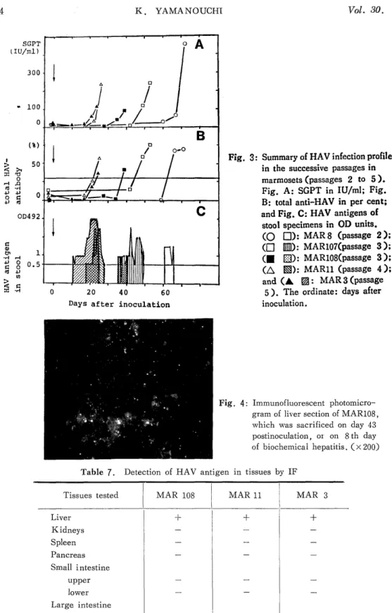
Hepatitis A virus (HAV) originally obtained from stool specimens of a human patient with an acute phase of hepatitis type A was successively passaged in marmosets, Saguinus labiatus, by oral administrations. The pattern of infection was as follows: after a certain incubation period, the appearance of HAV antigen in stools was the first sign of infection, followed by the simultaneous elevations of transaminases and anti-HAV antibody with ap- proximately 10-day intervals. This sequence was consistently observed throughout four successive passages. More passages resulted in less incubation time: the incubation period at passage 3 was 49 and 35 days in two animals, and those at passage 4 and 5 were 26 and 21 days, respectively. Because the inoculation dosis used beyond passage 2 was adjusted at 2,000 antigen units, the shorter incubation period in later passages suggested the adaptation of HAV in marmosets, Saguinus labiatus. Since the adapted virus showed reproducible incubation periods as well as clinical courses in marmosets, it is now possible to look for the primary target organs other than the liver of HAV, if any, and to use this system for the screening of vaccine candidates.

INTRODUCTION

Hepatitis type A, caused by hepatitis A virus (HAV), constitutes approximately 20 to 30 % of acute viral hepatitis a year in Japan.') HAV belongs to the enterovirus group of the family Picornaviridae.1) It has a single stranded RNA with a genome size of 2.3 X 10⁶ daltons,3>4> and the virion has a diameter of 27 nm with a density of 1.34 g/ml in CsCl.S> HAV is stable upon heating at 60°C for 1 hr. and by treatment with ether or acid at pH 3, but is inactivated by heating at 100°C for 5 min., or by treatment with chemicals or electromagnetic waves, such as formalin and ultraviolet irradiation.')

山之 内宏 一

(2)

HAV shows world-wide distributions but there are foci of highly endemic areas, especially in developing countries, such as New Guinea 7) and the Pacific Islands 8), Thai- land') and Africa10), where almost all of the population become anti-HAV positive by the age of 10. For example, the prevalence of anti-HAV positive population exceeds 90% in

11

late teens in Taiwan

In developed areas, such as North America"), Australia, 13) most European coun- tries") and Japan"), the prevalence of antibody positive population increases with age.

The seropositive rates in Japan are only 2.5 % for teens, and approximately 70 to 75 for those at 50 years of age or older"). Since most of seropositive population in the developed countries had been infected wtih HAV in their early lives, the apparent age- dependent increase of the seropositi ve rate has been considered as a dramatic decrease of HAV exposure in recent years due to the improvement of environmental and socio- economic conditions instead of continuous exposures to the virus in life"). In other words, the higher prevalence in the young ages of developing countries is caused by the higher density of HAV endemy. However, due to world-wide current transportation systems, people in non-endemic areas have increasing chances to be exposed to HAV in the en- demic areas16). Furthermore, hepatitis type A in adults is more often clinical and more serious than that in children"). Thus, continuous efforts should be paid to understand the ecology of HAV and to prevent hepatitis type A with active immunizing agents, such as live vaccines, even in the non-endemic areas.

Since the human being is the only natural host of HAV, the study of HAV had only been possible by help of volunteers in its early days")-"). The extent of such ex- periments has been obviously very limited. In 1967, DEINHARDT et al.2° first reported animal experiments utilizing two species of marmosets, as susceptible nonhuman primates.

The successful HAV infection in marmosets was confirmed by HOLMES et al.27) in 1969.

Similar results were obtained in "chimpanzees by MAYNARD et al. 28) , THORNTON et al .29) and DIENSTAG et al.") in 1975. Thereafter, marmosets and chimpanzees have been widely used as experimental animals for HAV transmission.

In 1973 FEINSTONE et al.") found virus-like particles in stool specimens obtained from the acute phase of hepatitis type A patients by immune elecron microscopy (IEM).

MAYNARD et al.32). LOCARNINI et al.") and PURCELL et al. 14) confirmed the pres-

ence of 27 nm particles in stool specimens of hepatitis type A patients. These observations

made it possible to use HAV purified from stool specimens obtained from the acute phase

of hepatitis type A of human and experimental animals as a virus source for infection or

as viral antigens for immunological reactions""". For detection of anti-HAV, MILLER

et al.") developed an immune adherence hemagglutination (IAHA) test in 1975 and

PURCELL39> developed radioimmunoassay (RIA) in 1976. BRADLEY et al. 40) established

RIA to detect immunoglobulin M class (IgM) anti-HAV in 1977, and MATHIESEN et

al.") developed the enzyme-linked immunosorbent assay (ELISA) for detection of HAV

and anti-HAV in 1978. Now these RIA and ELISA kits are widely used for antigen or

antibody detection.

(3)

To shed light on the natural history of HAV infection, the author intended to transmit HAV successively in marmosets (Saguinus labiatus). The results indicated that HAV was transmissible in marmosets by successive fecal-oral administrations and has adapted in marmosets. The postinfection profiles of virological and biochemical parameters were distinct and reproducible. Thus, the experimental system is potentially very useful to analyze the primary target and the natural history of HAV-infection, and further to

evaluate potential candidates of live HAV vaccine, in the near future.

MATERIAL AND METHODS

Marmosets : Five adult female marmosets (Saguinus labiatus), 518 to 575 gr in body weight, were purchased from Charles River Inc. Japan. Marmosets were numbered as MARS, MAR107, MAR108, MAR11 and MARS. Two marmosets, MAR8 and MAR107, were used to follow natural history of hepatitis type A, and the other three marmosets, MAR108, MAR11 and MAR3, were sacrificed at their acute phases of the biochemical hepatitis in order to locate early changes of HAV infection in various tissues. The in- fected marmosets were reared in the Infection Experiment Section of Laboratory Animal Center for Biomedical Research, Nagasaki University School of Medicine. All the ex- periments were performed under the strict regulations of Biosafety Level 2").

Virus : HAV used in these experiments was originated from stool specimens of a human patient with acute phase of hepatitis type A observed in a local epidemic at Kurume, Fukuoka Prefecture in 1982. For the successive transmission of HAV, the stock virus was made after one passage in a marmoset, MAR4. An unkown amount of HAV purified from the patient stool (dosage : 1 ml) was intravenously inoculated into the marmost. Transaminases started to rise on day 13, and MAR4 was sacrificed on day 17.

HAV was purified from liver extract as described below, and used as the stock virus.

Purification of HAV : HAV was purified by the method described by MORITSUGU et al.35) with minor modifications. Liver or stool specimens suspended in 10 volumes of 0.01 M phosphatebuffered saline (PBS) were homogenized with a Vertis blender for 3 min.

or freeze-thawed 5 times, respectively. After centrifugation at 3,000 rpm for 30 min. , the supernatant was ultracentrifuged at 27,000 rpm for 16 hr. with a Beckman SW27 rotor.

The pellet was resuspended with 2.0 ml of PBS containing 0.5% Nonidet p40. The suspension was sonicated at 20 kHz for 5 min. and centrifuged at 15,000 rpm for 15 min.

The supernatant was layered on a 10 - 40% linear sucrose gradient, and centrifuged at 24,000 rpm for 210 min. with a Beckman SW27 rotor. Fractions were col'.ected by bottom puncture, and HAV antigen concentration in each fraction was determined by ELISA.

The concentration of purified HAV was adjusted as 2, 000 HAV antigen units (AgU)/ 0.3 ml and freeze-stocked at -80°C until use.

Inoculation : Marmosets were lightly anesthetized by intramuscular injection of 0.2

ml/kg Ketamine hydrochloride (Veterinary Ketalar 50, Intramuscular, Sankyo Inc., Ja-

pan). The purified HAV was given into the oral cavity of a marmoset, drop by drop

(4)

using a 1 ml syringe with a metalic gastric canula for mice. The dose was 2000 HA\J AgU in 0.3 ml, except for MARE which was given 20 HAV AgU in 1 ml.

Sampling of specimens : Stool specimens of all marmosets were collected daily th- roughout the observation periods and stored at -80°C until use. Serum samples for detecting glutamic oxaloacetic transaminases (SGOT), glutamic pyruvic transaminase (SG- PT), and antibodies against HAV were obtained once a week and stored at -80'C . Liver, spleen, kidneys, pancreas, salivary glands, and small and large intestines were obtained from each sacrificed animal. Parts of those tissues embedded in Tissue-Tek II (Miles Lab. Inc., U . S . A .) were snap-frozen in liquid nitrogen for detecting HAV antigens by indirect immunofluorescence method.

Determination of liver enzyme levels ; The levels of SGOT and SGPT were measured within one week after sampling of sera using spectrophotometric rate assay modified from KARMEN's method 4a"44'. The normal values of SGOT and SGPT in another species of marmoset, Saguinus mystax, were reported as 97±26 and 31 ± 10 units/ml, respectively .41) In our laboratory, normal values for Saguinus labiatus are 118 ± 30 and 12.3 ± 7.4 IU/ml, respectively.

Measurement of anti-HAV antibodies : Levels o f total and IgM anti-HAV antibody in sera were determined by RIA kits, ANTI HARIA KIT and ANTI HAM-RIA KIT (Dainabot RI Inc., Japan), respectively. Briefly, for total anti-HAV, 10 p1 of serum sample, 200 pl of 125I-labelled antibody to HAV (Human) and a latex bead coated with HAV (Primate) were incubated for 18-24 hr. at 25°C. The bead was washed twice with distil ed water and counted in a autogammacounter. The degree of competition was calculated by the equation, [(average of negative controls) - (sample counts)]/((average of negative controls) -(average of positive controls)] X100, and the values are expressed in per cent. Sera under 30% or over 70% were scored as negative or positive, respectively.

Those between 70% and 30% were left unjudged.

To quantitate IgM anti-HAV, 10 pl of sample serum, 200ul of the specimen diluent and a latex bead coated with anti-human IgM (goat) antibody were incubated for 2 hr.

at 25'C. After washing twice with distilled water, the bead was incubated with 200 , el of HAV (Primate) for 18-22 hr. at 25°C. The bead was washed and incubated further

with 200 pl of '25I-labelled antibody to HAV (Human) for 4 hr. at 45°C. The bead was washed with distilled water and radioactivity was counted. Data are expressed by ratio of sample count of cut-off value. The cut off-value is calculated as 0.1 x (positive control count) + (negative control count). Sera which gave no less than 1.0 were scored as positive.

Detection of HAV antigen in stool specimens : 0.5 gr. of a stool, specimen was

suspended in 10 ml PBS, freeze-thawed 5 times, and centrifuged at 3,000 rpm for 30

min.. HAV antigen in the supernatant was detected by a ELISA Kit, Hepanostika HAV

Microelisa System (Organon Teknika, Holland). Briefly, 100 pl of fecal extract were

incubated in a well coated with anti-HAV at 25°C for 16-20 hr. and the well was washed

4 times with phosphate buffer. Each well received 100 al of human anti-HAV conjugated

(5)

with horse-radish peroxidase (HRP), and was incubated at 37°C for 120 min . within a humidified chamber in the dark. After 4 washes with phosphate buffer , the well was incubated with 100 gel of the mixture of urea peroxide and o-phenylene diamine hydrochloride (OPD) in the dark at 25°C for 45 min..; Enzyme reaction was stopped by adding 100 p1 of 2 M sulfuric acid. The optical density was determined at 492 nm by a spectrophotometer .

A cutoff value of 0.5 was used.

Detection of HAV antigen in tissue specimens by indirect immunofluorescene method . (IF) : The frozen tissues were sectioned at 4 pm by a cryostat and used for indirect IF.

The tissue sections mounted on glass slides and air-dried, were incubated at 25°C for 2 hr. with anti-HAV IgG obtained from a convalescent serum of a human patient with he- patitis type A. After washing with PBS three times, the tissues were stained by goat anti-human IgG conjugated with fluorescein isothiocyanate (MBL, Bethesda, MD) at 4 °C for 16 hr.. After 3 washes with PBS, the stained specimen was mounted by 90%

glycerol/ for PBS and immediately examinated by a ZEISS fluorescent microscope.

RESULTS

Establishment of oral infection of HAV in MAR 8 (passage 2) : MAR 8 was orally inoculated with the stock HAV (passage 1) obtained and purified from the liver extract of MAR 4 which had been sacrificed on day 4 of biochemical hepatitis, on day 17 after intravenous inoculation with HAV purified from the human patient. As shown in Table 1 and Fig. 1 , SGOT and SGPT began to elevate on day 67 postinoculation and stayed at high levels until day 81, i.e. biochemical hepatitis lasted for 15 days. The enzyme peak was on day 72, or the 6th day after the onset of biochemical hepatitis. IgM anti-HAV became positive on day 67 and returned negative on day 98. Total anti-HAV was first de- tected also on day 67, however, it remained at a high level beyond day 113. Thus, the elevation of detectable antibody started simultaneously with biochemical hepatitis. HAV antigen in stools was detected from day 61 to 66. The excretion of HAV in stools started

1 week and terminated a day before the onset of biochemical hepatitis.

Fecal-oral infection of HAV in MAR107 (passage 3 ): HAV was purified from po- oled stool specimens (61-63 days postinoculation) of the infected MAR 8 . HAV in an amount of 2,000 AgU was administered into the oral cavity of MAR107, and the clinical course was followed (Table 2 and Fig. 2) . SGOT and SGPT started to rise on day 49, and peaked on day 53. Total anti-HAV and IgM anti-HAV were first detected on day 49. The IgM anti-HAV was at a maximum on day 53 and returned to the preinocula- tion level on day 84. Total anti-HAV reached its plateau on day 78. HAV antigen in stools was detectable from day 37 to day 49. Although stool specimens were collected since the day of inoculation, HAV antigen were never detected in earlier stages.

Serial passages of HAV by fecal-oral administrations (passage 3 - 5) : HAV (pas-

sage 2 of MAR 8) was successively transmitted into 3 marmosets, MAR108, MAR11

and MAR 3 by the fecal-oral pathway. In order to see early pathological changes in the

(6)

Table 1. Clinical Profile of Hepatitis Type A of the Marmoset No. 8 (MAR 8) for Passage 2')

Days after GOT GPT anti-HAV antibody HAV antigen

inoculation totalb) I Me> in stool

IU/ml IU/ml % l S/CO OD492

0 103 11 0 0.3 -

59 -a) - - - 0.4

60 94 29 0 0.4 0.1

61*e) - - - - 1.5

62* - - - - 1.6

63* - - - - 1.7

64 - - - - 0.9

65 - - - - 0.5

66 - - - - 1.2

67 232 61 64 0.9 -

72 504 459 67 3.2 0.3

75 384 237 80 3.4 0

81 132 60 87 3.2 0

88 90 10 90 1.0 0

98 96 11 93 0.6 -

105 100 8 95 0.4 -

113 97 16 I 99 0.5 -

a) MARS was inoculated with 20 AgU of HAV purified from the liver extract of MAR4 sacrificed on day 17.

b) Per cent inhibition (%) = ((average of negative controls-sample counts)/

(average of negative controls-average of positive controls)) x100.

c) S/CO = (sample counts) / (cut off value). See text for detail. c

d) - : not tested.

e) HAV was purified from stool specimens on day 61 through 63, and used for inoculation into MAR107 and MAR108 (passage 3).

Fig. 1 : HAV infection profile in MAR 8 (passage 2). A shows liver enzymes, SGOT (0) and SGPT (E) in IU/ml; Fig. B shows IgM anti-HAV (0) and total anti-HAV (D) in

sera, both assayed by RIA, and HAV antigen in stool specimens assayed by ELISA

( ). See Materials and Methods for quantitation units. The ordinate shows days of

postinoculation

(7)

Table 2. Clinical Profile of Hepatitis Type A of the Marmoset No. 107 (MAR 107) for Passage 3a)

Days after I GOT GPT anti-HAV antibody HAV antigen

inoculation in stool

totalb) IgM,)

IU/ml IU/ml % S/CO OD 492

0 118 12 11 0.3 -d)

36 - - - - 0.4

37 - - - - 0.5

38 - - - - 0.8

39 - - - - 0.8

40 - - - - 0.8

41 - - - - 1.1

42 96 6 14 0.2 1.1

43 - - - - 2.5

44 - - - - 1.2

45 - - - - 0.3

46 - - - - 1.9

47 - - - - 1.8

48 - - - - 1.9

49 287 116 57 1.5 1.1

50 - - - - 0.2

53 356 246 75 3.0 0.5

58 123 46 83 2.5 0.1

63 114 22 87 1.8 0.2

69 88 12 93 1.1 0.4

78 93 11 96 0.6 0.4

84 101 16 96 0.4 0.1

a) MAR 107 was inoculated with 2000 AgU of HAV purified from the stool specimens of MAR 8 on day 61 through 63.

b-d) See foot notes of Table 1.

Fig. 2 : HAV infection profiles in MAR107 (passage 3 ). See figure

legeneds for Fig. 1 .

(8)

Table 3. Clinical Profile of Hepatitis Type A of the Marmoset No. 108 (MAR 108) for Passage 3a)

Days after GOT GRT anti-HAV antibody HAV antigen

inoculation in stool

totalb) IgM e)

iU/ml iU/-rnl % % S/CO OD492

0 132 10 0 0.3 -d)

28 88 3 6 0.3 0.2

35 240 43 0 0.2 0.2

36*e) - - - - 1.0

37* - - - - 1.1

38 - - - - 0.1

39 264 93 41 0.6 0.3

40 - - - - 0.2

41 - - - - 1.2

42 - - - - 0.2

430 255 86 - 1.5 -

a) MAR 108 was inoculated with 2000 AgU of HAV purified from the stool specimens of MAR 8 on days 61 throught 63.

b-d) See foot notes of Table 1.

e) HAV was purified from stool specimens on days 36 and 37, and inoculated into MAR 11 (passage 4)

f) MAR 108 was sacrificed on day 43.

Table 4. Clinical Profile of Hepatitis Type A of the Marmoset No. 11 (MAR11) for Passage 48

Days after GOT GPT anti-HAV antibody HAV antigen

inoculation in stool

totalb) IgMC}

IU/ml IU/ml % S/CO OD492

0 99 11 3 0.3 -d)

17 58 0 6 0.2 0.1

18 - - - - 0.5

19 - - - - 0.6

20 - - - - 0.4

21 - - - - 0.9

22*e 126 37 26 0.3 1.2

23* - - - 2.2

24 - - - - 0.9

25 - - - - 1.8

26 282 234 57 0.9 1.7

27 - - - - 0.6

28 - - - - 0.5

290 344 202 80 4.0 0.3

a) MAR 11 was inoculated with 2000 AgU of HAV purified from stool specimens of MAR 108 on days 36 and 37.

b-d) See footnotes of Table 1.

e) HAV was purified from stool specimens on days 22 and 23, and inoculated into MAR 3 (Passage 5 ).

f) MAR 11 was sacrificed on day 29.

(9)

liver and possible viral replication(s) site other than the liver, these three marmosets were sacrificed in acute phases of hepatitis, within 8 days after the rise of liver enzymes.

MAR108, as well as MAR107, was inoculated with passage 2 HAV purified from the pooled stool specimens of MAR 8 (Table 3 ). HAV antigen was detected in stools on days 36, 37 and 41 of post inoculation. Abnormal elevations of SGOT and SGPT occurred on day 35, followed by elevation of total anti-HAV and IgM anti-HAV on day 39. On day 43, MAR 108 was sacrificed.

Table 5. Clinical Profile of Hepatitis Type A of the Marmoset No. 3 (MAR 3 ) for Passage 5 a)

Days after GOT GPT anti-HAV antibody HAV antigen

inoculation in stool

totalb) We)

IU/ml IU/ml % S/CO OD 492

0 74 14 12 0.2 - d)

10 66 13 0 0.2 0.4

11 - - - - 0.8

12 - - - - 0.5

13 - - - - 0.7

14 - - - - 0.6

15 - - - - 0.7

16 - - - - 1.0

17 62 13 1 0.2 1.4

18 - - - - 1.4

19 - - - - 1.8

20 - - - - 2.1

21 129 56 17 0.2 2.9

22 - - - - 2.6

23 - - - - 3.0

24 174 111 29 0.3 2.8

25e) 198 112 36 0.4 2.3

a) MAR 3 was inoculated with 2000 AgU of HAV purified from stool specimens of MAR 11 on days 22 and 23.

b-d) See footnotes of Table 1.

e) MAR 3 was sacrificed on day 25.

Table 6. Summary of Succesive Fecal-Oral Passages of HAV in Marmosets

Passage HAV Onset of GPT Onset of HAV

levels inoculated elevation Ag detection

AgU days p.i. days p.i.

2 20 67 61

3 2000 49 37

3 2000 35 36

4 2000 26 18

5 2000 21 11

(10)

Fig. 3: Summary of HAV infection profiles in the successive passages in

marmosets (passages 2 to 5 ).

Fig. A: SGPT in IU/ml; Fig.

B: total anti-HAV in per cent;

and Fig. C: HAV antigens of stool specimens in OD units.

(0 0): MAR 8 (passage 2 );

([] ®): MAR107(passage 3 );

Q): MAR108(passage 3 );

(Q ®): MAR11 (passage 4);

and (A ®: MAR 3 (passage

5 ). The ordinate: days after

inoculation.

Fig. 4 : Immunof luorescent photomicro- gram of liver section of MAR108,

which was sacrificed on day 43

postinoculation, oI' on 8 th day

of biochemical hepatitis. (x 200)

Table 7. Detection of HAV antigen in tissues by IF

Tissues tested MAR 108 MAR 11 MAR 3

Liver + + +

Kidneys - - -

Spleen - - -

Pancreas - - -

Small intestine

upper - - -

lower - - -

Large intestine

upper - - -

lower - - -

Salivary glands - - -

(11)

The marmoset, MAR11, was inoculated with HAV (passage 3) purified from the stool specimens (36 -37 days postinoculation) of MAR108 (Table 4 ). The HAV antigen was detected in stools from 18 to 28 days postinoculation and rises of liver enzymes and anti-HAV developed on day 26 postinoculation. MAR11 was sacrificed on day 29.

MAR 3 was inoculated with HAV (passage 4) purified from stool specimens of MAR11 (22-23 days postinoculation). As shown in Table 5, hepatitis became apparent on day 21 with the elevations of SGOT and SGPT. HAV antigen was recognized in stool specimens collected from days 11 to 25. MAR 3 was sacrified on day 25, or 4 days after the elevation of liver enzymes. Total anti-HAV apparetly increased into the equivocal level on day 24, but the increase of IgM anti-HAV was not detectable by day 25.

Adaptation of HAV in marmosets : Shifts of parameters of HAV infection according to the serial passages in marmosets, including incubation periods for liver enzyme eleva- tion, anti-HAV production and fecal excretion of HAV, are summarized in Fig. 3 and Table 6 . The sequence of HAV excretion into the stool after certain incubation periods, followed by almost simultaneous elevation of liver enzymes and antibodies against HAV with an interval of approximately 10 days, was consistently shown in each passage. How- ever, the incubation periods were shortened by successive transmission in marmosets, from

5 to 7 weeks at passage 3 , to 3 weeks at passages 4 and 5 . Since the inoculation doses were constant throughout the experiments, the shorter incubation period in the later passages suggests the adaptation of HAV in the marmoset.

Attempts to localize replication site(s) of HAV : Marmosets, MAR108, MAR11 and MAR 3 , were sacrificed within the acute phase of hepatitis, and tissues of these animals served for IF study in an attempt to localize primary replication site(s) of HAV. The HAV antigen was detected in the liver of each marmoset (Fig. 4 ), with very fine gra- nular pattern in cytoplasm. However, none of the tissues other than the liver showed po- sitive evidince for HAV replication (Table 7) .

DISCUSSION

The purpose of this study was to establish a reproducible experimental model for fecal-oral infection of HAV to simulate the natural infection pathway of hepatitis type A in the human species. HAV obtained from stool specimens of a human hepatitis type A patient was serially passaged and adapted in marmosets (Saguinus labiatus) by fecal-oral inoculations.

In human hepatitis type A induced by natural infection, the incubation period is approximately 30 days followed by increase in transaminases, a hallmark for diagnosis").

Clinical manifestations other than increased transaminases appear in half of the cases in-

cluding fever, nausea, anorexia and general malaise. The elevation of transaminases re-

aches a maximum within 1 week when jaundice manifests itself in some cases. The

transaminase levels return to normal within 1 to 2 weeks. IgM anti-HAV usually be-

comes detectable in association with the onset of increase in transaminases and reaches its

(12)

peak-levels a week later46) . YANO and KOGA followed IgM anti-HAV profiles in 31 cases using the mothod described in this paper 47), and found that IgM anti-HAV is detectable up to 9 weeks after infection, but undetectable beyond 5 months. However, in another report, IgM anti-HAV stays at detectable levels for more than 12 month for some patients 48) . IgG anti-HAV appears approximately a week after the onset of hepatitis type A and reaches its plateau within 1 to 2 months. HAV antigen in stools usually returns to a nondetectable level at the onset of clinical hepatitis or the appearance of anti-HAV46).

In these experiments, SGOT and SGPT of marmoset increased up to 500 IU/ml and reached their peak approximately 5 days after the onset of their rise, and the increased enzyme 'evels continued for approximately 2 weeks. Since these patterns are consistent with that of human hepatitis, the marmosets were diagnosed to have hepatitis type A.

However, hepatitis type A in marmosets is apparently less severe than that in human species as suggested by (1) the elevation of liver enzymes was limited up to 500 IU/ml com- pared with more than 1,000 IU/ml in some clinical cases"), especially adults suffered from more severe hepatitis. Moreover, the normal level of SGOT in marmosets is approximately 100 IU/ml, higher than that in normal human beings ; (2) the level of SGPT was usually lower than that of SGOT in marmosets, however the SGPT/SGOT ratio is inverted in human hepatitis ;49) (3) clinical jaundice was never observed in our experiments ; and (4) histological damages in liver cells are less severe in the marmoset50).

The rise of IgM anti-HAV and total anti-HAV was associated in most cases. Since total anti-HAV includes IgM anti-HAV, this was not unexpected. Howerer, total anti- HAV usually became positive 1 to 4 days earlier than IgM anti-HAV. This was probably due to the sensitivity of the assays used. Although both assays were RIAs, total anti- HAV is based on a competition assay system, and IgM anti-HAV on a sandwich binding assay system. The IgM anti-HAV reached its peak at 4 to 9 days after the onset of the elevation, and became undetectable within 6 weeks, consistent with the duration of detectable IgM anti-HAV in most human hepatitis type A patients").

The HAV antigen in stools was detected for 7 to 15 days, and became undetectable within one week after the onset of biochemical hepatitis. Since anti-HAV will appear in association with biochemical hepatitis, the disappearance of HAV in stools seems to be associated with elimination of free virus by anti-HAV. In 4 marmosets out of the 5 used in the experiments, HAV in stools appeared in 2 peaks : since the remaining MAR3 was sacrificed after the first peak, the second peak might have been seen if the marmoset was not sacrificed at day 25. BRADLEY et al.51) observed a similar pattern and indicated that virions in the first peak are of empty particles. However, since the virus specimens used for passage in these experiments were collected from the first peak, the virus in the first peak was found infectious.

HAV antigen was first detected on day 61 in MAR8 (passage 2), on days 37 and

36 in MAR107 and MAR108, respectively (passage 3), on day 18 in MAR11 (passage

4), and on day 11 in MAR3 (passage 5). The longer incubation period in passage 2

(13)

could be explained by the lower titer of inoculum. The incubation period in passage 5 was 1/3 of that in passage 3, indicating the adaptation of HAV in marmosets. However, since the animals in passages 3 through 5 were killed in their early course of acute hepatitis, the potential increase of virulence by adaptation was not clear.

Adaptation of HAV in marmosets by serial passages has been suggested. DEIN- HARDT26) successively inoculated sera of infected marmosets, and found that the incuba- tion periods were shortened to approximately 20 days after 5 serial passages. PROVOST52 inoculated diluted liver extracts obtained from infected marmosets intravenously, and found that the incubation periods decreased from 21 to 7 days after 5 serial passages. EBERT et al. 53) successively inoculated serum samples of infected marmosets, pooled between 7 and 14 days prior to elevated enzyme activities. The incubation period decreased from 49-63 days to 25-34 days in the forth passage. BRADLEY 14) also found that the origi- nal incubation period of 49-63 days decreased and reached the level of 3-7 days after 9 serial passages of liver extracts. All of these data indicated the incubation period is shortened by serial passages. However, it should be pointed out that the inoculum sizes were not controlled due to lack of appropriate titration systems, and that the virus was passaged by an artificial pathway, intravenous injection, instead of the natural transmis- sion pathway, fecal-oral infection. In this study, the 2000 AgU of virus recovered from stool specimens were successively inoculated into oral cavities. Thus, the shortened in- cubation period after serial passages presented in this paper suggested the adaptation of HAV in marmosets by successive fecal-oral transmissions.

Since the incubation period of hepatitis type A is much longer than the eclipse time period of most enteroviruses, and since the ultimate target tissues of enteroviruses are often different from the primary replication sites, primary replication site(s) other than the liver have been looked for. HAV antigen has been found in liver, spleen, lymph nodes and kidneys, but not in intestines of the marmoset during acute hepatitis type All). HAV antigens found in the spleen and lymph nodes were believed as mere trapping of HAV instead of replicated viruses in these tissues. Since HAV antigen ap- pears earlier in the liver than in the stool, HAV has been considered to be excreted from the liver to the stool")-"). Although HAV was inoculated intravenously in these studies, MATHIESEN et al.") inoculated HAV orally into marmosets. They found HAV antigen only in the liver and bile juice by the time they detected HAV antigen in stools.

In this paper, collection of stool specimens was started on the day of inoculation, but HAV antigen was not found in the early phase of expected incubation peridos.

Furthermore, tissues obtained from the marmosets sacrificed at early acute phase of hepatitis

showed an accumulation of viral antigens only in the livers. The results are consistent

with the current concept that the liver per se is the primary replication site. However,

the sensitivity of the assay for HAV antigen may not be high enough to detect localized

replications of HAV during the phase of putative primary replication, and the animals

were sacrificed only after the biochemical hepatitis started. To shed light on this problem,

tissues of infected animals in very early stages of infection, probably much before the rise

(14)

of liver enzymes, should be examined extensively. However, a reproducible experimental system with known incubation periods and the natural infection pathway is inevitable for this purpose. This study showed that marmosets suffered from acute hepatitis type A with reproducible incubation periods of approximately 3 weeks by oral inoculation of 2,000 AgU HAV adapted in the marmosets, i. e. the search for the primary replication site for HAV is now possible.

Since this system, marmoset infected orally with adapted HAV, is reproducible in the length of incubation periods and in the incidence of biochemical hepatitis, it may be also useful for certification tests of putative live vaccine of HAV, when it is available.

ACKNOWLEDGEMENTS

The accomplishment of this study and thus the basis for this thesis was only made possible through enthusiastic and skilled collaboration of several investigators, technicians, and secretarial assistants.

For the development of this report, the author is particularly indebted to the following Professor Tsutomu MIYAMOTO, M. D., the Department of Bacteriology, Nagasaki University School of Medicine was my adviser for this thesis. His extensive scientfiic experience, his positive critique and support has been invaluable for this report.

Shigeo HINO, M. D., Associate Professor of the Department Bacteriology, Nagasaki University of Medicine gave me valuable advice regarding the study and revision of this manuscript.

Professor Hideyo ITAKURA, M. D., Institute for Tropical Medicine, Nagasaki University, and Yohko K. SHIMIZU, Ph. D., Nihon University School of Medicine, inspired and encouraged me and gave me the best possible working conditions throughout the project.

Yasuo MORITSUGU, M. D., National Institute of Health, Tokyo, kindly helped me in the purificaton of the virus.

Michitami YANO, M.D., and Miss Naoko MORI, Nagasaki Chuo National Hospital helped me in the measurements of anti-HAV antibody.

Dr. O. INOUE, Mr. N. KUBO, Mr. N. YASUKAWA, Miss R. YOSHIFUKU and Miss Y. ITOKI must be thanked for their technical assistance, and Ms. Carol ALBRIGHT for preparation of the English manuscript. Finally, I am especially indebted to my family, whose tolerance and love, have always been my strength.

Part of this study was supported by Grants-in-Aid from the graduates association of Nagasaki University School of Medicine.

REFERENCES

1) TANIKAWA, K., SADA, M. (1981): Epidemiology of type A hepatitis. Igaku No Ayumi 118: 503-509. (Jpn)

2) COULEPIS, A. G., LOCARNINI, S. A., WESTAWAY, E. G., TANNOCK, G. A.,

GUST, I, D. (1982): Biophysical and Biochemical Characterization of Hepatitis A

(15)

Virus. Intervirology 18: 107-127.

3) COULEPIS, A. G., TANNOCK, G. A., LOCARNINI, S. A., GUST, I. D. (1981):

Evidence that the genome of hapatitis A virus consists of single-stranded RNA.

J. Virol. 37: 473-477.

4) SIEGL, G., FROSNER, G. G., GAUSS-MULLER, V., TRATSCHIN, J. D. , DEINHARDT, F. (1981): The physicochemical properties of infectious hepatitis A virions. J. Gen. Virol. 57: 331-341.

5) SIEGL, G., FROSNER, G. G. (1978): Characterization and classification of virus particles associated with hepatitis A. I. Size, density, and sedimentation. J. Virol.

26: 40-47.

6) PROVOST, P. J., WOLANSKI, B. S., MILLER, W. J., ITTENSOHN, O. L., McALEER, W. J., HILLEMAN, M. R. (1975): Physical, chemical and morphologic

dimensions of human hepatitis A virus strain CR326 (38578). Proc. Soc. Exp. Biol.

Med. 148: 532-539.

7) HAWKES, R. A., BOUGHTON, C. R., FERGUSON, V, (1981): The seroepidemiology

of hepatitis in Papua, New Guinea. I. A long-term study of hepatitis A, Am. J.

Epidem. 114: 554-573.

8) GUST, I. D., LEHMANN, N. I., DIMITRAKAKIS, M. (1979): A seroepidemiologic

study of infection with HAV and HBV in five Pacific Islands. Am. J. Epidem. 110:

237-242.

9) BURKE, D. S., SNITBHAN, R., JOHNSON, D. E., SCOTT, R. McN. (1981):

Age-specific prevalence of hepatitis A virus antibodyy in Thailand. Am. J. Epidem.

113: 245-249.

10) BAYLET, R., LEMAIRE, J. M., RIDET, J. (1981): Seroepidemiologie de 1'hepatite A. Resultats obtenus au Senegal. Pathol, Biol. 29: 217-221.

11) HWANG, L., BEASLEY, R.. P., YANG, C., Hsu, L., CHEN, K. (1983) : Incidence of hepatitis A virus infection in children in Taipei, Taiwan. Intervirology 20: 149-154.

12) MOSLEY, J. W. (1978): Epidemiology of HAV infection. In viral hepatitis. Ed. by Vyas, G. N . , Cohen, S. N . , Schmid, R. Franklin Institute Press, Philadelphia,

pp. 85-104.

13) GUST, I. D., LEHMANN, N. I., LUCAS, C. R., FERRIS, A. A., LOCARNINI,

S. A. (1978): studies on the epidemiology of hepatitis A in Melbourne, In Viral hepatitis. Ed. by Vyas, G. N . , Cohen, S. N . , Schmid, R. Franklin Institute Press,

Philadelphia, pp. 105-112.

14) FROSNER, G. G., PAPAEvANGELOU, G., BUTLER, S., IWARSON, A., ALINDHOLM,

A., COUROUCE-PANTY, H., DEINHARDT, F. (1979): Antibodies against hepatitis A in seven European countries. Am. J. Epidem. 110: 63-69.

15) MORITSUGU, Y., TANAKA, T., SHIKATA, T. (1978): A preliminary Serologic Study of Hepatitis A Virus Infection in Japan. Jpen J. Med. Sci. Biol. 31: 325-338.

16) OBATA, H., HISAMITSU, T., TAKADA, K. (1979): Imported hepatitis. Jpn. J' Clin, Med. 38: 340-347. (Jpn)

17) TANIKAWA, K., KUBO, Y., ABE, H., SADA, M. (1979): Type A Hepatitis. Jpn.

J. Clin. Exp. Med. 56: 677-682. (Jpn)

18) VOEGT, H. (1942): Zur Aetiologie der Hepatitis epidemica. Munchen. med. Wchns-

chr. 89: 76.

(16)

19) CAMERON, J. D. S. (1943): Infective hepatitis. Quart. J. Med. 12: 139-155.

20) HAVENS, W. P., Jr. (1946): Period of infectivity of patients with experimentally

induced infectious hepatitis. J. Exp. Med. 83: 251-258.

21) NEEFE, J. R., GELLIS, S. S., STOKES, J., Jr. (1946): Homologous serum hepatitis.

Studies in volunteers bearing on immunological and other characteristics of etiological

agents. Am. J. Med. 1: 3-22.

22) PAUL, J. R., HAVENS, W. P., Jr., SABIN, A. B., PHILIP, C. B. (1945): Trans- mission experiments in serum jaundice and infectious hepatitis. J. A. M. A. 128:

911-915.

23) FINDLAY, G. M., WILLCOX, R. R. (1945): Transmission of infective hepatitis by faeces and urine. Lancet 1: 212.

24) FRANCIS, T., Jr., FTISCH, A. W., QUILLIGAN, J. J., Jr. (1946): Demonstration

of infectious hepatitis virus in presymptomatic period after transfer by transfusion.

Proc. Soc. Exp. Biol. Med. 61: 276-280.

25) KRUGMAN, S., WARD, R., GILES, J. P. (1962): The natural history of infectious hepatitis. Am. J. Med. 32: 717-728.

26) DEINHARDT, F., HOLMES, A. W., CAPPS R. B., POPPER, H. (1967): Studies on the transmission of human viral hepatitis to marmoset monkeys. I. Transmission of

disease, serial passages, and description of liver lesions. J. Exp. Med. 125: 673-688.

27) HOLMES, A. W . , WOLFE, L., ROSENBLATE, H., DEINHARDT, F. (1969) : Hepatitis in marmosets: induction of disease with coded.) specimens from a human volunteer

study. Science 165: 816-817.

28) MAYNARD, J. E., BRADIEY, D. W., GRAVELLE, C. R., EBERT, J. W., KRUSHAK,

D. H.(1975): Preliminary studies of hepatitis A in chimpanzees. J. Infect. Dis. 131:

194-196.

29) THORONTON, A., ZUCKERMAN, A. J., ALMEIDA, J. D. (1975): Chimpanzee model for hepatitis-A virus. Lancet 2: 226.

30) DIENSTAG, J. L., FEINSTONE, S. M., PURCELL, R. H., HOOFNAGLE, J. H., BARKER, L. F., LONDON, W. T., POPPER, H., PETERSON, J. M., KAPIKIAN,

A. Z. (1975): Experimental infection of chimpanzees with hepatitis A virus. J. Infect.

Dis. 132: 532-545.

31) FEINSTONE, S. M., KAPIKIAN, A. Z., PURCELL, R. H. (1973): Hepatitis A:

detedction by immune electron microscopy of a viruslike antigen associated with acute

illness. Science 182: 1026-1028.

32) MAYNARD, J. E., BRADLEY, D. W . , GRAVELLE, C. R., HORNBECK, C. L., COOK, E. H. (1974): Virus-Like Particles in Hepatitis A. Lancet 2: 1207.

33) LOCARNINI, S. A., FERRIS, A. A., STOTT, A. C., GUST, I. D. (1974): Pitfalls in Hepatitis A ? Lancet 2: 1007.

34) PURCELL, R. H., DIENSTAG, J. L., FEINSTONE, S. M., KAPIKIAN, A. (1975):

Relationship of hepatitis A antigen to viral hepatitis. Am. J. Med. Sci. 270: 61-71.

35) MORITSUGU, Y., DIENTSTAG, J. L., VALDESUSO, J., WONG, D. C., WAGNER, J., ROUTENBERG, J. A., PURCELL, R. H. (1976): Purification of hepatitis A antigen

from feces and detection of antigen and antibody by immune adherence hamagglutina-

tion. Infect. Immunity 13: 898-908.

36) BRADLEY, D. W., HOLLINGER, F. B., HORNBECK, C. L., MAYNARD, J. E, (19

(17)

76): Isolation and characterization of hepatitis A virus. Am. J. Clin. Pathol. 65:

876-889.

37) BRADLEY, D. W., HORNBECK, C. L., COOK, E. H., MAYNARD, J. E. (1977):

Purification of hepatitis A virus from chimpanzee stools. J. Virol. 22: 228-231.

38) MILLER, W. J., PROVOST, P. J., MCALEER, W. J., ITTENSOHN, 0. L., VILLAREJOS, V. M., HILLEMAN, M. R. (1975): Specific immune adherence assay

for human hepatitis A antibody. Application to diagnostic and epidemiologic investiga-

tions. (38783) Proc. Soc. Exp. Biol. Med. 149: 254-261.

39) PURCELL, R. H., WONG, D. C., MORITSUGU, Y. DIENSTAG, J. L., ROUTENBERG,

J. A., BOGGS, J. D. (1976): A microtitier solid-phase radioimmunoassay for hepatitis A antigen and antibody. J. Immun. 116: 349-356.

40) BRADLEY, D. W., MAYNARD, J. E., HINDMAN, S. H., HORNBECK, C. L., FIELDS, H. A., MCCAUSTLAND, K. A., COOK, E. H., Jr. (1977): Serodiagnosis of viral

hepatitis A : Detection of acute-phase immunoglobulin M anti-hepatitis A virus by

radioimmunoassay. J. Clin. Microbiol. 5: 521-530.

41) MATHIESEN, L. R., FEINSTONE, S. M,, WONG, D. C., SKINHOEJ, P., PURCELL, R. H. (1978): Enzyme-linked immunosorbent assay for detection of hepatitis

A antigen in stool and antibody to hepatitis A antigen in sera : comparison with solid-

phase radioimmunoassay, immune electron microscopy, and immune adherence hemag-

glutination assay. J. Clin. Microbiol. 7: 184-193.

42) Biosafety in microbiological and biomedical laboratories, by U.S. Department of Health and Human Service, Pubic Health Service, Centers for Disease Control and N.I.H.

(July, 1982)

43) KARMEN, A., WROBLEWSKI, F., and LADUE, J. S. (1955): Transaminase activity in human blood. Appendix: note on spectrophotometric assay of glutamic-oxal-acetic

transaminase in human serum. J. Clin. Invest. 34: 126-133.

44) WROBLEWSKI, F., LADUE, J. S. (1956): Serum glutamic pyruvic transaminase in cardiac and hepatic disease. Proc. Soc. Exp. Biol. Med. 91: 569-571.

45) LORENZ, D., BARKER, L., STEVENS, D., PETERSON, M., KIRSCHSTEIN, R. (197 0): Hepatitis in the marmoset, Saguinus mystax (35049). Proc. Soc. Exp. Biol. Med.

135: 348-354.

46) KRUGMAN, S., GoCKE, D. J. (1978): Acute viral hepatitis. In Viral hepatitis XV, Ed. by J. P. Giles and R. Ward. W. B. Saunders Co., Philadelphia, pp. 30-47.

47) YANO, M., KOGA, M., (1981): Diagnosis and profile of hepatitis type A. Kan- Tan-Sui (Japan) 2: 467-474. (Jpn)

48) FLEHMING, B., RANKE,T M. , BERTHOLD, H., and GERTH, H. (1979): A solid-phase

radioimmunoassay for detection of IgM antibodies. J. Infect. Dis. 140: 169-175.

49) DUBLIN, R., WROBLEWSKI, F. (1968): In Laboratory diagnosis of liver diseases.

Ed. by Sunderman, F. W . , Sunderman, F. W. Jr., W. H. Green, Inc., St. Louis

(translated into Japanese in 1973; Hirokawa Pub. Co., Tokyo), pp. 70-74.

50) INOUE, 0. (1985): Morphological studies on the mechanism of hepatocellular injury during acute phase of infection in marmosets inoculated with hepatitis A virus. Acta

Pathol. Jpn. (in press)

51) BRADLEY, D. W., GRAVELLE, C. R., COOK, E. H., FIELDS, R. M., MAYNARD, J.

E. (1977): Cyclic excretion of hepatitis A virus in experimentally infected chimpanz-

(18)

ees: Biophysical characterization of the associated HAV particles. J. Med. Virol. 1:

133-138.

52) PROVOST, P. J., VILLAREJOS, V. M., HILLEMAN, M. R. (1977): Suitability of the rufiventer marmoset as a host animal for human hepatitis A virus (39790). Pro. Soc.

Exp. Biol. Med. 155: 283-286.

53) EBERT, J. W., MAYNARD, J. E., BRADLEY, D. W., LORENZ, -D., KRUSHAK,

D. H. (1978): Experimental infection of marmosets with hepatitis A virus. Prim.

Med. 10: 295-299.

54) BRAELEY, D. W., SCHABLE, C; A., MCCAUSTLAND, K. A., COOK, E. H., MURPHY, B. L., FIELDS, H. A., EBERT, J. W., WHEELER, C., MAYNARD, J.

E. (1984): Hepatitis A virus: growth characteristics of in vivo and in vitro propagated

wild and attenuated virus strains. J. Med. 'vitol. 14: 373-386.

55) MATHIESEN, L. R., DRUCKER, J., LORENZ, D., MAGNER, J., GERETY, R. J., PURCELL, R. H. (1978): Localization of hepatitis A antigen in marmoest organs

during acute infection with hepatitis A virus. J. Infect. Dis. 138: 369-377.

56) SCHULMAN, A. N., DIENSTAG, J. L., JACKSON, D. R., HOOFNAGLE, J. H. , GERETY, R. J., PURCELL, R. H., BARKER, L. F. (1976): Hepatitis A antigen

particles in liver, bile, and stool of chimpanzees. J. Inject. Dis. 134: 80-84.

57) MATHIESEN, L. R., FEINSTONE, S. M., PURCELL, R. H., WAGNER, J. A. (19 77): Detection of hepatitis A antigen by immunofluorescence. Infect. Immunity 18:

524-530.

58) MURPHY, B. L., MAYNARD, J. E., BRADLEY, D. W., EBERT, J. W., MATHIESEN,

L. R., PURCELL, R. H. (1978): Immunofluorescence of hepatitis A virus antigen in chimpanzees. Infect. Immunity 21: 663-665.

59) MATHIESEN, L. R., MOLLER, A. M., PURCELL, R. H., LONDON, W. T., FEINSTONE, S. M. (1980): Hepatitis A virus in the liver and intestine of marmosets

after oral inoculation. Infect. Immunity 28: 45-48.

Table  1.  Clinical  Profile  of  Hepatitis  Type  A  of  the  Marmoset  No.  8  (MAR  8)  for  Passage  2')
Fig.  2  :  HAV  infection  profiles  in  MAR107  (passage  3 ).  See  figure          legeneds  for  Fig
Fig.  3:  Summary of HAV infection profiles        in  the  successive passages in

参照

関連したドキュメント

To capture the variation of effective control reproduction number (R c (t)), the control process are divided into three periods, the average of R c (t) are calculated for each stage

An example of a database state in the lextensive category of finite sets, for the EA sketch of our school data specification is provided by any database which models the

In this realization, the indecomposable objects of the cluster category correspond to certain homotopy classes of paths between two vertices.. Keywords Cluster category ·

In SLBRS model, all the computers connected to the Internet are partitioned into four compartments: uninfected computers having no immunity S computers, infected computers that

The commutative case is treated in chapter I, where we recall the notions of a privileged exponent of a polynomial or a power series with respect to a convenient ordering,

As in 4 , four performance metrics are considered: i the stationary workload of the queue, ii the queueing delay, that is, the delay of a “packet” a fluid particle that arrives at

Then the strongly mixed variational-hemivariational inequality SMVHVI is strongly (resp., weakly) well posed in the generalized sense if and only if the corresponding inclusion

L´evy V´ehel, Large deviation spectrum of a class of additive processes with correlated non-stationary increments.. L´evy V´ehel, Multifractality of