Acta Med. Nagasaki 46 : 19-24
Significance of MDR1-Gene and P-Glycoprotein (P-gp) Expressions
in the Lesional Skin of Psoriasis Vulgaris
Yoko ABE, Kazuhiro SHIMIZU, Ichiro KATAYAMA
Department of Dermatology, Nagasaki University School of Medicine
We have examined P-gp (P-glycoprotein) and multi-drug resistance 1 (MDR1) gene expressions in the lesional skin of psoriasis vulgaris and atopic dermatitis some of those showed a decreased clinical response to topical corticosteroid ointment during the clinical course.
The patients were subdivided into four groups; S (-): re- sponder to topical steroid without steroid ointment for one month at the time of biopsy, S (+): responder to topical ster- oid and under steroid therapy at the time of biopsy, R (-):
low or non- responder to topical steroid without steroid therapy for one month at the time of biopsy, R (+): low-or non-responder to topical steroid and under steroid therapy at the time of biopsy.
P-gp was mainly expressed in the cytoplasm of some epi- dermal keratinocytes and most of infiltrating cells in the dermis of the lesional skin of psoriasis or atopic dermatitis.
Scores of P-gp protein-expression was significantly higher in the patients under steroid ointment both in psoriasis vulgaris and atopic dermatitis. While R (+) group showed much more intense expression of P-gp than S (+) group in psoriasis vulgaris but this was not the case for atopic der- matitis.
MDR1 gene was expressed in the lesional skin of R (+) psoriasis but not in S (+) psoriasis or normal skin.
These results suggest that steroid-resistance occasionally experienced in psoriasis vulgaris might be related to the overexpression of P-gp which is possibly induced after topi- cal steroid ointment. This might provide a new insight for the mechanism of steroid -insensitivity in inflammatory skin disorders especially in psoriasis vulgaris.
ACTA MEDICA NAGASAKIENSIA 46 : 19-24, 2001
Key Words: P-glycoprotein, multi-drug resistance 1, topical corticosteroid ointment, psoriasis vulgaris,
atopic dermatitis
Address Correspondence: Ichiro Katayama, M.D.
Department of Dermatology, Nagasaki University School of Medicine, 1-7-1 Sakamoto, Nagasaki 852-8501, Japan TEL: +81-95-849-7333 FAX: +81-95-849-7335 E-mail: [email protected]
Reprint requests to: Yoko Abe, Department of Dermatology, Nagasaki University School of Medicine, 1-7-1 Sakamoto, Nagasaki 852-8501, Japan
Introduction
Although several side effects were recognized, topi- cal glucocorticosteroids (GC) ointment is the most widely used medication in the treatment of skin disor- ders; psoriasis vulgaris or atopic dermatitis.
Cellular factors that have significant roles in chemo- therapy resistance include at least the following five mechanism : (1) changes in drug uptake or drug efflux across cell membrane or between cytoplasm and nu- cleus (transport-mediated resistance), (2) changes in the activation or inactivation of drugs within the cell (metabolic resistance), (3) changes in targeted enzymes through altered levels of those targets within cells or through the altered affinity of cellular enzymes for the drug (target resistance), (4) changes in DNA repair processes, (5) changes in the ability of cells to execute programmed cell death or apoptotic mechanisms'.
P-Glycoprotein (P-gp), encoded by multi-drug resis- tance 1 (MDR 1) z gene, is a transmembrane efflux pump for different lipophilic drugs including GC'.
Thus upregulation of P-gp may provide a mechanism for reduced GC responses and GC induced cell apoptosis. To clarify whether P-gp plays a part in low responses to ordinal GC ointment in the patients with inflammatory skin disorders, we have examined P-gp expression and apoptosis in skin specimens from chronic inflammatory skin disorders such as psoriasis vulgaris or atopic dermatitis using a standard immunohistochemical technique. Furthermore, we ex- amined MDR 1 gene expression using RT-PCR tech- nique in skin specimens from psoriasis vulgaris.
Patients and Methods
Patients
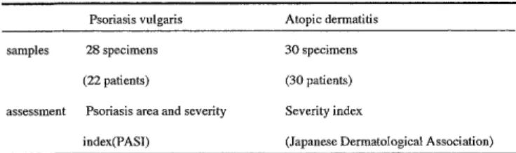
Twenty-eight patients with psoriasis vulgaris and 30 patients with atopic dermatitis were enrolled in this study. The samples and their assessment were shown in Table 1. Clinical assessment of psoriasis was
Table 1. Numbers of samples and thier assessment Psoriasis vulgaris Atopic dermatitis
samples 28 specimens 30 specimens
(22 patients) (30 patients)
assessment Psoriasis area and severity Severity index
index(PASI) (Japanese Dermatological Association)
made using PASI score' and atopic dermatitis using Severity Index proposed by Japanese Dermatological Association.
Nobody but one patient had received vitamin D3 ointment therapy at biopsy time in psoriasis vulgaris patients. Before and after one month ointment with group I or II class GCS, clinical assessment was made.
Responders were defined as patients with reducing scores below 75% compared with pretreatment scores', retrospectively. Low responders were defined as pa- tients not corresponded to above. Patients were classi- fied into four groups as described in summary.
Biopsied specimens were obtained from the patients under the informed consent before and after the treat- ment with topical GC. Paraffin-embedded tissues for immunostaining had been routinely fixed in 10% neu- tral. buffered formalin for 8-48 hours. Cryosections were preserved in -30°C until RNA extraction.
Antibodies
JSB-1 (NOVOCASTRA, Newcastle upon Tyne, UK), anti-P-gp antibody; it recognizes an internal epitope of PGP molecule'; the ascites was used in a dilution of 1:20. Anti CD3 antibody (DAKO, Copenhagen, Denmark) or anti CD20 antibody (DAKO) was used in a dilution of 1:50 or 1:100. Terminal deoxynucleotidyl transferase (Tdt) enzyme (INTERGEN, Oxford,UK) was used for labelling apoptotic cells'.
Phosphate-buffered saline(PBS) (pH 7.4) containing bovine serum albumin (BSA) (1%) was used for the di- lution of JSB-1.
Immunohistochemistry
Paraffin tissue sections of 5 p m were placed on siliconized glass slide and deparaffinized in xylene, 100% ethanol, 90% ethanol for P-gp, anti CD3, anti CD20 staining or Tdt-mediated Unscheduled Nick End Labelling (TUNEL).
Staining with anti P-gp staining: The antigen re- trieval was performed by treatment with the slides in 90V citrate buffer for 20 minutes. After washing in aqua for 10 minutes, endogenous peroxidase activities were blocked in aqua bidest, containing 3% H202 for
5 minutes. Then the slides were washed in PBS for 10 minutes. All tissue sections and positive controls' were incubated with JSB-1 for overnight at 4°C; nega- tive controls, however, were incubated only with 1 % BSA. After washing in PBS for 10 minutes, all slides were covered with biotinylated gout antimouse IgG (DAKO), for 10 minutes at room temperature. The slides were then washed again for 10 minutes in PBS.
Finally they were incubated for 10 minutes at room temperature with a streptavidin- biotin-conjugated peroxidase complex (DAKO).
Staining with anti-CD3 or CD20 staining: The anti- gen retrieval was performed by treatment in 0.1%
trypsin for 30 minutes at room temperature for anti CD3. After washing in PBS for 10 minutes, endoge- nous peroxidase activities were blocked in aqua bidest, containing 3% H202 for 5 minutes. After wash- ing in PBS for 10 minutes, the slides were incubated in 5% normal goat serum for 5minutes. All tissue sec- tions and positive controls were incubated with anti CD3 or CD20 antibody for lh at 4°C; negative con- trols, however, were incubated only with 1% BSA.
After washing in PBS for 10 minutes, all slides were treated same as P-gp staining.
TUNEL method: The slides were treated with 20,u g/ml proteinase K in PBS for 1 hour at room tempera- ture. Endogenous peroxidase activities were blocked in aqua bidest, containing 3% H202 for 5 minutes, same as P-gp staining. Then the slides were washed in PBS for 10 minutes and then treated with EQUILIBRATION BUFFER (INTERGEN, New York, USA) for 10 min- utes. Then the slides were incubated with Tdt enzyme for 1 hour at 37 °C and further incubated with
WORKING STRENGTH STOP/WASH BUFFER
(INTERGEN) for 10 minutes at room temperature.
Finally they were incubated for 30 minutes at room temperature with anti -digoxigenin peroxidase conju- gate (INTERGEN).
The peroxidase label was developed for tissues using 0.03% H202 and 3,3'-diaminobenzidine tetrachlo- ride (DAB), which results in a brown staining product.
The tissue sections were mounted with Permount (Fisher Scientific, New Jersey, USA), and counterstaining with hematoxylin and methyl green for immunohistochemistry and TUNEL method, re- spectively.
Semiquantitative immunohistochemical scores were defined; 0: no staining, 1: weakly positive, 2: moder- ately positive, 3: strongly positive. Sweat gland of nor- mal skin defined as score 16 for P-gp.
R T-PCR
Total cellular RNA was isolated using RNeasy Mini Kit (QIAGEN, California, USA) as described by Meller et al' RNA concentration and purity were determined spectrophotometrically
Total cellular RNA (500 ng) was amplified by RT PCR using Ready To GoTM RT-PCR Beads (Amersham Pharmacia Biotech, New Jersey, USA) with primers de scribed by Chen et al' (421C for 30 minutes to re verse transcribe and PCR cycles, an initial step of 95
°C fo
r 5 minutes, 45 cycles of a 3-temperature PCR [95°C for 1 minute, 62°C for 1 minute, 72°C for 1 min ute] and end with 72°C for 5 minutes Each PCR product was then size-fractionated through a 18% agarose gel The amplified cellular fragment (target) was 544 by
Further, RT PCR products were extracted and di gested with VSP I (Takara, Kyoto, Japan), digested al- most 400bp and 150bp (GENE BANK ACCESSION M14758)
Results
Immunostazning
In immunohistochemically, only sweat gland cells showed weakly positive staining of P gp in the normal skin (Fig la) In contrast, P gp was mainly expressed in the cytoplasm of infiltrated cells as well as keratinocyte and vascular endothelial cells in each samples obtained from psoriasis vulgaris or atopic der matitis patients The TUNEL positive cells showed a nuclear staining pattern in the lesinal skin of psoriasis vulgaris and atopic dermatitis
Patients profiles of psoriasis vulgaris are summa razed in Table 2 The mean age of S () group younger than other groups, but no significant differences were seen in other parameters
There was no significant correlation between immunohistochemical scores of P gp and ages, PASI scores, PUVA therapy, biopsy site nor complication of
Figure 1. This figure showed that a part of P gp positive cells were CD3 positive cells (a)Only sweat gland cells showed weakly positive staining of P gp in the normal skin (x100) (b) Low magnification of P gp immunostaining of the sample from psoriasis vulgaris patient P gp mainly expressed in the cytoplasm, was observed in infiltrating cells as well as keratinocyte and vascular endothelial cells (x50) (c) P gp immunostaining from the another patient which showed a low intensity of P gp (x50) (d) P gp immunostaining of the sample from psoriasis vulgaris patient Mononuclear cells showed positive (x100) (e) CD3 staining of the same pa tients (x100)
Table 2. Patients profiles of psoriasis vulgaris
Parameters S(-) S(+) R(-) R(+)
Number of subjects* 6 5 5 12
Age (years) ± S.D.** 38.5±16.3 50.8±16.9 62.8±21.7 61.6±16.2
Sex (m/f) 6/0 8/1 3/2 8/4
DM or hyperlipidemia (+/-) 3/3 0/5 2/3 4/8
Biopsy site (SEA***/non SEA) 1/6 2/3 4/1 1/11
PUVA (+/-) 1/5 2/3 0/5 5/7
*: mean value of subjects
, **: p<0.05, ***: SEA=sun exposured skin
S (-): responder to topical steroid without steroid ointment for one month at the time of biopsy.
S (+): responder to topical steroid and under steroid therapy at the time of biopsy, R (-): low or non- responder to topical steroid without steroid therapy for one month at the time of biopsy, R (+): low-or non-responder to topical steroid and under steroid therapy at the time of biopsy.
Table 3. Atopic dermatitis paients characteristics
Parameter S(-) S(+) R(-) R(+)
Number of subjects* 8 13 0 9
Age (years)* ± S.D. 20.1±4.5 22.8±7.3 26.7±12.5
Sex (m/f) 4/4 8/5 7/2
Number of eosinophils ± S.D. 899.4±743.0 969.6±705.7 801.7±522.9
Biopsy site (SEA***/non SEA) 4/4 7/6 5/4
Red face** (+/-) 0/7 0/13 8/1
*: mean value of subjects, **: p<0.05***: SEA=sun exposured skin
S (-): responder to topical steroid without steroid ointment for one month at the time of biopsy, S (+):
responder to topical steroid and under steroid therapy at the time of biopsy, R (-): low or non- responder to topical steroid without steroid therapy for one month at the time of biopsy, R (+): low-or non-responder to topical steroid and under steroid therapy at the time of biopsy.
Figure 3. TUNEL scores of the samples from psoriasis vulgaris patients. There was no differences between 4 groups S (-): responder to topical steroid without steroid ointment for one month at the time of biopsy, S (+): responder to topi- cal steroid and under steroid therapy at the time of biopsy, R (-): low or non- responder to topical steroid without steroid therapy for one month at the time of biopsy, R (+): low-or non-responder to topical steroid and under steroid therapy at the time of biopsy, in TUNEL scores.
Figure 2. In psoriasis vulgaris patients, immunohistochemical scores of P-gp were significantly higher in R(+) group than in other groups.
DM or hyperlipidemia at biopsy times (data not shown).
The immunohistochemical scores of P-gp and GC ointment duration were significantly higher in R(+) group than in other groups (Fig.2). There was no sig- nificant differences between R(-) samples and S(+) nor S(-) samples. Expression of PGP was correlated with GC ointment duration (Correlation coefficient=0.584, p<0.05), but not ages at biopsy times.
A part of P-gp positive cells were mast cells or CD3
Figure 4. In atopic dermatitis patients, immunohistochemical scores of P-gp were significantly higher in R (+) group than S (-) group, but not S (+) group.
positive cells (Fig.lb-e) but not CD20 positive cell (data not shown).
There was no differences between 4 groups in TUNEL scores in psoriasis vulgaris (Fig.3). TUNEL scores of symptomless margin of the lesion had not
differences from lesional scores.
In case of atopic dermatitis, clinical profiles are summarized in Table 3. No other parameters but atopic red face had significant differences between 4 groups.
There was no significant correlation between immunohistochmical scores and severity index, num- ber of peripheral blood eosinophils, total IgE nor bi- opsy site (data not shown).
The immunohistochemical scores of P-gp were sig- nificantly higher in R (+) group than in S (-) group (Fig.4). There was no differences of P-gp scores be- tween S (+) samples and R (-) nor R (+) samples. There was no significant differences between S (+) and R (+) group in GC ointment duration. Expression of P-gp was not correlated with GC ointment duration.
Same as in psoriasis vulgaris patients, a part of P-gp positive cells were mast cells or CD3 positive cells but not CD20 positive cell (data not shown).
There was no significant differences between 4 groups in TUNEL scores in atopic dermatitis same as psoriasis vulgaris (data not shown).
Expression of MDR-1 mRNA
Samples from which showed strong P-gp intensity in immunohischemistry, demonstrated MDR 1 gene ex- pression in RT-PCR (Fig. 5). RT-PCR products were di- gested into almost 400bp and 150bp fragments by VSP-I (data not shown). MDR1 mRNA expression was upregulated in the patients with R (+) group but not in S (+) group of psoriasis vulgaris.
Figure 5. MDR 1 gene expression of the lesional skin from the patients with psoriasis vulgaris by RT-PCR. Samples with strong P-gp intensity demonstrated significant MDR-1 gene expression. No.1,2,4: Sample from R (+) group, No. 3:
Sample from S (+) group, No. 5: normal human non sun- exposed skin, No. 6: negative control, M: Bio Marker
Discussion
Although several side effects were recognizedl0, topical GC ointment is the most widely used medica- tion in the treatment of skin disorders; psoriasis vulgaris" or atopic dermatitis". In the last decade, new type of skin manifestations, i.e., persistent erythema of the face (atopic red face) 12 or
"poikiloderma -like lesion on the neck"" , have been rec- ognized in patients with atopic dermatitis, especially in Japan. Poikiloderma-like lesion on the neck could be attribute to chronic inflammation and delay of wound healing process, possibly caused by long- standing topical GC therapy". Topical GC preparations
are categorized into different potency groups by vaso- constrictor (i.e., skin blanching) assay. However, this is subjective and dose not correlate perfectly with clini- cal antiinflammatory effects, percutaneous absorption, or propensity for adverse effects". No correlation was seen between Lymphocyte stertoid sensitivity (LSS) parameters: maximal percentage inhibition of thymidine incorporation achived at the highest con- centration of dexamethason (Imax) and the concentra- tion of steroid at which thymidine incorporation is re- duced to 50% of Imax value, and cutaenous vasoconstrictor response; suggested tissue differences in steroid sensitivity". Currently, there is no reliable and accessible method available for expressing GC po- tency in a better way".
Other problems have been implicated about topical GC ointment, i.e., GC insensitivity. GC insensitivity was observed in the subset of asthmatic' or leukemic"
patients : they showed low responses to ordinal GC therapy, but others showed good responses. Low re- sponders are resistant to the antiinflammatory effect of GC while simultaneously showing several other side effects known to reflect normal sensitivity".
P-gp is a energy-deriven plasma membrane trans- porter which pumps certain drugs including GC out of the cells". The physiologic role of P-gp is speculative, and the possibilities include the protection of normal tissues from environmental and endogenous toxins, steroid secretion in the adrenal gland, secretion of bile salts in the bile canaliculi, and secretary functions in the kidney'. Prolonged exposure of tumor cells to natural hydrophobic cytotoxic drugs may result in the acquisition of resistance by cells not only to the drugs used but also to a series of structurally and function- ally unrelated drugs".
Recently, several reports have described relation be- tween GC and P-gp in patients with asthma or nasal polyp. Henriksson et al." reported that nasal polyps from 5 of 17 patients treated with clinical dose of a topical nasal steroid showed a stronger staining inten- sity for P-gp than polyps from 13 untreated patients.
Montano et al.' showed that surface P-gp expression and Rh 123 efflux in B cells were decreased in GC sensitive asthma than in GC resistant patients or mild asthma, significantly. There was no significant differ- ences between their GC sensitive and resistant pa- tients in total daily steroid dose. Furthermore, they
observed reduced surface functional P-gp expression by B cells from normal volunteers after 48-h exposure of cells to 10 nM dexamethasone.
In our observation, intensity of P-gp and MDR 1 gene expressions showed a positive correlation to the duration of GC application to the lesional skin in pso- riasis vulgaris which might suggest that P-gp plays some role in induction of GC insensitivity as well asthma or nasal polyp patients.
In contrast to psoriasis vulgaris, there was no sig- nificant colleration between P-gp scores and duration of steroid ointment in the patients with atopic derma- titis; suggested atopic dermatitis occurred not only by inflammation but by impaired barrier function".
Several reports showed GC induces cell apoptosis22. 23 However, in our patients, TUNEL scores were not sig- nificantly different between 4 groups, and scores of most patients showed 0. Our data suggested that clini- cal response to GC ointment in inflammatory skin dis- eases was caused not by apoptotic but by another mechanism; i.e., downregulation of the expression of cytokines and adhesion molecules".
Maillefert et al. demonstrated that larger dose of GC induced higher expression of MDR 1 gene and P-gp in rheumatoid arthritis and suggested the possible resis- tance to other drugs". In a similar manner to their ob- servation, our data on psoriasis vulgaris suggested that GC ointment induces the expression of P-gp to re- duce the response to other drugs.
In conclusion, we should pay careful attention for the possible induction of GC insensitivity in the pa- tients with inflammatory skin disorders under GC ointment. Combination of phototherapy, vitamin D3 ointment, or immunosuppressive agent with GC might prevent the induction of drug resistance as demon- strated in combination chemotherapy for cancer pa- tients.
References
1. Bradshow DM, Arceci RJ: Clinical relevance of transmembrane
drug efflux as a mechanism of multidrug resistance. J Clin Oncol 16: 3674-90, 1998
2. Roninson IB, Chin JE, Choi KG, et al: Isolation of human mdr DNA sequences amplified in multidrug-resistant KB carcinoma
cells. Proc Natl Acad Sci USA 83: 4528-42, 1986
3. Montano E, Schmitz M, Blaser K and Simon HU: P-glycoprotein
expression in circulating blood leukocytes of patients with steroid resistant asthma. Invest Allergol Immunol 6: 14-21, 1996 4. Hofer A, Fink-puches R, Kerl H and Wolf P: Comparison of
phototherapy with near vs. far erythemogenic doses of narrow-
band ultraviolet B in patients with psoriasis. Br J Dermatol 138:
96-100, 1998
5. Trozak DJ: Topical corticosteroid therapy in psoriasis vulgaris.
Cutis 46: 341-50, 1990
6. Valk P, Kalken CK, Ketelaars H, et al: Distribution of multi-drug resistance-associated P-glycoprotein in normal and neoplastic
human tissues. Ann Oncol 1: 56-64, 1990
7. Megyesi J, Safirstein RL, Price PM: Induction of p21 WAF1/CIP1/SDI1 in kidney tubule cells affects to course of
cisplatin-induced acute renal failure. J Clin Invest 101: 777-82,
1998
8. Meller VH, Wu KH, Roman G, Kuroda MI and Davis RA: RoX1 RNA paints the X chromosome of male drosophila and is regu-
lated by the dosage compensation system. Cell 88: 445-57, 1997 9. Chen CJ, Clark D, Ueda K, Pastan 1, Gottesman MM, Roninson IB:
Genomic organization of the human multidrug resistance 1
(MDR1) gene and origin of P-glycoprotein. J Biol Chem 265: 506-
14, 1990
10. Drake LA, Ceilley RI, Cornelison RL, et al : Guidelines of care for psoriasis. Am J Acad Dermatol 1993; 28: 632-7
11. Kawashima M, Takigawa M, Nakagawa H, et al : Guidelines for therapy for atopic dermatitis. Jap J Dermatol 110: 1099-1104 (in
Japanese), 2000
12. Katayama I, Igawa K, Minatohara K, Nishioka K: Topical glucocorticoid augments IgE-mediated passive cutaneous anaphy-
laxis in Balb/C mice and mast cell deficient WBB6F1 v/v mice.
Clin Exp Allergy 27: 1477-83, 1997
13. Mukai H, Nishioka K, Kamimura K, Katayama I, Nishiyama S:
Poikiloderma-like lesions on the neck in atopic dermatitis: a histopathological study. J Dermatology 17: 85-91, 1990
14. E
llison JA, Patel L, Ray DW, David TJ, Clayton PE: Hypothalamic-
pituitary-adrenal function and glucocorticoid sensitivity in atopic dermatitis. Pediatrics 105: 794-9, 2000
15. Hearing SD, Norman M, Smyth C, Foy C, Dayan CM: Wide varia- tion in lymphocyte steroid sensitivity among healthy human vol-
unteers. J Clin Endocrinol metab 84: 4149-54, 1999
16. Kasper GJ, Kardos G, Pieters R, van Zantwijk CH, van Wering ER, veerman AJ: Clinical and cell biological features related to cellular
drug resistance of childhood acute lymphoblastic leukemia cells.
Leuk Lymph 19: 407-16, 1995
17. Ebrecht M, Buske-Kirschbaum A, Hellhanner D, et al: Tissue speci- ficity of glucocorticoid sensitivity in healthy adults. J Clin
Endocrinol metab 85: 3733-9, 2000
18. Chiele E, Santoni-Rugiu E, Cervelli F, et al: Differential modula- tion of P-glycoprotein expression by dexamethasone and 3-
methycholanthrene in rat hepatocyte primary cultures.
Carcinogenesis 15: 335-41, 1994
19. Kobayashi H, Takemura Y, Miyachi H, et al: Quantitative analysis of human multidrug resistance 1 (MDR1) gene expression by
nonisotopic competitive reverse transcriptase polymerase chain re-
action assay. J Clin Lab Anal 11; 258-66, 1997
20. Henriksson G, Norlander T, Zheng X, Stierna P and Westrin KM:
Expression of P-glycoprotein 170 in nasal mucosa may be in-
creased with topical steroids. Am J Rhinol 11: 317-21, 1997 21. Di nardo A, Werts P, Giannetti A, Seidenari S: Ceramide and cho-
lesterol composition of the skin of patients with atopic dermatitis.
Acta Derm Venereol 78: 27-30, 1998
22. Tuosuto L, Cundari E, Gilardini MM, Picollera E: Analysis of sus- ceptibility of mature human T lymphocytes to dexamethasone-
induced apoptosis. Eur J Immunol 24: 1061-5, 1994
23. Bourgeois S, Gruol DJ, Newby RF, Rajah F: Expression of an mdr gene is associated with a new form of resistance to
dexamethasone-induced apoptosis. Mol Endo 7: 840-51, 1993 24. Prakash A, Benfield P: Topical mometasone. A review of its phar-
macological properties and therapeutic use in the treatment of
dermatological disorders. Drugs 55: 145-63, 1998
25. Maillefert JF, Maynadie M, Tebib JG Aho S, et al: Expression of the multidrug resistance glycoprotein 170 in the peripheral blood
lymphocytes of rheumatoid arthritis patients. The percentage of
lymphocytes expressing glycoprotein 170 is increased in patients
treated with predonisolone. Br J Rheumatol 35: 430-5, 1996