Organic and Peptidyl Constituents of Snake Venoms: The Picture Is Vastly More Complex Than We Imagined
Author Alejandro Villar‑Briones, Steven Aird journal or
publication title
Toxins
volume 10
number 10
page range 392
year 2018‑09‑26
Publisher MDPI AG, Basel, Switzerland Rights (C) 2018 The Author(s).
Author's flag publisher
URL http://id.nii.ac.jp/1394/00000872/
doi: info:doi/10.3390/toxins10100392
Creative Commons?
Attribution 4.0 International?
(https://creativecommons.org/licenses/by/4.0/)
toxins
Article
Organic and Peptidyl Constituents of Snake Venoms:
The Picture Is Vastly More Complex Than We Imagined
Alejandro Villar-Briones
1and Steven D. Aird
2,*
1
Division of Research Support, Okinawa Institute of Science and Technology, 1919-1 Tancha, Onna-son, Kunigami-gun, Okinawa 904-0495, Japan; [email protected]
2
Division of Faculty Affairs and Ecology and Evolution Unit, Okinawa Institute of Science and Technology, 1919-1 Tancha, Onna-son, Kunigami-gun, Okinawa 904-0495, Japan
*
Correspondence: [email protected]; Tel.: +81-98-982-3584
Received: 29 August 2018; Accepted: 20 September 2018; Published: 26 September 2018
Abstract: Small metabolites and peptides in 17 snake venoms (Elapidae, Viperinae, and Crotalinae), were quantified using liquid chromatography-mass spectrometry. Each venom contains >900 metabolites and peptides. Many small organic compounds are present at levels that are probably significant in prey envenomation, given that their known pharmacologies are consistent with snake envenomation strategies. Metabolites included purine nucleosides and their bases, neurotransmitters, neuromodulators, guanidino compounds, carboxylic acids, amines, mono- and disaccharides, and amino acids. Peptides of 2–15 amino acids are also present in significant quantities, particularly in crotaline and viperine venoms. Some constituents are specific to individual taxa, while others are broadly distributed. Some of the latter appear to support high anabolic activity in the gland, rather than having toxic functions. Overall, the most abundant organic metabolite was citric acid, owing to its predominance in viperine and crotaline venoms, where it chelates divalent cations to prevent venom degradation by venom metalloproteases and damage to glandular tissue by phospholipases. However, in terms of their concentrations in individual venoms, adenosine, adenine, were most abundant, owing to their high titers in Dendroaspis polylepis venom, although hypoxanthine, guanosine, inosine, and guanine all numbered among the 50 most abundant organic constituents.
A purine not previously reported in venoms, ethyl adenosine carboxylate, was discovered in D. polylepis venom, where it probably contributes to the profound hypotension caused by this venom. Acetylcholine was present in significant quantities only in this highly excitotoxic venom, while 4-guanidinobutyric acid and 5-guanidino-2-oxopentanoic acid were present in all venoms.
Keywords: snake venoms; metabolites; peptides; purine nucleosides and bases; neurotransmitters;
neuromodulators; guanidinium compounds; carboxylic acids; amines; mono- and disaccharides;
amino acids
Key Contribution: This paper presents the first comprehensive analysis of small metabolites in snake venoms and shows that some of these undoubtedly contribute to prey immobilization. Many novel di-, tri-, and tetrapeptide sequences have been determined.
1. Introduction
In contrast to insect, arachnid, and anuran venoms, snake venom chemistry is dominated by proteins. As a result, relatively little attention has been paid to small organic constituents thereof.
Ganguly and Malkana [1] detected cholesterol and lecithin in cobra venom, and Devi [2] claimed that glycerophosphate was present in cobra, viper, and pitviper venoms. Monosaccharides and free amino acids have also been reported, but no role in envenomation for these non-proteinaceous components
Toxins2018,10, 392; doi:10.3390/toxins10100392 www.mdpi.com/journal/toxins
Toxins2018,10, 392 2 of 49
has ever been suggested. Bieber [3] published the last thorough review of non-proteinaceous snake venom constituents.
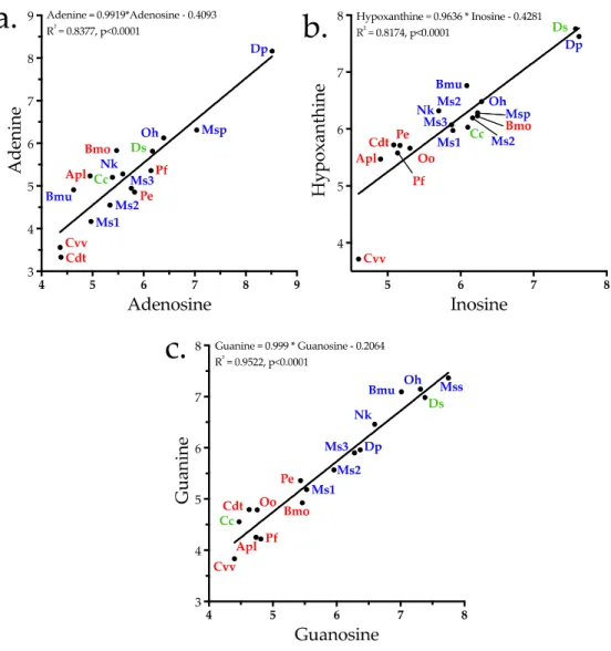
Aird [4] proposed that the purine nucleosides adenosine, inosine, and guanosine, documented in various venoms [5–11], actually occupy a central position in the envenomation strategies of all venomous snakes, whether as venom constituents, or when released from prey tissues by venom proteins and the biochemical cascade they unleash. Later Aird [12,13] documented the purine levels in both snake and helodermatid venoms, confirming that some ophidian venoms comprise as much as 8.7% nucleosides by mass, exceeding the levels of many proteinaceous toxins and lending further credence to his earlier hypothesis about their strategic importance.
Recently, after deproteinating snake venom samples to serve as controls in another study, we were surprised to discover that the small metabolite content of snake venoms is vastly richer than we imagined. While it might have been reasonable to expect trace levels of a broad array of compounds, due to death and replacement of venom gland cells, many of these compounds are 3–6 orders of magnitude more abundant than such anticipated background levels. Accordingly, we identified and semi-quantified small metabolites and peptides in 17 snake venoms representing the families Elapidae (8), Viperinae (2), and Crotalinae (7). Elapid venoms include three individual samples from Micrurus surinamensis, in order to assess levels of individual variation; thus, 15 snake taxa were examined.
The diversity of small metabolites seen in these venoms greatly exceeds what has been reported previously. While many of the more abundant compounds have well-known pharmacologies, some, like adenosine, can exert opposing effects, depending upon their tissue concentrations. Like the proteinaceous constituents, titers of these metabolites vary between individuals and between taxa.
Undoubtedly, some of them act synergistically with proteinaceous venom components to achieve rapid immobilization of prey.
2. Results and Discussion
2.1. Compounds Isolated from Snake Venoms
Deproteinated samples of 17 snake venoms (Supplementary Table S1) were analyzed by liquid chromatography-mass spectrometry, with simultaneous detection of positive and negative ions (Figure 1). Both positive and negative ions were combined into a master table containing all 17 datasets.
Ions were identified on the basis of mass, chromatographic retention time, and fragmentation pattern.
Each venom contained roughly 900 LC-MS peaks containing small organic molecules and peptides (<2 kDa). Not all of these peaks represent unique compounds. Some metabolites interact with others during LC, with the result that the same compound occasionally elutes in more than one location. In addition to these duplicate peaks, highly concentrated metabolites, such as adenosine, citric acid, and guanosine, sometimes produced fragments. Still others, such as a couple of tripeptides, were identified not only as monomers, but produced a secondary dimeric peak. Data from these
“duplicate peaks” were manually combined. It is safe to say that each venom contained in excess
of 850 small organic molecules and peptides. Unquestionably, many more are present at trace
levels. For example, we found xanthosine in preliminary experiments, but it was not detected
automatically in the final dataset. A manual search in the venom of Bungarus multicinctus, where it
had previously been most abundant, revealed that it was indeed present, but at levels only slightly
above background noise. Accordingly, we have tried to be conservative here. All but one of the 50
most abundant metabolites (Figure 2, Supplementary Table S1) were positively identified, or at least,
well characterized. Identifications beyond the top 50 compounds are considered tentative for small
metabolites. Peptide sequences provided are confirmed.
Toxins2018,10, 392 3 of 49
Toxins 2018, 10, x FOR PEER REVIEW 3 of 51
Supplementary Table S1). Based upon their adjusted areas (the sum of positive and negative ion peak areas for all venoms), the most abundant small venom constituents included carboxylic acids (some of which possessed guanidino groups), purine nucleosides and their bases, neurotransmitters and neuromodulators, amines (mostly tertiary amines), amino acids, and peptides ranging from 2 to 15 amino acids.
Metabolites identified are discussed below by class and within each class they are presented in order of their decreasing maximum abundance in the venoms examined. That sequence reflects their possible strategic importance in the venoms in which they achieved their highest concentrations, and not necessarily their mean abundance across all venoms examined.
Figure 1. Total ion chromatograms of negative and positive ions of metabolites and peptides from Agkistrodon piscivorus leucostoma venom. The negative ion peak that dwarfs all others is citric acid.
Assuming no metabolite loss during deproteination, the metabolites and peptides separated here represent the small molecule component of ~69 µg of crude venom. Metabolites were separated on a SeQuant ZIC-pHILIC HPLC 2.1 × 150 mm column, flow rate 120 µL/min, using acetonitrile as solvent A, and 10 mM ammonium carbonate, 0.1% ammonium hydroxide in water as solvent B. Separation was done in HILIC mode, with a linear gradient from 20% to 80% solvent B in 30 min, followed by a wash for 20 min with 20% acetonitrile, 0.5 M sodium chloride in water (solvent C) and, finally, column re-equilibration with starting conditions for 15 min.
0 5 10 15
0 1×10
82×10
83×10
84×10
8Run Time (min)
Pe ak In te ns ity
Positive Ions Negative Ions
Figure 1.
Total ion chromatograms of negative and positive ions of metabolites and peptides from Agkistrodon piscivorus leucostoma venom. The negative ion peak that dwarfs all others is citric acid.
Assuming no metabolite loss during deproteination, the metabolites and peptides separated here represent the small molecule component of ~69
µg of crude venom. Metabolites were separated on aSeQuant ZIC-pHILIC HPLC 2.1 × 150 mm column, flow rate 120
µL/min, using acetonitrile as solventA, and 10 mM ammonium carbonate, 0.1% ammonium hydroxide in water as solvent B. Separation was done in HILIC mode, with a linear gradient from 20% to 80% solvent B in 30 min, followed by a wash for 20 min with 20% acetonitrile, 0.5 M sodium chloride in water (solvent C) and, finally, column re-equilibration with starting conditions for 15 min.
Quantities of these small molecules present in each venom differed by orders of magnitude, within and across species, a pattern mimicking that of snake venom proteins [14–16] (Figure 2, Supplementary Table S1). Based upon their adjusted areas (the sum of positive and negative ion peak areas for all venoms), the most abundant small venom constituents included carboxylic acids (some of which possessed guanidino groups), purine nucleosides and their bases, neurotransmitters and neuromodulators, amines (mostly tertiary amines), amino acids, and peptides ranging from 2 to 15 amino acids.
Metabolites identified are discussed below by class and within each class they are presented
in order of their decreasing maximum abundance in the venoms examined. That sequence reflects
their possible strategic importance in the venoms in which they achieved their highest concentrations,
and not necessarily their mean abundance across all venoms examined.
Toxins2018,10, 392 4 of 49
Toxins 2018, 10, x FOR PEER REVIEW 4 of 51
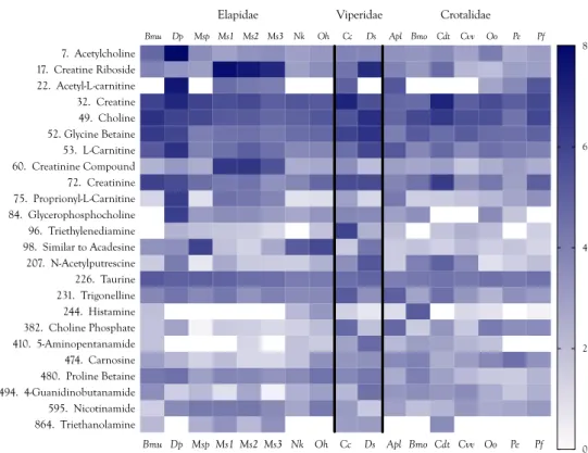
Figure 2. Heat map of the 50 most abundant metabolites and peptides found in 17 snake venoms, arranged in decreasing order of the maximum concentrations found among the species examined.
Compound abundances represent the log10 of peak intensities of positive and negative ions combined, after subtraction of respective baselines. Logarithmic representations have the effect of compressing apparent differences, so these venoms are compositionally much more divergent than can be shown graphically. Taxonomic names: Bmu, Bungarus multicinctus; Dp, Dendroaspis polylepis; Msp, Micrurus spixii; Ms1–3, Micrurus surinamensis, 3 individuals; Nk, Naja kaouthia; Oh, Ophiophagus hannah; Cc,
Bm u D p M sp M s1 M s2 M s3 N k O h C c D s A pl Bm o C dt C vv O o Pe Pf 1. Adenosine
2. Adenine 3. Citric acid 4. pEKW 5. TPPAGPDVGPR 6. 4-Guanidinobutyric acid 7. Acetylcholine 8. pEIWPRPH 9. pENW 10. VPPDH 11. pE(VSMAHAAWAL)DIPP 12. pEWQ 13. pEGRPR 14. pEEWPPGHHIPP 15. pENWPAPK 16. N-Acetylneuraminic Acid 17. Creatine Riboside 18. RPPHP 19. Hypoxanthine 20. VP 21. Guanosine 22. Acetyl-L-carnitine 23. 5-guanidino-2-oxopentanoic Acid 24. pEGGPR 25. pEGRP 26. Inosine 27. pEKWV(GP)G(PE)PIPP 28. Proline 29. Ethyl Adenosine Carboxylate 30. (QPQG)RPPHVPP 31. HPPRPQIPP 32. Creatine 33. Glycosylated Peptide 34. pEH 35. WPPGPPIPP 36. PP 37. Cytidine 38. Imidazoleacetic acid 39. pEQ 40. Pyroglutamate 41. pESKPGRSPPISP 42. pEE 43. Guanine 44. pE(NH) 45. VPPGPPGPPP 46. Contains Methylpyrimidine 47. pER 48. PGP(GC)P(R)G(PE)PIPP 49. Choline 50. Unidentified
0 1 2 3 4 5 6 7 Elapidae
Bm o Pf
Bm u D p M sp M s1 M s2 M s3 N k O h C c D s A pl C dt C vv O o Pe Crotalidae Viperidae
8
Figure 2.
Heat map of the 50 most abundant metabolites and peptides found in 17 snake venoms, arranged in decreasing order of the maximum concentrations found among the species examined.
Compound abundances represent the log
10of peak intensities of positive and negative ions combined, after subtraction of respective baselines. Logarithmic representations have the effect of compressing apparent differences, so these venoms are compositionally much more divergent than can be shown graphically. Taxonomic names: Bmu, Bungarus multicinctus; Dp, Dendroaspis polylepis; Msp, Micrurus spixii; Ms1–3, Micrurus surinamensis, 3 individuals; Nk, Naja kaouthia; Oh, Ophiophagus hannah;
Cc, Cerastes cerastes; Ds, Daboia siamensis; Apl, Agkistrodon piscivorus leucostoma; Bmo, Bothrops moojeni;
Cdt, Crotalus durissus terrificus; Cvv, Crotalus viridis viridis; Oo, Ovophis okinavensis; Pe, Protobothrops
elegans; Pf, Protobothrops flavoviridis.
Toxins2018,10, 392 5 of 49
2.2. Carboxylic Acids That Chelate Divalent Cations
2.2.1. Citric Acid
Citric acid was present in in all venoms and was the first-ranked metabolite overall (Figures 3 and 4, Supplementary Table S1). Its concentration was 2–3 orders of magnitude greater in viperine and crotaline venoms than in elapid venoms. Citrate was first discovered in snake venoms in the laboratory of Ivan I. Kaiser [17,18]. Francis et al. [18] found citrate concentrations ranging from 95 to 150 mM in viperine venoms, from 63 to 142 mM in crotalines, and from 17 to 163 mM in elapids.
They found that in Bothrops asper venom, Ca
2+concentrations ranged from 2.5 to 3.6 mM. At those Ca
2+concentrations, a phospholipase A
2from Bothrops asper venom was completely inhibited by only 20 mM citrate. They further reported that Crotalus adamanteus 5
0-nucleotidase and phosphodiesterase were inhibited 100% and 75%, respectively, by 100 mM citrate [18]. They suggested that citrate may inactivate metalloenzymes in the venom gland by chelating essential metal cofactors. Both phosphodiesterase and 5
0-nucleotidase are Mg
2+-dependent enzymes. It appears that citrate chelates Ca
2+more effectively than Mg
2+, a possibility also implied by Maguire and Cowan [19], who note that a 10 × excess of EGTA over Ca
2+in a given system would also chelate 20% of the Mg
2+. Nonetheless, citrate does chelate Mg
2+effectively, and many bacterial citrate transporters preferentially transport its Mg
2+salt [20].
Toxins 2018, 10, x FOR PEER REVIEW 5 of 51
Cerastes cerastes; Ds, Daboia siamensis; Apl, Agkistrodon piscivorus leucostoma; Bmo, Bothrops moojeni;
Cdt, Crotalus durissus terrificus; Cvv, Crotalus viridis viridis; Oo, Ovophis okinavensis; Pe, Protobothrops elegans; Pf, Protobothrops flavoviridis.
2.2. Carboxylic Acids That Chelate Divalent Cations
2.2.1. Citric Acid
Citric acid was present in in all venoms and was the first-ranked metabolite overall (Figures 3 and 4, Supplementary Table S1). Its concentration was 2–3 orders of magnitude greater in viperine and crotaline venoms than in elapid venoms. Citrate was first discovered in snake venoms in the laboratory of Ivan I. Kaiser [17,18]. Francis et al. [18] found citrate concentrations ranging from 95 to 150 mM in viperine venoms, from 63 to 142 mM in crotalines, and from 17 to 163 mM in elapids. They found that in
Bothrops asper venom, Ca2+concentrations ranged from 2.5 to 3.6 mM. At those Ca
2+concentrations, a phospholipase A
2from Bothrops asper venom was completely inhibited by only 20 mM citrate. They further reported that
Crotalus adamanteus 5′-nucleotidase and phosphodiesterasewere inhibited 100% and 75%, respectively, by 100 mM citrate [18]. They suggested that citrate may inactivate metalloenzymes in the venom gland by chelating essential metal cofactors. Both phosphodiesterase and 5′-nucleotidase are Mg
2+-dependent enzymes. It appears that citrate chelates Ca
2+more effectively than Mg
2+, a possibility also implied by Maguire and Cowan [19], who note that a 10× excess of EGTA over Ca
2+in a given system would also chelate 20% of the Mg
2+. Nonetheless, citrate does chelate Mg
2+effectively, and many bacterial citrate transporters preferentially transport its Mg
2+salt [20].
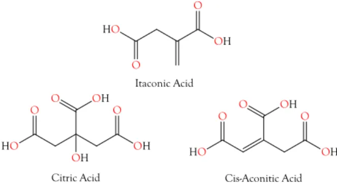
Figure 3. Structures of citric acid, cis-aconitic acid, and itaconic acid. cis-Aconitate is an intermediate between itaconic acid and citric acid, in the citric acid cycle. It seems probable that itaconic acid and cis-aconitic acid exist to support citric acid production in the venom glands. These tricarboxylic acids chelate divalent cations to inactivate phospholipases, metalloproteases, nucleases, and other metalloenzymes in the venom gland; however, upon injection into prey tissues, these components are immediately activated.
Odell et al. [21] reported citrate concentrations ranging from 42 to 154 mM (3.6–12.9%) in various elapid, viperine, and crotaline venoms, including a value of 10.3% in Dendroaspis polylepis venom.
They also found that protease activity of
Crotalus atrox venom against hide powder azure andazocasein was inhibited 7.5%, and that of Bothrops picadoi venom was inhibited 78% with the addition of 18–27 mM exogenous citrate. Thus, the citrate concentrations occurring in venoms should be more than ample to inactivate metalloenzymes, especially considering that venom serine proteases, which do not require metal cofactors, would not have been affected by this treatment. While citrate serves primarily to protect the venom gland from metalloenzymes, given its high concentration in various venoms documented in the foregoing studies and in the present one, it is likely that citrate also functions in envenomation as an anticoagulant, by scavenging Ca
2+required by coagulation factors [22,23] and for platelet aggregation [24,25].
OH O
OH O OH O
HO
O HO
O OH O
OH Itaconic Acid
Citric Acid Cis-Aconitic Acid
O
OH O
HO
Figure 3.
Structures of citric acid, cis-aconitic acid, and itaconic acid. cis-Aconitate is an intermediate between itaconic acid and citric acid, in the citric acid cycle. It seems probable that itaconic acid and cis-aconitic acid exist to support citric acid production in the venom glands. These tricarboxylic acids chelate divalent cations to inactivate phospholipases, metalloproteases, nucleases, and other metalloenzymes in the venom gland; however, upon injection into prey tissues, these components are immediately activated.
Odell et al. [21] reported citrate concentrations ranging from 42 to 154 mM (3.6–12.9%) in various elapid, viperine, and crotaline venoms, including a value of 10.3% in Dendroaspis polylepis venom.
They also found that protease activity of Crotalus atrox venom against hide powder azure and azocasein
was inhibited 7.5%, and that of Bothrops picadoi venom was inhibited 78% with the addition of 18–27 mM
exogenous citrate. Thus, the citrate concentrations occurring in venoms should be more than ample
to inactivate metalloenzymes, especially considering that venom serine proteases, which do not
require metal cofactors, would not have been affected by this treatment. While citrate serves primarily
to protect the venom gland from metalloenzymes, given its high concentration in various venoms
documented in the foregoing studies and in the present one, it is likely that citrate also functions in
envenomation as an anticoagulant, by scavenging Ca
2+required by coagulation factors [22,23] and for
platelet aggregation [24,25].
Toxins2018,10, 392 6 of 49
Toxins 2018, 10, x FOR PEER REVIEW 7 of 51
Figure 4. Organic acid abundances in snake venoms span nearly 8 orders of magnitude, based on combined positive and negative ion intensities, after subtraction of the blanks. The vast majority are unquestionably accidental venom constituents, probably resulting from cellular degradation.
However, compounds with peak intensities above E06, are probably sufficiently concentrated to make substantive contributions to venom pharmacology. Baseline (noise) has been subtracted from all ion intensities. Taxonomic names: Bmu, Bungarus multicinctus; Dp, Dendroaspis polylepis; Msp, Micrurus spixii; Ms1–3, Micrurus surinamensis, 3 individuals; Nk, Naja kaouthia; Oh, Ophiophagus hannah; Cc, Cerastes cerastes; Ds, Daboia siamensis; Apl, Agkistrodon piscivorus leucostoma; Bmo, Bothrops moojeni;
Cdt, Crotalus durissus terrificus; Cvv, Crotalus viridis viridis; Oo, Ovophis okinavensis; Pe, Protobothrops elegans; Pf, Protobothrops flavoviridis.
2.3. Other Carboxylic Acids
In all, 37 organic acids were identified in the venoms we surveyed. Many more are undoubtedly present, although probably at levels irrelevant to envenomation. Various fatty acids feature prominently among these. However, other organic acids may serve toxic functions in the prey, rather than protective or housekeeping functions in the venom gland. Nearly six decades ago, Curtis and Watkins [27] demonstrated that various carboxylic acids and their α-decarboxylation products have opposing actions on spinal neurons (Table 1), with the former being excitatory and the latter being inhibitory.
Figure 4.
Organic acid abundances in snake venoms span nearly 8 orders of magnitude, based on combined positive and negative ion intensities, after subtraction of the blanks. The vast majority are unquestionably accidental venom constituents, probably resulting from cellular degradation.
However, compounds with peak intensities above E
06, are probably sufficiently concentrated to make substantive contributions to venom pharmacology. Baseline (noise) has been subtracted from all ion intensities. Taxonomic names: Bmu, Bungarus multicinctus; Dp, Dendroaspis polylepis; Msp, Micrurus spixii; Ms1–3, Micrurus surinamensis, 3 individuals; Nk, Naja kaouthia; Oh, Ophiophagus hannah;
Cc, Cerastes cerastes; Ds, Daboia siamensis; Apl, Agkistrodon piscivorus leucostoma; Bmo, Bothrops moojeni;
Cdt, Crotalus durissus terrificus; Cvv, Crotalus viridis viridis; Oo, Ovophis okinavensis; Pe, Protobothrops elegans; Pf, Protobothrops flavoviridis.
2.2.2. Itaconic and cis-Aconitic Acids
Itaconic acid was also quite abundant in most venoms, reaching its highest concentrations in viperine and crotaline venoms (Figures 3 and 4, Supplementary Table S1). Like citric acid, for which it serves as a precursor via cis-aconitate, itaconic acid is also an excellent chelator of alkali and alkaline earth metals, with divalent cations being bound more strongly than monovalent cations.
Using a methylene succinic linker, itaconic acid has been used with a polymeric matrix to make chromatographic columns capable of resolving even very similar ion pairs, such as Ca
2+with Sr
2+or Mn
2+[26]. The literature has nothing to say about the metal chelation capacity of cis-aconitate,
but it is probably similar to that of citric acid. Venom itaconate levels are only slightly more highly
correlated with citrate levels (r
2= 0.9965) than with cis-aconitate (r
2= 0.9706). cis-Aconitate is also
Toxins2018,10, 392 7 of 49
highly correlated with citrate (r
2= 0.9663). The high titers of venom citrate suggest that in venom gland cells, C5-branched dibasic acid and citric acid pathways are being used in unusual ways. Itaconic acid is converted to cis-aconitate by aconitate decarboxylase (EC 4.1.1.6). Normally, in the citric acid cycle, aconitate hydratase (EC 4.2.1.3) catalyzes both the conversion of citrate to cis-aconitate and cis-aconitate to isocitrate. It is not clear how the backward reaction could be promoted and the forward reaction inhibited. However, the mass spectrometer cannot distinguish between citrate and isocitrate, so perhaps much of the citrate is actually isocitrate. From the snake’s standpoint, this probably makes no functional difference, as long as the subsequent step in the citric acid cycle, the conversion of isocitrate to oxalosuccinate by isocitrate dehydrogenase (EC 1.1.1.42), is blocked.
2.3. Other Carboxylic Acids
In all, 37 organic acids were identified in the venoms we surveyed. Many more are undoubtedly present, although probably at levels irrelevant to envenomation. Various fatty acids feature prominently among these. However, other organic acids may serve toxic functions in the prey, rather than protective or housekeeping functions in the venom gland. Nearly six decades ago, Curtis and Watkins [27]
demonstrated that various carboxylic acids and their α-decarboxylation products have opposing actions on spinal neurons (Table 1), with the former being excitatory and the latter being inhibitory.
Table 1.
Excitatory amino acids and their inhibitory
α-decarboxylated derivatives, as determined onrat spinal neurons [27]. Relative potency of the metabolite in question is indicated by the number of + or − symbols. Most importantly, Curtis and Watkins [27] reported that when the
α-carboxyl groupof the excitatory acidic amino acids is removed (lacking), the pharmacological action was completely reversed, not merely diminished or abolished. Several of each were identified in venoms in this study.
Taurine, an aminosulfonic acid, is one of the most abundant amino acids in animal cells, and is a significant constituent of all venoms investigated herein, particularly that of Bungarus multicinctus. See Figure 13.
Acidic Amino Acid Excitation α-Decarboxylation Product Inhibition
Aspartic Acid
+ + + β-Alanine− − −
Glutamic Acid
+ + +GABA − − −
Cysteic Acid
+ + +Taurine − − −
β-Hydroxyglutamic Acid + + γ-Amino-β-hydroxy-n-butyric Acid
− −
N-Methylaspartic Acid
+ +N-Methyl-β-Alanine − −
Aminomalonic Acid
+Glycine − −
α-Aminoadipic Acid + δ-Aminoadipic Acid
− −
α-Aminopimelic Acid + ε-Aminocaproic Acid
−
N,N-Dimethylaspartic Acid
+N,N-Dimethyl-β-Alanine 0
N-Methylglutamic Acid 0 N-Methyl-γ-Amino-n-butyric Acid 0
2.3.1. 4-Guanidinobutyric Acid
Three reactions are required to convert
L-arginine to 4-guanidinobutyric acid (4GBA), also known as 4-guanidinobutanoic acid (Supplementary Table S1, ID 6) and γ-guanidinobutyric acid (Figure 5);
thus, the latter is only two enzymatic reactions removed from 5-guanidino-2-oxopentanoic acid
(5G2OA). Overall, this was the second most abundant organic acid (Figures 4 and 5). Compounds
with imidazole or guanidinium groups inhibit the vasodilatory actions of K
ATPchannel openers [28],
but since a hypertensive function would make little sense in envenomation by most snakes, like 5G2OA,
the pharmacological function of 4GBA is probably its capacity to induce seizures. Hiramatsu [29] and
Tachikawa and Hosoya [30] reported that accumulation of guanidino compounds in the brain may
induce epileptic discharges and convulsions. Jinnai et al. [31] found that cisternal injection of rabbits
with 5 mg/kg of 4GBA caused both tonic and clonic seizures, although intravenous injection of 5 or
25 mg/kg of 4GBA did not. The epileptogenicity of guanidino compounds apparently stems from their
inhibition of the inhibitory actions of GABA
Aand, possibly also, glycine receptors [32,33]. The failure
Toxins2018,10, 392 8 of 49
of the nonbenzodiazepine anxiolytic drug, CGS 9896, to reverse the antagonism of GABA activity by guanidino compounds, indicated that they act at a site distinct from the GABA binding site.
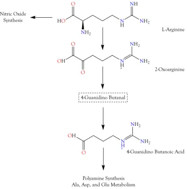
Toxins 2018, 10, x FOR PEER REVIEW 9 of 51Figure 5. The guanidino group of L-arginine is utilized in the production of nitric oxide (NO).
However, L-arginine can also be oxidized to 2-oxoarginine (2OA), a potent convulsant that exerts its effects by blocking chloride channels of GABAA and glycine receptors. With two additional enzymatic reactions, 2OA can be converted to γ-guanidinobutyric acid, which is also a convulsant. Some guanidino compounds also reduce blood pressure and suppress “fight or flight” responses in rats and aggressive behavior in cats. All of these pharmacological effects are consistent with snake envenomation strategies [4].
Why do snakes employ both 5G2OA and 4GBA? De Deyn et al. [34] found that inhibitory effects of guanidine and methylguanidine on GABA were additive, and it is possible that 5G2OA and 4GBA also act synergistically, likely by stimulating distinct sites on the GABAR (i.e., on the Cl
−channel and on an allosteric site), or on different subclasses of GABARs [33,35,36]. Both pharmacological activities are capable of inducing epileptic seizures by reducing GABAergic inhibition in the central nervous system [37–39].
The superior cervical ganglion is a part of the autonomic nervous system and is specifically responsible for “fight-or-flight” responses. Antagonism of GABAergic neurotransmission in the posterior hypothalamus elicits escape locomotor behavior in rats [40], whereas stimulation of GABAergic signaling suppresses such behavior, which would be to the advantage of the snake. Kása et al. [41] and Wolff et al. [42] found that GABAergic axons are distributed unevenly within the superior cervical ganglion. GABAergic innervation of the soma and the stem dendrites of a subpopulation of principal ganglion cells was especially significant, suggesting that GABAergic innervation is critical to the function of this ganglion. Two enzymes involved in GABA metabolism, glutamate decarboxylase and GABA-transaminase, have also been localized in neurons of sympathetic ganglia [43,44]. Galvan et al. [45] reported that in the rat isolated superior cervical ganglion, in 4-aminopyridine solution (200–300 µM), 100 µM GABA, evoked “bursts” of spikes and EPSPs, in addition to neuronal depolarization.
Relative to envenomation, the importance of 4GBA’s action in the superior cervical ganglion may pertain to its agonism of GABA receptors. 4GBA depolarized rat superior cervical ganglia in the same manner as GABA, but with only about 1% the potency thereof [46]. Siegel and Schubert [47]
reported that a GABAergic pathway from medial to lateral hypothalamus suppresses aggressive
NH2
NH NH
NH2 O
HO
O
OH O
NH +
NH2 NH2
O OH
NH +
NH2 NH2 Nitric Oxide
Synthesis
L-Arginine
2-Oxoarginine
4-Guanidino Butanoic Acid 4-Guanidino Butanal
Polyamine Synthesis Ala, Asp, and Glu Metabolism
Figure 5.
The guanidino group of L-arginine is utilized in the production of nitric oxide (NO).
However,
L-arginine can also be oxidized to 2-oxoarginine (2OA), a potent convulsant that exerts its effects by blocking chloride channels of GABA
Aand glycine receptors. With two additional enzymatic reactions, 2OA can be converted to
γ-guanidinobutyric acid, which is also a convulsant.Some guanidino compounds also reduce blood pressure and suppress “fight or flight” responses in rats and aggressive behavior in cats. All of these pharmacological effects are consistent with snake envenomation strategies [4].
Why do snakes employ both 5G2OA and 4GBA? De Deyn et al. [34] found that inhibitory effects of guanidine and methylguanidine on GABA were additive, and it is possible that 5G2OA and 4GBA also act synergistically, likely by stimulating distinct sites on the GABAR (i.e., on the Cl
−channel and on an allosteric site), or on different subclasses of GABARs [33,35,36]. Both pharmacological activities are capable of inducing epileptic seizures by reducing GABAergic inhibition in the central nervous system [37–39].
The superior cervical ganglion is a part of the autonomic nervous system and is specifically responsible for “fight-or-flight” responses. Antagonism of GABAergic neurotransmission in the posterior hypothalamus elicits escape locomotor behavior in rats [40], whereas stimulation of GABAergic signaling suppresses such behavior, which would be to the advantage of the snake.
Kása et al. [41] and Wolff et al. [42] found that GABAergic axons are distributed unevenly within the superior cervical ganglion. GABAergic innervation of the soma and the stem dendrites of a subpopulation of principal ganglion cells was especially significant, suggesting that GABAergic innervation is critical to the function of this ganglion. Two enzymes involved in GABA metabolism, glutamate decarboxylase and GABA-transaminase, have also been localized in neurons of sympathetic ganglia [43,44]. Galvan et al. [45] reported that in the rat isolated superior cervical ganglion, in 4-aminopyridine solution (200–300 µ M), 100 µ M GABA, evoked “bursts” of spikes and EPSPs, in addition to neuronal depolarization.
Relative to envenomation, the importance of 4GBA’s action in the superior cervical ganglion may
pertain to its agonism of GABA receptors. 4GBA depolarized rat superior cervical ganglia in the same
manner as GABA, but with only about 1% the potency thereof [46]. Siegel and Schubert [47] reported
Toxins2018,10, 392 9 of 49
that a GABAergic pathway from medial to lateral hypothalamus suppresses aggressive behavior in cats. Nonetheless, it is unclear whether the quantities of 4GBA detected here would be sufficient to affect the superior cervical ganglia, even if it does act as suggested.
Takahashi et al. [48] reported that iv injections of GABA in anesthetized rabbits produced hypotension and bradycardia. Similar results were obtained with GABA injections into dogs, cats, and rats [49–51]. Serafin et al. [52] found that 2-guanidino-benzimidazole and 2-guanidino-5-aminobenzimidazole dihydrochloride had hypotensive activity, comparable to that of aminophylline. Thomas et al. [53] reported that two guanidino compounds,
L-homoarginine and
L
-amino-tau-guanidino butyric acid, behave like
L-arginine in reversing the vasoconstrictor effect of
L
-NMMA, in a stereospecific and concentration-dependent manner. However,
L-amino guanidino propionic acid and guanidine were ineffective. Thus, as guanidino compounds, both 5G2OA and 4GBA may be hypotensive, which would be consistent with snake envenomation strategies [4].
4GBA may also have antimicrobial actions in the venom gland, given that 4GBA and other guanidino acids are effective at suppressing bacillus and coccus infections [54]. However, such action would likely be secondary to a role in envenomation.
2.3.2. 5-Guanidino-2-oxopentanoic Acid
5-Guanidino-2-oxopentanoic acid (5G2OA), also known as 2-oxoarginine, is the first metabolite of arginine catabolism (Figure 5). 5G2OA was the third most abundant organic acid among the venoms we surveyed, based upon their maximal concentrations (Supplementary Table S1, ID 22). 5G2OA levels are increased in patients with argininemia, a deficiency of the enzyme arginase (EC 3.5.3.1).
Among arginine metabolites, 5G2OA has been especially implicated in the central nervous system damage that occurs in that disease [55]. De Deyn et al. [35,36] first suggested that the convulsant effects of 5G2OA in rabbits might be due to a blockade of chloride channels associated with GABA and glycine receptors, thus inhibiting responses to these inhibitory neurotransmitters. Later, GABA
Areceptors were specifically implicated [33] in its convulsant effects.
2.3.3. Imidazole-4-acetic Acid
Imidazole-4-acetic acid (I4AA) (Figure 6) is the fifth most abundant carboxylic acid occurring in the venoms we tested. Found in the brains of mammals, it is a naturally occurring histidine metabolite that is structurally similar to GABA [56–61]. Numerous studies have reported I4AA pharmacology that is consonant with snake envenomation. When applied iontophoretically to cat cortical neurons stimulated with glutamate, I4AA inhibited neuronal firing in a manner similar to GABA [62], but with a slower onset [63]. Roberts and Simonsen [64] found that I4AA had sedative and analgesic effects when injected ip (4 µ g/g) into mice. A subsequent study from the same group reported that mice injected with increasing doses from 1 to 3 µ mol/g displayed hyperactivity, ataxia, catalepsy and, finally, complete loss of a righting reflex [65]. Similar results were obtained in rats. Tunnicliff et al. [66]
discovered that I4AA injected ip into mice at 3 µmol/g caused body temperature to decrease steadily over a 2 h time course. Sooriyamoorthy et al. [67] found that I4AA (500 mg/kg) in conscious rats decreased cerebral blood flow by 42%.
Toxins 2018, 10, x FOR PEER REVIEW 11 of 51
whereupon it decreases blood pressure and heart rate by agonizing GABA
Areceptors in the CNS [81].
Both effects accord perfectly with the envenomation strategies identified by Aird [4].
Figure 6. Imidazole-4-acetic acid, (left) an agonist of mammalian GABAA receptors. The natural agonist, γ-amino butyric acid, or GABA, is shown on the right.
Roberts and Simonsen [82] discovered that I4AA activates 3′,5′-nucleotide phosphodiesterase by binding to an allosteric site. Since this enzyme degrades cAMP to AMP, the latter, which is somewhat hypotensive, can be dephosphorylated to release adenosine, which is strongly so. Thinking that a decrease in cAMP might reduce blood pressure, Walland [83] injected I4AA into the lateral ventricle of the brain in cats, and found that it elicited dose-dependent hypotension.
In vertebrate retina, the taurine transporter (TAUT) is largely responsible for retinal transport of GABA, in contrast to the brain, where the GABA transporter has a larger functional role [84]. Retinal taurine influx is significantly inhibited in a concentration-dependent manner by both GABA and by I4AA [85]. GABA transporter 2 (GAT-2, also known as BGT-1) is also a taurine transporter [86], although its affinity for GABA is ~30× higher than its affinity for taurine [87,88]. It is unclear whether I4AA also inhibits GAT2, although this would not be surprising.
2.3.4. 4-Hydroxyphenylacetic and 4-Hydroxyphenylpyruvic Acids
4-Hydroxyphenylpyruvic acid (4HPPA) (Figure 7), like 4-hydroxyphenylacetic acid (4HPAA), occurs at moderate levels in all crotaline venoms (Figure 4). It is essentially absent from Dendroaspis
polylepis venom, and is minimal in most other elapid venoms. Very little is known about thiscompound. The biomedical literature is likewise all but silent on it. However, tyrosine can be catabolized by tyrosine aminotransferase (EC 2.6.1.5) to form 4HPPA and glutamate [89]. 4HPPA, in turn, can be converted to homogentisic acid by the action of 4-hydroxyphenylpyruvate dioxygenase (EC 1.13.11.27). Szwajgier [90] reported that of nine phenolic acids tested, homogentisic acid and 4HPPA were the most effective acetylcholinesterase inhibitors, using the spectrophotometric method of Ellman et al. [91]; however, many phenolic acids are inhibitors of both acetylcholinesterase and butylcholinesterase [92]. This inhibitory activity would be consistent with the mamba envenomation strategy, thus, its complete absence from D. polylepis venom is unexpected (Figure 4), raising the question of whether this really is its primary function.
Nucaro et al. [93] reported that in vitro, Taiwan cobra (Naja atra) venom is able to convert tyrosine into phenolic oxidation products via 4-hydroxyphenylpyruvate (Figure 7). They found that tyrosine was released from neuropeptides by oligopeptidases in the venom. Nucaro et al. [93]
determined that venom
L-amino acid oxidase (LAO) initially releases the keto form of 4- hydroxyphenylpyruvic acid and hydrogen peroxide using tyrosine as a substrate. They asserted that, thereafter, a venom tautomerase effects a partial conversion of the keto form of 4HPPA into an enol.
The latter is oxidized to 4-hydroxybenzaldehyde and 4-hydroxyphenol, while the keto form is oxidized to 4-hydroxyphenylacetic acid by H
2O
2co-released from tyrosine by LAO. Utilizing both of these oxidation routes, Naja atra venom generates still other phenolics [93]. Whether the venom titers of these compounds are sufficient to influence prey envenomation is debatable. Nonetheless, it seems likely that quantities of 4HPPA and 4HPAA released in the prey by venom enzymes may well be significant.
O– O
NH
N O–
O NH2
O O
O– HO
Figure 6.
Imidazole-4-acetic acid, (left) an agonist of mammalian GABA
Areceptors. The natural agonist,
γ-amino butyric acid, or GABA, is shown on the right.Roberts [68] reported that I4AA displaces
3H-GABA from receptor-related membrane sites with
an IC
50of 1.3 µ M (Figure 6). I4AA acts as an agonist at GABA
Areceptors [59,69,70], and has been
reported as everything from a partial agonist to a strong antagonist at GABA
Creceptors [56,61,70–75].
Toxins2018,10, 392 10 of 49
Binding of the ligand, GABA, to GABA
Aor GABA
Creceptors, opens a chloride channel [76].
Whereas GABA
Areceptors produce rapid, transient responses, GABA
Creceptors promote rapid, prolonged responses [77]. While GABA
Creceptors are widely distributed, they are much less abundant than GABA
Aand GABA
Breceptors [78], and their pharmacology and response speed do not seem particularly pertinent to envenomation.
On the other hand, GABA
Areceptors are ionotropic, and occur as heteropentamers [79].
GABA
Aagonists reduce neuronal excitability and exhibit sedative effects [80]. For instance, the partial GABA
Aagonist, MRK-409, induces sedation in humans at only ~10% receptor occupancy. I4AA inhibits the firing of CNS neurons [62]. It readily crosses the blood–brain barrier when administered systemically, whereupon it decreases blood pressure and heart rate by agonizing GABA
Areceptors in the CNS [81]. Both effects accord perfectly with the envenomation strategies identified by Aird [4].
Roberts and Simonsen [82] discovered that I4AA activates 3
0,5
0-nucleotide phosphodiesterase by binding to an allosteric site. Since this enzyme degrades cAMP to AMP, the latter, which is somewhat hypotensive, can be dephosphorylated to release adenosine, which is strongly so. Thinking that a decrease in cAMP might reduce blood pressure, Walland [83] injected I4AA into the lateral ventricle of the brain in cats, and found that it elicited dose-dependent hypotension.
In vertebrate retina, the taurine transporter (TAUT) is largely responsible for retinal transport of GABA, in contrast to the brain, where the GABA transporter has a larger functional role [84].
Retinal taurine influx is significantly inhibited in a concentration-dependent manner by both GABA and by I4AA [85]. GABA transporter 2 (GAT-2, also known as BGT-1) is also a taurine transporter [86], although its affinity for GABA is ~30 × higher than its affinity for taurine [87,88]. It is unclear whether I4AA also inhibits GAT2, although this would not be surprising.
2.3.4. 4-Hydroxyphenylacetic and 4-Hydroxyphenylpyruvic Acids
4-Hydroxyphenylpyruvic acid (4HPPA) (Figure 7), like 4-hydroxyphenylacetic acid (4HPAA), occurs at moderate levels in all crotaline venoms (Figure 4). It is essentially absent from Dendroaspis polylepis venom, and is minimal in most other elapid venoms. Very little is known about this compound.
The biomedical literature is likewise all but silent on it. However, tyrosine can be catabolized by tyrosine aminotransferase (EC 2.6.1.5) to form 4HPPA and glutamate [89]. 4HPPA, in turn, can be converted to homogentisic acid by the action of 4-hydroxyphenylpyruvate dioxygenase (EC 1.13.11.27).
Szwajgier [90] reported that of nine phenolic acids tested, homogentisic acid and 4HPPA were the most effective acetylcholinesterase inhibitors, using the spectrophotometric method of Ellman et al. [91];
however, many phenolic acids are inhibitors of both acetylcholinesterase and butylcholinesterase [92].
This inhibitory activity would be consistent with the mamba envenomation strategy, thus, its complete absence from D. polylepis venom is unexpected (Figure 4), raising the question of whether this really is its primary function.
Toxins 2018, 10, x FOR PEER REVIEW 11 of 51
whereupon it decreases blood pressure and heart rate by agonizing GABA
Areceptors in the CNS [81].
Both effects accord perfectly with the envenomation strategies identified by Aird [4].
Figure 6. Imidazole-4-acetic acid, (left) an agonist of mammalian GABAA receptors. The natural agonist, γ-amino butyric acid, or GABA, is shown on the right.
Roberts and Simonsen [82] discovered that I4AA activates 3′,5′-nucleotide phosphodiesterase by binding to an allosteric site. Since this enzyme degrades cAMP to AMP, the latter, which is somewhat hypotensive, can be dephosphorylated to release adenosine, which is strongly so. Thinking that a decrease in cAMP might reduce blood pressure, Walland [83] injected I4AA into the lateral ventricle of the brain in cats, and found that it elicited dose-dependent hypotension.
In vertebrate retina, the taurine transporter (TAUT) is largely responsible for retinal transport of GABA, in contrast to the brain, where the GABA transporter has a larger functional role [84]. Retinal taurine influx is significantly inhibited in a concentration-dependent manner by both GABA and by I4AA [85]. GABA transporter 2 (GAT-2, also known as BGT-1) is also a taurine transporter [86], although its affinity for GABA is ~30× higher than its affinity for taurine [87,88]. It is unclear whether I4AA also inhibits GAT2, although this would not be surprising.
2.3.4. 4-Hydroxyphenylacetic and 4-Hydroxyphenylpyruvic Acids
4-Hydroxyphenylpyruvic acid (4HPPA) (Figure 7), like 4-hydroxyphenylacetic acid (4HPAA), occurs at moderate levels in all crotaline venoms (Figure 4). It is essentially absent from Dendroaspis
polylepis venom, and is minimal in most other elapid venoms. Very little is known about thiscompound. The biomedical literature is likewise all but silent on it. However, tyrosine can be catabolized by tyrosine aminotransferase (EC 2.6.1.5) to form 4HPPA and glutamate [89]. 4HPPA, in turn, can be converted to homogentisic acid by the action of 4-hydroxyphenylpyruvate dioxygenase (EC 1.13.11.27). Szwajgier [90] reported that of nine phenolic acids tested, homogentisic acid and 4HPPA were the most effective acetylcholinesterase inhibitors, using the spectrophotometric method of Ellman et al. [91]; however, many phenolic acids are inhibitors of both acetylcholinesterase and butylcholinesterase [92]. This inhibitory activity would be consistent with the mamba envenomation strategy, thus, its complete absence from D. polylepis venom is unexpected (Figure 4), raising the question of whether this really is its primary function.
Nucaro et al. [93] reported that in vitro, Taiwan cobra (Naja atra) venom is able to convert tyrosine into phenolic oxidation products via 4-hydroxyphenylpyruvate (Figure 7). They found that tyrosine was released from neuropeptides by oligopeptidases in the venom. Nucaro et al. [93]
determined that venom
L-amino acid oxidase (LAO) initially releases the keto form of 4- hydroxyphenylpyruvic acid and hydrogen peroxide using tyrosine as a substrate. They asserted that, thereafter, a venom tautomerase effects a partial conversion of the keto form of 4HPPA into an enol.
The latter is oxidized to 4-hydroxybenzaldehyde and 4-hydroxyphenol, while the keto form is oxidized to 4-hydroxyphenylacetic acid by H
2O
2co-released from tyrosine by LAO. Utilizing both of these oxidation routes, Naja atra venom generates still other phenolics [93]. Whether the venom titers of these compounds are sufficient to influence prey envenomation is debatable. Nonetheless, it seems likely that quantities of 4HPPA and 4HPAA released in the prey by venom enzymes may well be significant.
O– O
NH
N O–
O NH2
O O
O–
HO
Figure 7.
Structure of 4-hydroxyphenylpyruvic acid, an inhibitor of acetylcholinesterase produced by the action of venom
L-amino acid oxidase on tyrosine [93].
Nucaro et al. [93] reported that in vitro, Taiwan cobra (Naja atra) venom is able to convert tyrosine
into phenolic oxidation products via 4-hydroxyphenylpyruvate (Figure 7). They found that tyrosine
was released from neuropeptides by oligopeptidases in the venom. Nucaro et al. [93] determined
that venom
L-amino acid oxidase (LAO) initially releases the keto form of 4-hydroxyphenylpyruvic
acid and hydrogen peroxide using tyrosine as a substrate. They asserted that, thereafter, a venom
tautomerase effects a partial conversion of the keto form of 4HPPA into an enol. The latter is
oxidized to 4-hydroxybenzaldehyde and 4-hydroxyphenol, while the keto form is oxidized to
Toxins2018,10, 392 11 of 49
4-hydroxyphenylacetic acid by H
2O
2co-released from tyrosine by LAO. Utilizing both of these oxidation routes, Naja atra venom generates still other phenolics [93]. Whether the venom titers of these compounds are sufficient to influence prey envenomation is debatable. Nonetheless, it seems likely that quantities of 4HPPA and 4HPAA released in the prey by venom enzymes may well be significant.
2.3.5. Indole-3-acrylic Acid
Xanthine oxidase oxidizes hypoxanthine to xanthine for subsequent conversion to uric acid, but degradation of hypoxanthine is blocked by indole-3-acrylic acid (I3AA), which inhibits xanthine oxidase with an IC
50of 136 µM [94]. Therefore, indole-3-acrylic acid could potentially contribute to inosine accumulation in the venom gland, or it may block degradation of hypoxanthine to xanthine by prey xanthine oxidase, driving conversion of hypoxanthine to inosine by prey enzymes.
Kynurenine aminotransferase (KAT1) converts kynurenine into kynurenic acid, an inhibitor of NMDA glutamate receptors [95–98] and α7-nicotinic (neuronal) acetylcholine receptors [96,97,99].
I3AA inhibits human KAT1, thereby blocking formation of kynurenic acid [100]. As a result, the net effect of I3AA on NMDA iGluRs and α 7 nAChRs would depend upon the balance between exogenous and endogenous agonists and antagonists, a balance that could differ depending upon the snake involved. Interestingly, mamba (Dendroaspis) venoms, arguably the most excitatory snake venoms known, do not contain I3AA. However, this is probably because they already contain dendrotoxins [101,102], fasciculins [103–105], and acetylcholine [106]. This combination presumably floods nicotinic synapses with neurotransmitter, causing fasciculations and, also, promotes hypotension via vascular M3 muscarinic AChRs [107–110]. For a review of muscarinic receptors in snake envenomation, see Aird et al. [16].
I3AA (Figure 8) is also a potent inhibitor of mammalian tryptophan 2,3-dioxygenase, although it does not inhibit indoleamine 2,3-dioxygenase, an enzyme that degrades 5-hydroxytryptamine and serotonin [111]. Tryptophan 2,3-dioxygenase deficiency results in neuroprotection because it degrades tryptophan to kynurenine, a precursor to kynurenic acid (Figure 9). Interestingly, the two related compounds have opposing pharmacologies [112]. Kynurenines, such as quinolinic acid are excitatory, while kynurenic acid is inhibitory.
Toxins 2018, 10, x FOR PEER REVIEW 12 of 51
Figure 7. Structure of 4-hydroxyphenylpyruvic acid, an inhibitor of acetylcholinesterase produced by the action of venom L-amino acid oxidase on tyrosine [93].
2.3.5. Indole-3-acrylic Acid
Xanthine oxidase oxidizes hypoxanthine to xanthine for subsequent conversion to uric acid, but degradation of hypoxanthine is blocked by indole-3-acrylic acid (I3AA), which inhibits xanthine oxidase with an IC
50of 136 µM [94]. Therefore, indole-3-acrylic acid could potentially contribute to inosine accumulation in the venom gland, or it may block degradation of hypoxanthine to xanthine by prey xanthine oxidase, driving conversion of hypoxanthine to inosine by prey enzymes.
Kynurenine aminotransferase (KAT1) converts kynurenine into kynurenic acid, an inhibitor of NMDA glutamate receptors [95–98] and α7-nicotinic (neuronal) acetylcholine receptors [96,97,99].
I3AA inhibits human KAT1, thereby blocking formation of kynurenic acid [100]. As a result, the net effect of I3AA on NMDA iGluRs and α7 nAChRs would depend upon the balance between exogenous and endogenous agonists and antagonists, a balance that could differ depending upon the snake involved. Interestingly, mamba (Dendroaspis) venoms, arguably the most excitatory snake venoms known, do not contain I3AA. However, this is probably because they already contain dendrotoxins [101,102], fasciculins [103–105], and acetylcholine [106]. This combination presumably floods nicotinic synapses with neurotransmitter, causing fasciculations and, also, promotes hypotension via vascular M3 muscarinic AChRs [107–110]. For a review of muscarinic receptors in snake envenomation, see Aird et al. [16].
I3AA (Figure 8) is also a potent inhibitor of mammalian tryptophan 2,3-dioxygenase, although it does not inhibit indoleamine 2,3-dioxygenase, an enzyme that degrades 5-hydroxytryptamine and serotonin [111]. Tryptophan 2,3-dioxygenase deficiency results in neuroprotection because it degrades tryptophan to kynurenine, a precursor to kynurenic acid (Figure 9). Interestingly, the two related compounds have opposing pharmacologies [112]. Kynurenines, such as quinolinic acid are excitatory, while kynurenic acid is inhibitory.
Figure 8. Structure of indole-3-acrylic acid, an inhibitor of xanthine oxidase, kynurenine aminotransferase, and D-dopachrome tautomerase.
Figure 9. Structures of L-kynurenine (left) and kynurenic acid (right). L-kynurenines such as quinolinic acid are excitatory, but kynurenic acid, produced from L-kynurenine by the action of kynurenine aminotransferase (KCAT1), is an inhibitor of NMDA iGluRs and α7 nAChRs. I3AA inhibits KCAT1, blocking production of both molecules by this pathway.
Lastly, I3AA also potently inhibits D-dopachrome tautomerase, an isomer of macrophage migration inhibitory factor (MIF) [113]. Despite its discovery decades ago, the natural substrate of MIF remains unidentified [114]; hence, it is impossible to say exactly what effects its blockade by I3AA might have.
2.3.6. 5-Aminolevulinic Acid
Hermes-Lima [115] reported that 5-aminolevulinic acid (Figure 10) generates oxygen radicals in vitro and, possibly, in vivo during pathologic situations in which its concentration is elevated. The
HN
O O–
NH2 O
O
O– N
O– O
OH NH2
Figure 8.
Structure of indole-3-acrylic acid, an inhibitor of xanthine oxidase, kynurenine aminotransferase, and
D-dopachrome tautomerase.
Toxins 2018, 10, x FOR PEER REVIEW 12 of 51
Figure 7. Structure of 4-hydroxyphenylpyruvic acid, an inhibitor of acetylcholinesterase produced by the action of venom L-amino acid oxidase on tyrosine [93].
2.3.5. Indole-3-acrylic Acid
Xanthine oxidase oxidizes hypoxanthine to xanthine for subsequent conversion to uric acid, but degradation of hypoxanthine is blocked by indole-3-acrylic acid (I3AA), which inhibits xanthine oxidase with an IC
50of 136 µM [94]. Therefore, indole-3-acrylic acid could potentially contribute to inosine accumulation in the venom gland, or it may block degradation of hypoxanthine to xanthine by prey xanthine oxidase, driving conversion of hypoxanthine to inosine by prey enzymes.
Kynurenine aminotransferase (KAT1) converts kynurenine into kynurenic acid, an inhibitor of NMDA glutamate receptors [95–98] and α7-nicotinic (neuronal) acetylcholine receptors [96,97,99].
I3AA inhibits human KAT1, thereby blocking formation of kynurenic acid [100]. As a result, the net effect of I3AA on NMDA iGluRs and α7 nAChRs would depend upon the balance between exogenous and endogenous agonists and antagonists, a balance that could differ depending upon the snake involved. Interestingly, mamba (Dendroaspis) venoms, arguably the most excitatory snake venoms known, do not contain I3AA. However, this is probably because they already contain dendrotoxins [101,102], fasciculins [103–105], and acetylcholine [106]. This combination presumably floods nicotinic synapses with neurotransmitter, causing fasciculations and, also, promotes hypotension via vascular M3 muscarinic AChRs [107–110]. For a review of muscarinic receptors in snake envenomation, see Aird et al. [16].
I3AA (Figure 8) is also a potent inhibitor of mammalian tryptophan 2,3-dioxygenase, although it does not inhibit indoleamine 2,3-dioxygenase, an enzyme that degrades 5-hydroxytryptamine and serotonin [111]. Tryptophan 2,3-dioxygenase deficiency results in neuroprotection because it degrades tryptophan to kynurenine, a precursor to kynurenic acid (Figure 9). Interestingly, the two related compounds have opposing pharmacologies [112]. Kynurenines, such as quinolinic acid are excitatory, while kynurenic acid is inhibitory.
Figure 8. Structure of indole-3-acrylic acid, an inhibitor of xanthine oxidase, kynurenine aminotransferase, and D-dopachrome tautomerase.
Figure 9. Structures of L-kynurenine (left) and kynurenic acid (right). L-kynurenines such as quinolinic acid are excitatory, but kynurenic acid, produced from L-kynurenine by the action of kynurenine aminotransferase (KCAT1), is an inhibitor of NMDA iGluRs and α7 nAChRs. I3AA inhibits KCAT1, blocking production of both molecules by this pathway.
Lastly, I3AA also potently inhibits D-dopachrome tautomerase, an isomer of macrophage migration inhibitory factor (MIF) [113]. Despite its discovery decades ago, the natural substrate of MIF remains unidentified [114]; hence, it is impossible to say exactly what effects its blockade by I3AA might have.
2.3.6. 5-Aminolevulinic Acid
Hermes-Lima [115] reported that 5-aminolevulinic acid (Figure 10) generates oxygen radicals in vitro and, possibly, in vivo during pathologic situations in which its concentration is elevated. The
HN
O O–
NH2 O
O
O– N
O– O
OH NH2
Figure 9.
Structures of
L-kynurenine (left) and kynurenic acid (right).
L-kynurenines such as quinolinic acid are excitatory, but kynurenic acid, produced from
L-kynurenine by the action of kynurenine aminotransferase (KCAT1), is an inhibitor of NMDA iGluRs and
α7 nAChRs. I3AA inhibits KCAT1,blocking production of both molecules by this pathway.
Lastly, I3AA also potently inhibits D-dopachrome tautomerase, an isomer of macrophage
migration inhibitory factor (MIF) [113]. Despite its discovery decades ago, the natural substrate
of MIF remains unidentified [114]; hence, it is impossible to say exactly what effects its blockade by
I3AA might have.
Toxins2018,10, 392 12 of 49
2.3.6. 5-Aminolevulinic Acid
Hermes-Lima [115] reported that 5-aminolevulinic acid (Figure 10) generates oxygen radicals in vitro and, possibly, in vivo during pathologic situations in which its concentration is elevated.
The damage is ascribed primarily to –OH radicals. Bechara [116] found that 5-aminolevulinic acid undergoes transition metal-catalyzed oxidation to yield O
−2, H
2O
2, and HO
−. 5-Aminolevulinic acid was present in all venoms at low to very low concentrations (Figure 4). Oddly, it was most abundant in venoms of B. multicinctus and D. siamensis.
Toxins 2018, 10, x FOR PEER REVIEW 13 of 51
damage is ascribed primarily to –OH radicals. Bechara [116] found that 5-aminolevulinic acid undergoes transition metal-catalyzed oxidation to yield O
−2, H
2O
2, and HO
−. 5-Aminolevulinic acid was present in all venoms at low to very low concentrations (Figure 4). Oddly, it was most abundant in venoms of B. multicinctus and D. siamensis.
Figure 10. Structure of 5-aminolevulinic acid.
2.4. Purine Nucleosides
2.4.1. Adenosine
Adenosine contributes to prey immobilization by activation of neuronal adenosine A
1receptors [117–124], suppressing acetylcholine release from motor neurons [125–130] and excitatory neurotransmitters from central sites [131–133]. It also exacerbates venom-induced hypotension by activating A
2receptors in the vasculature, and by depressing cardiac output and causing atrioventricular block [134–141]. Inosine potentiates the coronary vasodilatory effects of adenosine [142,143]. Aird [144] also reviewed numerous secondary effects of adenosine that are germane to snake envenomation.
The most abundant small organic compounds overall were adenosine and adenine, owing principally to their extremely high concentrations in Dendroaspis polylepis venom, which exceeded the levels found in the remaining 16 venoms by 1–4 orders of magnitude (Figures 2 and 11, Supplementary Table S1). High levels of adenosine have previously been reported for Dendroaspis
angusticeps venom [12,145]. Aird [4,144] proposed that purine nucleosides (adenosine, inosine, andguanosine) are central to the envenomation strategies of nearly all venomous snakes, either as exogenous components co-injected with proteinaceous toxins, or as endogenous secondary messengers released from prey tissues by the action of enzymatic venom constituents. The first part of that hypothesis has been amply documented [12,145–149], and several recent studies have also provided strong support for the second part.
Figure 11. Purine nucleosides and their bases are significant constituents of elapid, viperine, and crotaline venoms. Elapid and viperine venoms contain greater quantities of them than crotaline venoms. The purine strategy of D. polylepis is particularly noteworthy. Taxonomic names: Bmu, Bungarus multicinctus; Dp, Dendroaspis polylepis; MSP, Micrurus spixii; Ms1–3, Micrurus surinamensis, 3
NH2 O
O– O
Bm u D p M sp M s1 M s2 M s3 N k O h C c D s A pl Bm o C dt C vv O o Pe Pf 1. Adenosine
2. Adenine 19. Hypoxanthine 21. Guanosine 26. Inosine 29. Ethyl Adenosine Carboxylate 43. Guanine 56. Probable Purine lacking sugar 221. Xanthine 450. Uric Acid 560. 3'-Adenosine monophosphate (3’-AMP) 712. N2-Methylguanosine 801. 1-Methylguanine 853. N6-Methyladenine 889. 2’-Deoxyadenosine
Bm o
Elapidae Viperidae Crotalidae
Pf Bm u D p M sp M s1 M s2 M s3 N k O h C c D s A pl C dt C vv O o Pe
0 1 2 3 4 5 6 7 8
Figure 10.


![Table 1. Excitatory amino acids and their inhibitory α-decarboxylated derivatives, as determined on rat spinal neurons [27]](https://thumb-ap.123doks.com/thumbv2/123deta/6948702.2271400/8.892.124.772.661.858/table-excitatory-inhibitory-decarboxylated-derivatives-determined-spinal-neurons.webp)