Laser Induced Fluorescence and Transient Absorption Studies on the Excitation Energy Transfer of Free Radicals and Carbenes
著者 甲谷 繁
year 1997‑03‑25
URL http://hdl.handle.net/2297/30577
tS E :ma sc
Laser Induced Fluorescence and Transient Absorption Studies on the Excitation Energy Transfer of Free Radicals and Carbenes.
L/ b-••V"--•IilEiiEILlj2i)Ejl!ft.UiiiiEiijuti]llipJkLJIslLtfE>•J)ie}!kez J •6 7 iLJ v-- •lii7 .yN is71/ ,}r
>ij iL/hCl )/ (1) hih ilZ =L 2N, 7V )EEN •-- liZlllb ec EIE] -3A g> IHI P,yitL
rp z} pt
博 士 論 文
L a s e r l n d u c e d F l u o r e s c e n c e and T r a n s i e n t A b s o r p t i o n S t u d i e s on t h e E x c i t a t i o n Energy T r a n s f e r o f F r e e R a d i c a l s and C a r b e n e s .
レーザー誘起蛍光及び過渡吸収分光法によるフリーラジカルとカルベンの 励起エネルギー移動に関する研究
金沢大学大学院自然科学研究科
甲 谷 繁
CHAPTER 1 Referenees
CONTENTS
lNTRODUCTJON
15
CHAPTER 2
Resonance Energy Transfer from the '
' Molecules to a Stable Free Radical
tt
' 2.1 Abstract'
'
2.2 Introduction
'
' 2.3
Experimental Section 2.4 Results and Discussion 2.4.1 Critical energy transfer
,.. from spectroscopic data
2.4.2 Fluorescence quenching of Figures and Tables
References
LIST OF COMPOUNDS
Excited Singlet State of Dye
distances calculated
dye molecules
7 8 10 12 12
13 18 26 28
CHAPTER 3
Resonance Energy Transfer from the Lowest Excited Triplet State
of Diphenylcarbene to Dye Molecules: Utilization to
Characterization of the Triplet-Triplet Fluorescence
3.3 Experimental Section 3.4 Results and Discussion 3.4.1 Steady-state spectra 3.4.2 Fluorescence decays
3.4.3 Estimated values of fluorescence radiative and nonradiative rate for the fast and slow components
Figures and Tables References
APPENDIX
LIST OF COMPOUNDS
quantum eonstants
yields,
32 34 34 36 38
45 54 56 58
CHAPTER 4
Lase.r Flash Photolysis 'Study of the
1 , 8-bist4-( a-diazobenzoyOphenoxy7octane : Magnetic Fie1d
4.1 Abstraet 4.2 Introduction
4.3 Experimental Seetion 4.4 Results and Discussion
5.4.1 Steady state photolysis at 77 K 5.4.2 Laser flash photolysis
Figures and Tables References and Notes APPENDIX
Primary
Effects
Process of Laser
in and
59
60
61
64
64
64
71
78
80
LIST OF COMPOUNDS 82
SnmRY 83
ACKNOWLEDGMENT LIST OF PUBLICATIONS LIST OF SUB-PUBLICATIONS
85
86
87
t
CIIAPTER 1
INTRODUCTION
Introduction
Free radicals and carbenes are typical transient species in photochemical reactions. Free radicals are atoms or molecules that possess one or more unpaired electrons. Carbenes are the species with a divalent carbon atom which consists of two
different electronic configurations for two nonbonding electrons,
i.e,, singlet and triplet. Paramagnetism is an important
characteristic of free radicals and triplet carbenes.
7T 7T
Rl 7///z
R2
sing1et
With the development numerous investigations short-lived intermediates methods for detecting such
lasers with nano- and picosecond and triplet carbenes
e1ectron paramagnetic recent time-resolved ESR us with a great amount electronic structures and
Rl
7!//,
R2 triplet
of laser flash photolysis in 1960's, of direct spectral observation for have been reported. There are various short-lived species by using pulsed time duration.1'2 Free radicais
have been also extensively studied by resonance (EPR) spectroscopy including spectroscopy.3'4 These methods provide
of informat i' on about molecular geometry,
reaction dynamics.4)5
Recent interest is directed toward investigating physical and chemical interactions of free radicals and carbenes with other molecules. In photochemistry, numerous investigations of bimolecular processes such as electronic energy transfer6-9, the excited-state complex formationslO'11, and photoinduced electron 12
have been reported. In the case where free radicals transfer
and/or carbenes involved, however, there are a few studies on photophysical bimolecular processes, especially for electronic energy transfer, because of instability of these species in
solution.
,
Electronic energy transfer is one of the important process in photochemistry. Most energy transfer can be described by
D* +A -> D+ A*. (1)
This process is observed at the earlier stage of photosynthetic reaction in plants or photosynthetic bacteria.13 solar energy conversion begins with the capture of sunlight by hundreds of chlorophyll array and the efficient energy transfer toward a reaction center is followed. The transfer mechanism is mainly due - to Coulombic dipole-dipole interaction which was theoretically developed by F6rster and others.6-9 In this theory, critical transfer distance, Ro, is defined as a mean distance between donor and acceptor where the rate constant for resonance energy transfer is equal to the sum of rate constants for all other
donor deexcitation processes in the absence of acceptors. The
quantity Ro is useful in evaluating the effectiveness of the
resonance energy transfer.
Triplet-triplet fluorescence of diphenylcarbene (DPC) and its derivatives in low-temperature matrix have attracted special interest. The fluorescence of these carbenes clearly shows nonexponential decay curves and they are modified in the
14 presence of a magnetic field.
Owing to paramagnetism of triplet
carbenes, the fluorescent state (Tl) of DPC is influenced by internal and external magnetic fields. In order to understand the interaction between electron spin and magnetic fields, i.e.
spin-orbit coupling and external magnetic field effects in the excited DPC, the characterization of the Tl sublevels is an important matter. Further, magnetic field effects on a coupling reaction of two carbenes seem to be an interesting subject but have not been reported at all. Contrary to this situation, there are so many reports for the magnetic field effects on radical 15--17 recombination reactions.
In this thesis, the author deals with the three interesting
and unsolved topics eoncerning with aromatic free radicals and
carbenes: (1) quantitative evaluation of intermolecular
electronic energy transfer on the basis of the F6rster theory, (2) spin-orbit coupling induced intersystem crossing process in the excited states of DPC, and (3) magnetic field effects on a pair of diphenylcarbene moieties generated in the bichromophoric system.
In order to demonstrate the validity of Fb'rster theory for
the energy transfer participating a free radical as energy
acceptor, in chapter 2, fluorescence quenching of dye molecules by 2,4,6-tri-tert-butylphenoxyl radical (TBPR) is presented. TBPR ,
is a suitable free radical for studying resonance energy transfer quantitatively, because this radical is stable even in solutions
at room temperature18 and has relatively strong and broad absorp- tion band in visible region.
In chapter 3, resonance energy transfer from the excited triplet state Tl of DPC to dye molecules is described. Here, the author suggests that the resonance energy transfer is a powerful tool for characterization of the Tl sublevels of DPC which cannot be obtained by an ordinary spectroscopy. The energy transfer analysis applied for the quenched fluorescence decay curves of DPC will be considered as an excellent method to investigate the rates of deactivation processes in the triplet excited state Tl.
In chapter 4, effects of pulsed laser intensity and
magnetic fields on the primary photochemical process in
1,8-bis[4--(a-diazobenzoyl)phenoxyl]octane (BDO) is considered.
On the high density pulsed laser excitation, two diphenylcarbene - moieties on the both ends of alkyl chain can be simultaneously generated from BDO. It is demonstrated that the biphotonic reaction are well controlled by pulsed laser intensity and
magnetic fields.
References
(1) Fleming, G. R. Chemical Applicatiofl of Ultrafast
spectroscopy, Oxford University Press, New York, 1986.
(2) BrUckner, V.; Feller, K. H. ; Grummt U. W. Applications of Time-resolved Optical Spectroscopy, Elsevier, Amsterdam, 1990.
(3) Advanced EPR - Applications in Biology and Biochemistry - (ed. Hoff, A. J.) Elsevier, Amsterdam, 1989.
(4) Spin Chemistry - Spin Polarization and Magnetic Field Effects
in Photochemical Reactions - (ed. I'Haya, Y. J.), The Oji
International Conference, 1991; references therein.
(5) Handbook of Organic Photochemistry (ed. Scaiano, J. C.) CRC Press, Boea Raton, 1989; Volume 1, 2.
(6) Fb'rster, T. Discuss. Faraday Soc. 1959, 27, 7.
(7) Dexter, D. L. J. Chem. Phys., 1953, 21, 836.
(8) Lamola, A. A. ; Turro N. J. Energy Transfer and Organic Photochemistry, Wiley, New York, 1969; Chapter 2.
(9) Berlman, I. B. Energy Transfer Parameters of Aromatic
Compounds, Academic Press, New York, 1973.
' (10)' Organic Molecular Photophysics (ed. Birks J. B.), Wiley, New York, 1975; Volume 2 Chapter 9.
(11) (a) Ware, W. R.; Watt, D.; Holmes, J. D. J. Am. Chem. Soc.
1974, 96, 7853. (b) Ware, W. R.; Holmes, J. D.; Arnold D. R. J.
Am. Chem. Soc. 1974, 96, 7861.
(12) Marcus, R. A.; Sutin N. Biochem. Biophys. Acta 1985, 811,
265.
(13) Barber J.; Andersson B. IVature 1994, 370, 31.
(14)(a) Haider, K. W.;Platz, M, S.;Despres, A.; Lejeune, V.;
Migirdicyan, E. J. Phys. Chem. 1990, 94, 142. (b) Despres, A.;Lejeune, V.;Migirdicyan, E.;Platz, M. S. J. Phys. Chem. 1992,
96, 2486. (c) Despres, A.;Lejeune, V.;Migirdicyan, E.;Admasu, A.;Platz, M. S.; Berthier, G.; Parisel, O.; Flament, J. P.;
Baraldi, I.; Momicchioli, F. J. Phys. Chem. 1993, 97, 13358.
(d) Kozankiewicz, B.;Despres, A.;Lejeune, V.;Migirdicyan, E.;
Olson, D.; Michalak, J.;Platz, M. S. J. Phys. Chem. 1994, 98,
10419.
(15) Spin Polarization and Magnetic Effects in Radical Reactions (ed. Molin, Yu. N.), Elsevier, Amsterdam, 1984.
(16) Doubleday Jr., C.; Turro N. J.; Wang J. F. Acc. Chem. Res.
1989, 22, 199.
(17) Steiner, U.; Ulrich, T. Chem. Rev., 1989, 89, 51
(18) Denisov, E. T.; Khudyakov I. V. Chem. Rev. 1987, 87, 1313.
THE
t
CHAPTER 2
RESONANCE ENERGY TRANSFER FROM
EXCITED SINGLET STATE OF DYE MOLECULES TO
A STABLE FREE RADICAL
2.1 Abstract
Fluorescence quenching of dye molecules such as Coumarin 153, t
DcM, and nile red by the stable 2,4,6-tri-tert-butylphenoxyl radical in an MTHF glass matrix at 77 K was studied by means of subnanosecond time-resolved fluorescence spectroscopy. The quenching process is ascribed to the dipole-dipole interaction
amenable to Fb'rster theory. The average critical energy transfer
distances were determined to be 2.0, 2.7, and 3.1 nm for Coumarin
153, DCM, and nile red, respectively. These values are consistent
with those evaluated from spectroscopic data.
2.2 Introduction
Free radicals possess great ability to interact with t
aromatic molecules which results in the quenching of their excited states. Several mechanisms have been considered in the excited state quenching by free radicals: e.g. energy transfer of Fdrster type or Dex'ter type, electron transfer, electron exchange induced relaxation processes (intersystem crossing and internal conversion). In the case of excited singlet state quenching, it is expected that the Fdrster energy transfer according to
ID* + 2A -,. ID + 2A*
efficiently occurs because both the radiative transition ID* -->
ID and the absorptive transition 2A -> 2A* are spin allowed.
However, there has been few reports to confirm this quenching
process quantitatively, because the high reactivity and
instability of the radicals prevent the exact determination of their concentrations. Therefore, stable free radicals are suited to the study of the F6rster energy transfer from an excited singlet state molecule to a free radical.
' Nitroxyl radieals sueh as 2,2,6,6-tetramethyl-1-piperidinyloxy
and di--tert-butyl nitroxide which are stable even at room
temperature are the best known quenchers of excited singlet
state molecules.1-6 However, a F6rster-type energy transfer has
been suggested to be insignificant in these systems because of
the low extinction coeffjcients of nitroxyls (460 nm, E tv 10
M-lcm-1). To the best of the author's knowledge, only the
stable diphenylpicrylhydrazyl radical has been proven to act as the fluorescence quencher in the F6rster-type energy transfer by 7, means of steady-state fluorescence measurements.
In this study, fluorescence decay curves of dye molecules, 2,3,5,6-IH,4H-tetrahydro-8-trifluoromethylquino1izino-<9,9a,1-gh>
coumarin (coumarin 153), 4-dicyanomethylene-2-methyl-6-(p-
dimetylaminostylyl)-4H-pyran (DCM), and 5-amino-9- diethyliminobenzo(a)phenoxazone (nile red) quenched by
2,4,6-tri-tert-butylphenoxyl radical (TBPR) in 2-
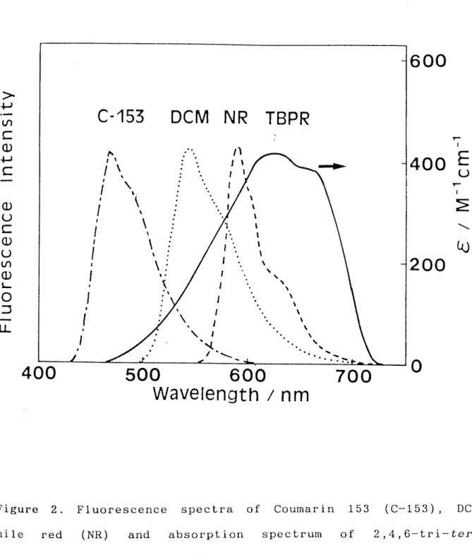
methyltetrahydrofuran (MTHF) at 77 K were measured by means of subnanosecond time-resolved fluorescence spectroscopy. The stable TBPR was chosen as an energy acceptor because it has a relatively strong and broad absorption band (Amax = 625 nm, Emax = 420 M-lcmkl) as shown in Fig. 2. The F6rster-type energy transfer
leads to non-exponential time dependence of the donor
fluoreseence decay which gives us the critical energy transfer
distance (Ro). The values of Ro. determined from fluorescenee
decay curves are compared with those estimated from a calculation
of spectral overlap.
2.3 Experimental Section
Coumarin 153 (Lambda Physik), DCM (Lambda Physik) and nile red L
(Aldrich) were used without further purification. MTHF (Tokyo
Kasei), washed with 100/o NaOH and dried over MgS04, was subjected to fractional distillation over CaH2 followed by distillation
over potassium metal. TBPR was prepared by treating 2,4,6-tri- tert-butylphenol (Tokyo Kasei) with Pb02 powder (O.15 g / 5 ml) in degassed MTHF under vigorous stirring. The solution was filtrated through a glass filter to remove Pb02 powder. The radical solution ('vO.1 mM) thus obtained was mixed with a degassed dye solution. Finally, the mixed solution was
transferred into a quartz cell (1 mm optical pathlength) and sealed off. All these proeedures were performed in the vacuum condition (tvlO-3 Torr), since TBPR is gradually oxidized in aerated solution. Concentrations of TBPR were calculated from the absorbance and molar extinetion coefficient at 625 nm.
The value of e62s in MTHF at 77K was determined to be
42oÅ}13 M-lcm-1 by the reported method.8 ' '
Figure 1 shows the experimental setup for a measurement of a
subnanosecond time-resolved fluorescence. The hydrogen Raman- shifted second harmonic (436 nm) of a pulsed Nd:YAG laser
(Coherent Antares 76-s/Continuum RGA 60; 100 ps pulse width, 10
Hz repetition rate) was used for photoexcitation. The
fluorescence of DCrvl and that of Coumarin 153 were observed
through a polychromator with 100 grooves/mm grating (Jobin Yvon
HR250). TimemResolved detection was performed with a photon- counting streak camera (Hamamatsu Photonics C2050/M1952)/CCD temporal analyzer (3140-69) system interfaced with a' Hamamatsu
c3366 I/F unit. The fluorescence of nile red, selected at 580 nm
by a monochromator (Ritsu MC-20L), was detected by a photo- multiplier (Hamamatsu H-3284). Signals from the photomultiplier were recorded on a digital storage oscilloscope (Tektronix TDS-
520) interfaced to a personal computer (NEC PC9801).
2.4 Results and Discussion
2.4.1 Critical energy transfer distances calculated from
, spectroscopic data
According to the F6rster theory9, Ro can be expressed by
gooo(inio) K2 ÅëD
R06 = 12s 7t s.4N J (1)
2
where K and ÅëD are the orientation factor and fluorescence quantum yield of donor in the absence of energy transfer,
respectively: n and N are the refractive index of the medium and
Avogadro's number, respectively. J is the F6rster overlap
integral as expressed by
J. J FD(V).--E4A( V) d il (2)
FD()) and EA( -y-) are the spectra of donor emission normalized to
unity and the molar extinction coefficient of acceptor,
respective1y.
The fluorescence spectra of the dye molecules (Coumarin 153, DCM-and nile red) and the absorption spectrum of TBPR are shown in Figure 2. The absorption band of TBPR around 625 nm is so broad that the spectral overlap between the fluorescence of the dye molecules and the absorption of TBPR is significant. The values of Ro calculated from eq. 1 and 2 are 1.9, 2.5, and 2.8 nm for Coumarin 153, DCM, and nile red, respectively. These values are summarized in Table 1.
The values of ÅëD for Coumarin 153, DCM, and nile red are
also listed in Table 1. These values are obtained by the relation of ÅëD = kr' 2rl, Where kr and z'1 are the radiative rate constant L
and the fluorescence lifetime of dye molecules. kr was calculated 10
and irl was determined by from the Strickler-Berg equation
analyzing the fluorescence decay of dye molecules in the absenee of TBPR as mentioned later. Calculated kr values are sufficiently reliable beeause these dye molecules satisfy the following two requirements described in reference 10: (1) large extinction
coefficient (more than sooo M-lcm-1); (2) unchanging
configuration in the excited state. The value K2 = O.476 is quoted for a rigid random orientation. The value of n for MTHF at 77 K has not been reported so far; therefore, this value is
determined to be 1.43 using n = 1.405 at 293 K and its
temperature dependence dn/dT = - lo-4 K-1.11
2.4.2 Fluorescence quenching of dye molecules
Since TBPR is non-fluorescent, the quenching mechanism could . not be determined by measuring the doublet-doublet fluorescence of TBPR. The author therefore tried to prove it by time-resolved
fluorescence measurements of the dye molecules. A 436 nm subnanosecond pulse was used as excitation light, because the absorption intensity of TBPR is negligible at this wavelength
(436 nm). However, the molar extinction coefficient of nile red
iS also small at 436 nm. Consequently, the fluorescence of nile
red was detected by a highly sensitive photomultiplier instead of the streak camera.
Fluorescence decay curves of Coumarin 153, DCM, and nile red in the presence of various concentrations of TBPR are shown in Figures 3 " 5, respectively. The fluorescence intensities at the time origin are nearly normalized. The fluorescence lifetimes
of Coumarin 153, DCM and nile red in the absence of TBPR were 5.3, 2.2 and 4.7 ns, respectively, as will be mentioned later.
Apparent fluorescence lifetime decreases with increasing TBPR concentration. These results clearly indicate that the lowest excited singlet state (Sl) of dye molecules are quenched by TBPR.
The fluorescence of DCM and nile red are efficiently
quenched by TBPR, whereas the fluorescence of Coumarin 153 is not so efficiently quenched even at the highest concentration (57.1
mM) of TBPR. This result is consistent with the fact that the Ro value of Coumarin 153 determined from spectroscopic data is the smallest of those of the three dye molecules. The Fdrster--type
energy transfer is therefore the most probable quenching
mechanism.
The fluorescence decay of the donor undergoing dipole-dipole resonance energy transfer in uniformly distributed and randomly oriented sysLem is expressed by12
I(t) = A exp[-t/Tl-x(t/Tl)1/2] (3)
Where z- 1 is the fluorescence lifetime of the donor in the
absence of TBPR and
x = g(4/3ooo) 7r 3/2NcARo3 (4)
in which g is the orientation factor, CA is the concentration of ,
acceptor, N is Avogadro's number and Ro is the critical energy transfer distance. However, the observed fluorescence signals involved rise and decay components, they could not be fitted by eq. 3 which involves only the decay process. The term reflecting fluorescence rise may be introduced by taking into account the vibrational relaxation from upper vibronic levels to the lowest
one in Sl of the donor. Considering such a vibrational
relaxation, the temporal behavior of the donor fluorescence is 13 expressed by
i(t) =Ajli.., [- ti", - ."i-x( ."1)iY ]du (s)
where u is an integration variable with the dimension of time and T2 is a time constant of the vibrational relaxation.
Calculated fluorescence curves were obtained by the
convolution method using the parameters A, irl, ?r2, and X. The
set '
of parameters was found by a non-linear least-squares method
so that the calculated curve makes the best fit to the observed curve. X can be set to zero for fluorescence curve measured in the absence of TBPR. All the parameters were determined for fluoreseence curves measured in the presence of TBPR. Figure 6
'
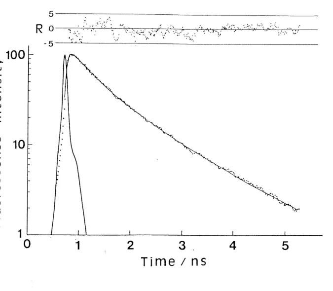
shows a logarithmic plot of the fluorescence signal (dotted
Curve) of DCM in the presence of 21.6 mM TBPR. The decay curve
clearly shows a non-exponential decay. A solid curve, calculated from eq. 5 by using parameters (7rl = 2.2 ns, z-2 = O.04 ns and X
= 1.80) fits the observed one well. Other fluoresc'ence decay curves in the presence of TBPR were also fitted well by using the parameters summarized in Table 2. Ro can be calculated from eq.
4, where g for rigid random orientation is O.845. The values of Ro are also listed in Table 2.
Three important features are found for fluorescence quenching of the three dye molecules in Table 2. (1) The values of 2rl in the presence of various concentrations of TBPR are in good agreement with the fluorescence lifetime of dye molecules in the absence of TBPR (CA = O mrvl). (2) The values of Ro derived from X were nearly constant, since X increases linearly with increasing TBPR concentration as indicated by eq. 4. (3) The average values
of Ro for Coumarin 153/TBPR, DCM/TBPR, and nile red/TBPR combinations (2.0, 2.7, and 3.1 nm) are in good agreement with
those (1.9, 2.5, and 2.8 nm) estimated from the spectral
over1ap.
•These three features clearly indicate that the resonance energy transfer with dipole-dipole interaction is responsible for
the fluorescence quenching of dye molecules (Coumarin 153, DCM
and Nile Red) by TBPR. Furthermore, they also show that other
processes such as electron transfer and/or electron exchange are
never included. If other quenching mechanisms play an important
role, the three characteristic feat•ures described above cannot be
Obtained. The fluorescence decay functions responsible for the
electron transfer and the eleetron exchange quenching are different from that responsible for the dipole-dipole energy 14 transfer quenching.
Empirically, the distanee dependence of the electron transfer rate constant in frozen media is expressed by
k(r) = koexp[-B(r-ro)] (6)
where r is the center to center distance between donor and acceptor, ro is a small connection for their radii, and the constant /3 is about n -- 12 nm-1 for aromatic systems.15 The largest rate (ko) is usually thought to be close to the frequency
of a single molecular vibration, lo13 s-1. In the systems studied, the value of r is 1.9 nm even in the highest
concentration (57.1 mM) of TBPR and the value of ro is about O.5
- O.7 nm. Consequently, the largest value of k(r) is
approximately estimated to be 1Å~lo7 s-1. Thus, the long--distance
electron transfer in the nanosecond time region has a small possibility. The electron exchange mechanism is also negligible, because the limiting distance for the occurrence of electron
- exchange is usually shorter than that of electron transfer.
Regenerative Amp1ifier
532nm 1OHz
Shifter •
cw-Mode-Locked Nd:YAG Laser
H2
1O64nm 76MHz
streak trlgger )n
Personal Computer
CCD
Interface
UnitCCD camera
Streak System
436nm '
FWHIvl :
1OHz 60ps
Samp1e/MTHF 77K
,
Figure 1.
subnanosecond
Experimental Setup for a
time-resolved fluorescence.
measurement of a
v >
t-nco
=
([1.)
v =
o o c o Q m o L
5 o
-e--
-
C-153
t"s
1 Ns
i Ns
i Ks
/ Ks
i
i
,l
'l
,1
.
DCM
tt- - ----
:: ---
--
-- --
-- -- e-
ii
: :
x: ":
N:'
t' K
:s
:s :N ll
: . '
N. 1/
NR
A IS tl ts
is ts
-i s l'• 1 )s t". 1
's t'• N
t :' l -"-
--- i--
.
'-hLÅ~
TBPR
:
-iml))pb-
N
NN N
N N
N s
--
•N
. ..N
.N
. ..N
-N
.
--. .N: ).
400 500 600 Wavelength / 700
6OO
N'i'
4oo E
rriU
s
Å~
(Lc.,)
2OO
nm
o
Figure 2. Fluorescence spectra of
nile red (NR) and
butylphenoxyl radical
absorption
(TBPR).
Coumarin 153 spectrum of
(C-153), DCM, 2 4 6-tri-tert-
))
]!.l 1.0
m
s:p o =
-
o v c o.5 o U m o L o
j
N -
o
a
b
C
d
o 1 t2 3 Time / ns 4 5
Figure 3. Fluoreseence
(a) O, (b) 9.7, (c)
decay curves of
25.4, and (d)
Coumarin 153 in
57.1 mM TBPR
MTHF with
at 77 K.
L
v >
'.-."
, 1.0
8
:t
2•
O.5
g
8-
i
o
a
b C
d e
o 1 2
Time
3
/ ns
4 5
Figure 4.
(b) 6.0,
Fluorescence (c) 12.4, (d)
decay 21.6
'curves and
of
(e)DCM 32
in
.1 mM
MTHF TBPR
with
at
(a)
77 K
o
y.
>
m 1.0
= o
p = -
U o
= oO.5
U m o L o =
E
o
a
b
C
,
o 2 4
Time/ns
10 12 14
Figure 5.
(a) O,
Fluorescence
(b) 9.6 and
decay curves
(c) 19.3
of Nile Red
mM TBPR
in at
MTHF
77
with
K.
5
Ro
-5
k:, ., t s.
, L{. t :1••-, . •..e•: :-:'• ;`':'
E ,,.. ••t• : ••
--- - t:
:.. : ".X.:t.M';'-..'l.1;.i.;r ..."t": ::.;:. ..:.1..;: '. '"s:t'
:• .' v
100 p >
t-(dt')
c
v o
s S 10 8
8 9
g z 1
.
.
.
.
.
.
.
. .
s
hs
:-t-
o
,
1 2
Time
3,
/ns
4 5
Figure 6. A logarithmic plot of as a function of time for 21.6 curve was calculated from eq.
nS, T2 = O.04 ns and X= 1.80.
the fluorescence intensity
mM TBPR in MTHF at 77 K. The 5 by using parameters: z"
1of DCrvl
solid
= 2.2
TABLE 1. Parameters for energy transfer from dye molecules 2,4,6-tri-tert--butylphenoxyl radical (TBPR).
to
donor molecule
lo-16J a) 6 -1 mo1 cm
io8k b) r s-1
c) TO
ns
Åë Dd)
Roe)
nm Coumarin 153
DCM Nile Red
4.6 28.4 48.4
1.75 3.32 2.00
5.3 2.2 4.7
O.93 O.74 O.94
1.9 2.5 2.8
a) The spectral overlap between fluorescence of dye and absorption of TBPR.
b) calculated by the Strickler-Berg equation.
C) Obtained form the fluorescenee decay curves in the of TBPR.
d)I ÅëD = kr'To'
e) calculated from Eq• 1•
mo1ecu1es
absence
TABLE 2. Parameters for analysis dye molecules undergoing the
butylphenoxyl radical (TBPR).
Donor:Coumarin 153
of fluorescence decay energy transfer to
icurveS Of 2,4,6-tert-
CA/mrvl
T 1/ns T 2/ns x Ro/nm
9
25 57
o .7 .4 .1
5.3 4.9 5.0 5.3
O.07 O.05 O.04 O.03
O.30 O.80 2.12
2.0 2.0 2.1
Donor : DCrvl
CA/mM T 1/ns T2/ns x Ro/nm
6 12 21 32
o .o .4 .6 .1
2.2 2.1 2.0 2.2 2.2
O.06 O.06 O.04 O.04 O.03
O.39 O.88 1.80 2.80
2.6 2.7 2.8 2.8
Donor:Nile Red
CA/mM T 1/ns T2/ns x Ro/nm
lg 9
o .6 .3
4.7 4.5 4.8
o.
o.
o.
03 03 02
O.93 2.30
3.0 3.2
CA is Ro is
the concentration of TBPR.
calculated from Eq. 4
References
(1) Green, S. A.; Simpson, D.J.: Zhou, G.; Ho, P. S.; Blough N.V. J. Am. Chem. Soc., 1990, 112, 7337.
(2) Darmanyan, A. P.; Tatikolov, A. S. J. Photochem., 1986, 32,
157.
(3) Chattopadhyay, S. K.; Das, P. K.; Zhou, G.; Ho, P. S•;
Blough, N. V. J. Am. Chem. Soc., 1983, 105, 6205.
(4) Kuzmin, V. A.; Tatikolov, S. A. Chem. Phys. Lett., 1977, 51,
45.
(5) Watkins, A. R. Chem. Phys. Lett., 1974, 29, 526.
(6) Green, J. A.; Singer, L. A.; Park, J. H. J. Chem. Phys.,
1973, 58, 2690.
(7) Lisovskaya, I. A.; Plotonikov, V. G.; Alfimov, M. V. Opt.
Spectrosc., 1973, 35, 634.
(8) Cook, C.D.; Norcross, B. E. J. Am. Chem. Soc., 1959, 81,
1176.
(9) Forster, T. Discuss. Faraday Soc., 1959, 27, 7.
(10) Strickler, S. J.; Berg, R. A. J. Chem. Phys., 1962, 37,
814.
(11) Perrin, D. D.; Armare'go, W. L. F.; Perrin, D. R.
Purification of laboratory Chemicals; Pergamon, Oxford, 1980.
(12) Lamola, A. A.; Turro, N. J. Energy Transfer and Organic Photochemistry, Wiley, New York, 1969; p 41.
(13) Lu, P. Y.; Yu, Z. X.; Alfano, R. R.; Gestern, J. I. Phys.
Rev. AL, lgs2, 26, 3610•
(14) (15)
265
Inokuti M.; Hirayama, F. J. Chem.
Marcus R. A.; Sutin, N. Biochem.
and references therein.
Phys., 1965, 43 Biophys. Acta,
1978
'
1985, 811
'LIST OF COMPOUNDS
N ,,,,.. O O
x1 ti CF3 C-153
(H3C)2N "cH==cH
CN NC
(C2Hs)2N
DCM
1:'/so
(donor)
6
"
l.
TBPR
(acceptor)
t
THE
CHAPTER 3
RESONANCE ENERGY TRANSFER FROM
LOWEST EXCITED STATE OF DIPHENYLCARBENE TO
DYE MOLECULES:
UTILIZATION FOR THE CHARACTERIZATION OF
THE TRIPLET-TRIPLET FLUORESCENCE
3.1 Abstract
Triplet'-triplet (Tl --> To) fluorescence spectra and decay ,
curves of diphenylcarbene (DPC) in the absence and in the
presence of dye molecules such as Rhodamine 6G and Rhodamine B were measured in several organie glasses at 77 K. In the absence
of dye molecules, the Tl -> To fluorescence signals exhibit
biexponential decay with the lifetimes of -v30 and -v140 ns,
which are attributable to the independent Tl --> To emission from
the individual Tl sublevels of DPC. In the presence of dye
molecules, fluorescence decay signals of both fast and slow
eomponents were well analyzed in terms of resonance energy
transfer of F6rster type from DPC to dye molecule. The reaction
scheme of the energy transfer and their various parameters are
discussed on the basis of the independent Tl -> To emission from
the individual Tl sublevels of DPC at low temperature.
3.2 Introduction
Diphenylearbene (DPC) has been extensively studied over 40 t
years. In the process, a considerable amount of information about the structure, chemical reactivity, and physical properties of this molecule has been obtained. It is well-known that the fluorescence of DPC is ascribed to the transition from the first excited triplet state (Tl) to the triplet ground state (To).
Fluorescence spectra of DPC were first reported by Gibbons and Trozzolol and observed in a single crysta12 and in liquid phase.3'4 Fluorescence lifetime and quantum yield were also measured for Dpc in organic glasses5 and in liquid phase.3,4
Before 1990, it had been considered that fluorescence decay
of DPC in low-temperature matrix was composed of only one component and exhibited single-exponential decay. Therefore, fluorescence quantum yield and radiative and nonradiative rate 5a constants of DPC were determined for only one component.
Resonance energy transfer from the excited DPC to fluorescein was also observed and analyzed on the basis of the idea of one
' component fluorescence.6 However, lvligirdicyan and co-workers found that fluorescence decay curves of DPC and its derivatives in low-temperature Shpol'skii matrix clearly show nonexponential
decay and are significantly modified in the presence of a magnetic field,7 which is similar to the decay of the Tl -> So
phosphorescence of aromatic compounds in a low-temperature
8 matrix.
I
!
In this study, it was confirmed that fluorescence decay curves show biexponential with the lifetimes of tv30 and tv140 ns
(initial intensity ratio, fast/slow -vO.5) at 77 K. Further, the
author attempted to estimate fluorescence quantum yields (Åë) and radiative (kr) and nonradiative (knr) rate constants for the
fast and slow components to examine the decay proeesses
quantitatively. Here, he will propose a novel method to estimate 9 these values. Resonance energy transfer of F6rster type from
the Tl of DPC to a suitable acceptor molecule gives information about Åë for each fluorescence components as will be mentioned later. The respective values of kr and knr will be obtained from
the corresponding Åë and fluorescence lifetimes. In order to obtain these values, fluorescence decay curves of DPC quenched by the energy transfer to Rhodamine 6G (Rh6G) or Rhodamine B
(RhB) were measured. The respective values of Åë, kr, and knr thus estimated will be compared between the fast and slow com- ponents and further discussed eoncerning to the triplet--triplet
fluorescence characteristics of DPC at low temperature.
3.3 Experimental Section 3.3.1 Materials.
L
The precursor of DPC, diphenyldiazomethane, was synthesized by the base-catalyzed oxidation of benzophenone hydrazone (Tokyo Kasei) as described in a previous paper.10 Rh6G (Tokyo Kasei) and RhB (Nacalai Tesque) were recrystallized from water/methanol mixed solvent. Solvents used in this study are ethanol, EPA
(diethyl ether:isopentane:ethanol = 5:5:2), 3-methylpentane (3MP), and 2--methyltetrahydrofuran (MTHF). Spectrograde ethanol (Nacalai Tesque) and Spectrograde diethyl ether (Nacalai Tesque) were used without further purification. Isopentane (Nacalai Tesque) and 3MP (Aldrich) were distilled over LiAIH4. MTHF (Tokyo Kasei), washed with 10 O/o NaOH and dried over MgS04, was subjected to fractional distillation over CaH2 followed by distillation
over potassium metal. The sample solutions were degassed by repeated freeze-pump-thaw cycles. The concentration range of diphenyldiazometane was 2Å~lo-4 to 2xlo-3 M.
3.3.2 Measurements.
Photolysis of diphenyldiazometane at 77 K was performed for about 30 seconds with a xenon arc lamp (Ushio, UI-501C) through glass filters (Toshiba UV-29 and Corning 7--54, 300--400 nm).
Fluorescence measurements were carried out using a conventional
Spectrophotometer(Hitachi,85O)
The fluoreseenee lifetime measurements were carried out with
xeCl excimer laser (Lambda Physik 53MSC) pumped dye laser (Lambda physik FL2002, Coumarin 47, 460 nm, fwhm •v 6 ns) for'excitation
pulse. In the case of obtaining time-resolved fluorescence speetra, the second harmonic (320 nm) of dye laser (Rhodamine 101, 640 nm) was used. The fluorescence signals at the wavem length selected by a monochromator (Ritsu MC-20L) were detected by a photomultiplier tube (Hamamatsu, R928). In the presence of
dye molecule, a bandpass filter (Corning 5-60) was placed
between the sample and the monochromator to prevent strong
emission from dye molecule. Signals from the photomultiplier were
recorded on a digital storage oscilloscope (Tektronix 2430)
interfaced to a personal computer (NEC PC9801).
3.4 Results and Discussion
The UV photolysis of diphenyldiazomethane dispersed in t
organic glasses at 77 K gives rise to randomly oriented DPC. DPC thus generated gradually reacts with organic solvents even in low-temperature matrix as pointed out by platz et al.11 For instance, about 200/o of DPC disappears within 30 minutes in ethanol glass at 77 K. However, it was confirmed that the emission from the product is not observed in the wavelength region of the fluorescence of DPC. Therefore, the product does not influence the experimental results.
3.4.1 Steady--State Spectra
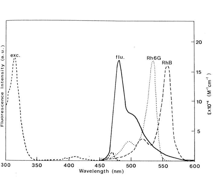
Fluorescence (solid) and excitation (broken) spectra of DPC in ethanol glass at 77 K are shown in Figure 1. The fluorescence peak at 480 nm is observed with a shoulder at longer wavelength.
This spectrum agrees well with that reported in previous
papers.1,5a The excitation spectruth consists of two weak bands at 468 and -v415 nm and a strong band at 316 nm. The two bands at
468' (weak) and 316 nm (strong) originate from DPC, which are consistent with those reported in the previous papers,1,5a whereas the band at tv415 nm is likely from impurities. Spectral
features of both fluorescence and excitation spectra in other
organic glasses (EPA, 3MP, and MTHF) are quite similar to those
in the ethanol one. The peak positions of these spectra are
listed in Table 1. The peak positions of fluorescence spectra are
observed nearly at 480 nm while those of excitation spectra are at 468 (weak) and 'v318 nm (strong) in these glasses. This result indicates that electronic spectra of DPC are not
influenced by properties of the medium (e.g., solvent polarity or viscosity).
According to the electronic spectra of DPC trapped in substitutional sites of a benzophenone single crystal,2 a sharp zero-phonon line (470.7 nm) is observed with a broad sideband extending to high energy in absorption (lmax 'v 465 nm) and to low energy in emission (Amax -v 476 nm) in the temperature range 2 - 20 K. The sharp zero-phonon line disappears with increasing temperature while the broad sideband is observed still above 20 K. The fluorescence and excitation spectra observed in this study closely resemble these broad sidebands. The geometry of DPC in the benzophenone single crystal was determined to be nonplanar with a central C-C-C angle of 1400 and dihedral angles of 29.90 and 26.70 by means of electron nuclear double resonance (ENDOR) spectroscopy.12 It is therefore suggested that the fluorescence
band. observed in this study is from the nonplanar geometry similar to DPC in the benzophenone single crystal.
On the other hand, Figure 1 also shows absorption spectra of
Rh6G (dotted) and RhB (dashed). The spectral overlaps between the
fluorescence of DPC and the absorption of dye molecules are
significant. Since resonance energy transfer efficiency from DPC
to dye molecule increases with increasing speetral overlap,9
this type of energy transfer from DPC to dye molecule is expected
to occur.
3.4.2 Fluorescence Decays. ,
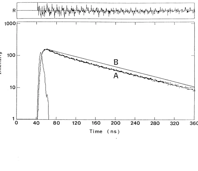
Fluorescence decay curve of DPC (A) in ethanol glass at 77 K is displayed on semilogarithmic scale in Figure 2. The solid curve (B) shows a caleulated decay curve of single exponential of 115 ns. Obviously the decay curve (A) is nonexponential. The decay eurve (A) is analyzed as a sum of two exponential decays with the lifetimes of 25 and 115 ns and with the corresponding
initial intensities of O.30 and O.70, respectively. The plot of the weighted residuals clearly shows that this biexponential analysis is valid. In other organic glasses at 77 K, fluorescence decay curves of DPC are also analyzed as biexponential decay.
The lifetimes of the two components, the corresponding initial intensity, and the ratio of initial intensity (Al/A2) are
summarized in Table 2. 0n the other hand, time-resolved fluorescence spectra in the organic glasses at 77 K remain unchanged as the time proceeds and agree well with steady state
fluo'
rescence spectrum.
As a result, three characteristic features are found in the
fluorescence decays of DPC regardless of the properties of the glasses: (a) The lifetimes of the fast component and the slow one are rv30 ns and tv140 ns (115 ns in ethanol), respectively. (b) The initial intensity ratio of two components (Al/A2) is "vO.5.
(c) Time-resolved fluorescence spectra almost remain unchanged as
the time proceeds.
The nonexponential fluoreseence decay of DPC in low- temperature matrix was also observed by Migirdicyan and co- workers.7 They have explained this result as follows: the
independent fluorescenee (Tlx -> Tox, Tly -> Toy, and Tlz '-' Toz) are emitted from the individual Tlu (u = x, y, z) sublevels at a
rate faster than the spin-lattice relaxation between the different sublevels. The depopulation rate of the isolated Tlu
sublevels should be equal in the absence of spin-orbit
interaction and nonradiative decay processes. However, the wave funetions describing the Tlu sublevels are mixed with those of singlet states possessing the same symmetry by spin--orbit coupling. Consequently, the fluorescence decay of DPC is composed of three components with distinet depopulation rates caused by different intersystem crossing rates from the Tlu sublevels.
The experimental results can be also explained by the idea of the independent emission from the individual Tl sublevels of DPC.
That is, the fast component is composed of the emission from only a specific sublevel of Tl whose depopulation rate is faster than the others, while the slow component is composed of those from two other sublevels. According to this interpretation, the
initial intensity ratio (Al/A2) reflects the population of the
corresponding sublevels in the ground state, To. Since the zero-- -1
and E/hc = O.O19 field splitting energy of To (D/hc = O.405 cm
cm-1 in the benzophenone single crystai13) is much smaner than
the energy of kT (53.4 cm-1) at 77 K, the populations of the
three Tou sublevels are equal. Consequently, the initial intensitY ratio (Al/A2) may be close to O.5. The sublevels of L
both To and Ti are almost degenerate so that the observation (c) is due to the same transition energy of the three Tlu -) Tou
transitions.
3.4.3 Estimated Values of Fluorescence Quantum Yields and
Radiative and Nonradiative Rate Constants for the Fast and Slow Components
The author proposes a novel method to estimate the values of fluorescence quantum yields and radiative and nonradiative rate constants for the fast and slow components of DPC in ethanol glass at 77 K. The method is to use the resonance energy
transfer from the excited DPC to a suitable acceptor molecule. In general, one needs to measure the fluorescence lifetime and quantum yield to determine the radiative and nonradiative rate constants experimentally. In the case of this study, however, the fluoreseence quantum yields for the fast and slow components
of DPC cannot be measured independently by means of a
conventional method using a standard such as quinine bisulfate in aqueous H2S04 solution.
According to F6rster theory,9 the critical transfer distance (Ro) of resonance energy transfer can be expressed by gooo(lnlo) K2 Åë
R06= 12s ,t sn4N J (1)
where K2 and qS are the orientation factor and fluorescence quantum yield of the donor in the absence of energy transfer, i
respectively; n and IV are refractive index of the medium and Avogadro's number, respectively. J is the spectral overlap
integral as expressed as follows:
J. j' FD(V)yE4A( VS')dv (2)
--' N where FD(v) and EA(y) are the spectra of donor emission normalized to unity and the molar extinction coefficient of acceptor, respectively. Accordingly, the values of Åë for each fluorescence component can be estimated from the corresponding values of Ro. In order to determine the values of Ro for both fluorescence components simultaneously, the fluorescence decay curves quenched by Rh6G or RhB were measured.
Fluorescence decay curves of DPC in the presence of Rh6G or RhB in ethanol glass at 77 K are shown in Figures 3 and 4, respectively. The fluorescence intensities are normalized at the time origin. The apparent fluorescence lifetime decreases with
'
incr'easing concentration of dye molecule. It is confirmed that
the Tl state of DPC is efficiently quenched by Rh6G or RhB. Wu and Trozzolo observed resonance energy transfer from DPC to fluorescein by means of fluorescence decay measurement and determined the value of Ro to be 6.1 nm6. The author used Rh6G or RhB as the energy acceptor instead of fluorescein, because a
eonsiderable amount of emission from fluorescein mixes into the
fluorescence of DPC. This emission prevents analyzing the
quenched decay curves of DPC strictly.
The fluorescenee deeay of the donor undergoing dipole-dipole resonance energy transfer in uniformly distributed and randomly oriented system is expressed as follows:14
i(t) =A exp[-t/ 'r -x(t/ 'r )1/2] (3)
where T' is the fluorescence lifetime of donor in the absence of acceptor and
x= g(3/4ooo) 7r 3/2NRo3cA (4)
in which g is the orientation factor, CA is the concentration of acceptor, N and Ro are denoted in eq. 1. In the case of this study, the fluorescence of DPC contains two components so that the fluorescence decay of DPC in the presence of dye molecule is expressed by
l( t) = 2 Ai exp[- t/ 'r i-- xi(t/ T i)1/2] (i = 1, 2) (s)
where 'ri and Xi are the fluorescence lifetime and X for each fluorescence component of DPC, and i = 1 and 2 represent the fast fluorescence component and the slow one, respectively.
The value of Ro for the fast and slow components can be
'
obtained by determining Xl and X2 by means of curve fitting procedure at several concentrations of dye molecule. The convolution and subsequent nonlinear least-squares method were carried out with the parameters (Xl and X2) and the fixed constant values (Al = O.3, A2 = O.7, Tl = 25 ns and 7r2 = 115 ns) which determined from the absence of dye molecule. A
calculated decay curve (solid) with the parameters (Xl = O.70 and
x2 = 1.57) and an observed decay curve in the presence of 4 mivl '
Rh6G are shown in Figure 5. The plot of the weighted residuals ,
clearly indicates that the two curves fit very well. Other
fluorescence decay curves in the presence of dye molecule were fitted in the same manner.
The plots of Xl (O, A) and X2 (-, A) vs concentration of
dye molecule are shown in Figure 6. Both Xl and X2 inerease linearly with inereasing concentration of dye molecules as predicted from eq. 4. The values of Rol and Ro2 are calculated from the slope of the plot, g(3/4ooo)7t3/2NRo3, where g for a rigid random orientation is O.845.15 The values of Rol and Ro2 for DPC/Rh6G combination are 3.6 and 4.7 nm, and those for DPC/RhB combination are 3.5 and 4.5 nm, respectively. Comparing the values of Roi between DPC/Rh6G and DPC/RhB, the values of Roi for DPC/Rh6G are slightly larger than those for DPC/RhB for both the fast and slow components. This result is consistent with the prediction from the spectral overlap J, in which the value of Dpc/Rh6G (2sxlo-14 cm6) is larger than that of Dpc/RhB (lgxlo-14 . cm6).
The value of Åëi for the fast and slow components can be estimated from the F6rster equation (1), where the respective Values of Rol and Ro2 are substituted into the F6rster equation
(1). It should be emphasized that the values of J for both
fluoreseence components are equal because time--resolved
fluoreseenee spectra remain unchanged as the time proceeds. The
value K2 = O•476 is quoted for a rigid random orientation.15 The value of n for ethanol at 77 K has not been reported so far;
therefore, we determined this value as 1.45 using n = 1.37 at 273
K and its temperature dependence dn/dT = 4 Å~ lo-4 K-1.16
consequently, the calculated values of Åë1 and Åë2 are O.09 and O.46 for DPC/Rh6G combination, and O.11 and O.47 for DPC/RhB combination, respectively. Comparing the values of Åëi between
DPC/Rh6G and DPC/RhB, these values agree well with each other.
Thus the similar values of Åëi are obtained from two different DPC/dye combinations. These values are listed in Table 3. The
average values of Åë1 and Åë2 are about O.1 and O.5,
respectively.
Considering the total fluorescence quantum yield ( Åëtot), this is expressed as follows:
Åëtot = Al Åë1 + A2 Åë2 (Al + A2 = 1),
where Al and A2 are the fractions of the initial intensities denoted in eq. 5. The value of Åëtot is estimated to be A- O.4.
Taking account of the uncertainty of the value of n at 77 K and experimental errors, this value is in reasonable agreement with the experimental value (O.2) in ethanol glass at 77 K reported by
Ono and ware.5a Therefore, the values of both Åë1 and Åë2
obtained from energy transfer analysis are reliable.
The respective values of kri and knri for the fast and slow components can be easily calculated from the relation of kri = Åëi/ iri and knri = (1-Åëi)/ 2ri (i = 1, 2). The values of kri and
Knri (i = 1, 2) are summarized in Table 3.
It is obvious that the values of kri for both the fast and
slow components are nearly equal (4 Å~ lo6 s-1) for both Dpc/dye ,
combinations. This result clearly indicates that the radiative processes from the individual Tl sublevels are not influenced by some kinds of interaction such as spin-orbit coupling. Since the radiative processes of the individual transitions (Tlx --> Tox, Tly " Toy, and Tlz -> Toz) of DPC are electronically allowed, the corresponding transition moments are not so perturbed.
On the other hand, the value of nonradiative rate constant for the fast component (knrl -v 36 x lo6 s-1) is about 7-s times as much as that for the slow one (knr2 -v s Å~ 106 s-1). The biexponential fluorescence decays of DPC in some organic glasses at 77 K are definitely caused by this difference between knrl
and knr2• This difference is ascribed to the distinct
intersystem crossing rates from the corresponding sublevels of Tl to lower energy singlet states (Sn) as reported by Migirdicyan and co-workers.7 In particular, i' t js probably suggested that there is a specific spin sublevel in Tl which strongly couples to
. Iower Sn by spin-orbit interaction. Migirdicyan and co-workers have also presented that three or four singlet states exist between To and Tl of DPC from a molecular orbital calculation 7c (CS-INDO CI method).
Here, let us consider the reason why fluorescenee decay
curves of DPC in some organic glasses at 77 K exhibit
biexponential with the initial intensity ratio, Al/A2 'v O.5. Two
possible explanations might be offered to account for the reason;
(1) Spin-orbit coupling interact only a specific spin sublevel but not two others of Tl. (2) Even if the three intersystem crossing rates are different, the depopulation rates of two spin sublevels of Tl except for that of the specific one are equalized by the spin-lattice relaxation process between the two sublevels
because it still remains a question whether spin--lattice
relaxation can sufficiently be suppressed at 77 K. At the present
stage, it is impossible to decide which explanation is proper.
,t-N
:
v
o•-
>
ut- :o
.
:oo co oco Åë
oL :
L exc.
t) ll
It tl Jl ee ee tt lt tl
11 tl
11 lt
t 1 1 ` s t , t t ,
s
t s , ss N
N
p- .
"e .t- -pep pt -
,
----
flu.
/s •. ,'
tA'
.. "A; -t
-- --/
/ - -
.. -
Rh6G "': RhB
:: tN l: ll :: tl
:: tl
i ': lt
:' :lt
:' :• l1 i :. lt
,.:: li. ll li
:• : l t
: :l t : :l t
:Lt
. /I 1 •" 1: t
1"'N
Ni l :. N
:. N
NN
--
N20
15 A
TE TO
g 9 10 1
,l5,
5
300 350 400 450
Wavelength (nm) 500 550 600
Figure 1. Fluorescence (solid) diphenylcarbene and absorption
Rhodamine B (RhB) in
and excitation (broken) spectra of Rhodamine 6G
ethanol glass at
spectra (Rh6G)
77
of and
K.
R
1000
is
, b•
rwiifNtxfVifi,S=Y•i*VfiSr•7
f->
ca
c
-oc
H
1OO
10
1
B
A
.Y.c
.`.--,fi
o 40 80 120 160 200
Time (ns)
240 280 320 360
Figure 2. Fluorescence decay curve of diphenylcarbene (A) in ethanol glass at 77 K. The solid curve (B) is a calculated decay eurve of single exponential (115 ns). The weighted residuals show the best fitting with the 1ifetimes of 25 and 115 ns and with
corresponding initial intensities of O.30 and O.70.
•:
>
ut
:di
-
cH
Rh6G
x \NY/Åéaj,XX"X-
ajs"
o 40 80 120 160 200
Time
240
(ns)
280 320 360 40O
Figure O, (B)
3
1
.
'
Fluorescence decay (C) 3, and (D) 5 mM
curves of Rhodamine
diphenylcarbene with 6G in ethanol glass at
(A)
77 K.
>
•.-- co
o:
-
=H
o 40 80
CI'i)rti
,
MgsK
/lll'liliil"'`lkN
•;u,;;:ie:lt
120
vt
xAB
Xs,/,y,,4k Fg,C,,,,,, X'Fi"v>{vts..
`""Lsk.,Y")ilt:I:rtttilllllllllllllllllllllll:$M{'s",hN LnxLk,1I-i][:tfy.--Mv"boN-r"
160 200
Time
24O
( ris)
280 320 360
tA
400
Figure O, (B)
4
1
.
'
Fluorescence decay (C) 3, and (D) 5 mM
curves of Rhodamine
diphenylcarbene with B in ethanol glass at
(A)
77 K.
R
1OOO
t
:>
co
c
co
-: H
1OO
10
1
>
:le-
- l-r---- -o 40 80 120
Time
160 (ns)
200 24O 280
Figure 5. Fluorescence decay curve of Rhodamine 6G in ethanol glass at 77 K.
Iated from eq. 5 by using the values Al ns, T2= 115 ns, Xl = O.70, and X2 = 1.
diphenylcarbene The solid curve = O.3, A2 = O.
57.
with 4 mM
was calcu-
7, Tl = 25
Å~
2
1
o
A
Rh6G RhB
Rh6G RhB
o
12 3
CA/mM
4 5 6
Figure 6. Plots
concentration of dye
of Xl (
mo1ecu1es.
ll A)
' and X2 (-,A) vs
TABLE 1 Organlc
. Peak Position of Glasses at 77 K
Fluorescence and Excitation Spectra in
g1ass f1uorescence
A max/nm
excitation X (weak)/nm
a
A(strong)/nm EtOH EPA
rvlTHF
3MP
480 479 477 482
468 468 468 468
316
318
320
317
a Excitation spectra are uncorrected.
t
TABLE 2. Fluorescence and Initial Intensity
Lifetimes ( 'zr Ratio (Al/A2)
i)
in
, Initial Intensity Organic Glasses at
a 77
(Ai), K
g1ass fast
Al
component T 1/ns
s1ow A2
component
T2/ns
Al/A2
EtOH EPA MTHF 3MP
O.30 O.31 O.28 O.34
25 29 32 25
O.70 O.69 O,72 O.66
115 146 141 141
o o o o
'
.
.
.
43
45
39
52
a Initial intensity are normalized: Al + A2 =1.
TABLE 3.
Radiative (knri) in
Estimated Values Rate Constanta Ethanol Glass at
of Fluorescence Quantum (kri), and Nonradiative 77K (i = 1, 2)
Yields Rate
(Åëi)' b Constant
acceptor
Åë1