論文 内 容の 要旨
IntroductionArsenic is a natural substance that has been used medicinally for over 2400 years. Arsenic was rekindled after it was identified as an active ingredient in traditional medicines in China. Arsenic drugs have been generally used for the treatment of malignant hematologic diseases. Arsenic trioxide (As2O3) has been
confirmed to be an effective treatment for acute promyelocytic leukemia (APL). It has been reported that the effects of As2O3 are not confined to APL cells but can
also be observed in various myeloid and lymphoid cells. The action mechanisms of As2O3 in APL and other malignancies are thought to involve inhibition of
growth and induction of apoptosis.
MOLT-4, a human T-lymphoblastoid leukemia cell line, has been used extensively for studies of leukemia cell biology and antileukemia therapy. We have established a daunorubicin- resistant MOLT-4 subline MOLT-4/DNR by exposing the parental MOLT-4 cells stepwise to increasing concentrations of daunorubicin. MOLT-4/DNR cells overexpress functional P-glycoprotein and MDR1 mRNA. Most of the drugs excreted via this efflux pump are hydrophobic organic compounds, and As2O3 may not be excluded from drug-resistant cells
expressing functional P-glycoprotein. However, whether As2O3 affects the
growth of lymphocytic leukemia cells expressing functional P-glycoprotein is unknown. Thus, in Chapter 1, the effects of As2O3 on the growth and apoptosis
of parental MOLT-4 and the resistant MOLT-4/DNR cells were investigated. Cancer cell sensitivity to As2O3 correlates with intracellular glutathione
こ し あ お め い 氏 名 ( 本 籍 ) 胡 晓梅 (中国) 学 位 の 種 類 博 士( 薬学) 学 位 記 番 号 論博 第329号 学位授与の日付 平成 26 年 3 月 20 日 学位授与の要件 学位 規則第 4 条 第 2 項該当 学 位 論 文 題 目
Action Mechanisms of Arsenic Compounds on Leukemia
levels. Cells expressing higher levels of glutathione or glutathione -associated enzymes are less sensitive to As2O3 than cells expressing lower levels of these
molecules. Arsenic- resistant cells are also reported to contain higher levels of glutathione. Moreover, cells with increased glutathione levels can be sensitized to As2O3 by agents that deplete intracellular glutathione. Thus, in Chapter 2, the
apoptosis-inducing effects of As2O3 in the presence of glutathione modulators in
MOLT-4 and MOLT-4/DNR cells were examined.
It has been recognized that benefit and risk of arsenic are strictly dependent on the individual chemical forms of arsenic. Although As2O3 has been confirmed
to be an effective treatment for APL, serious adverse drug reaction induced by As2O3 was occasionally reported. Arsenic disulfide (As2S2), the most
important component of Xiong huang, was a candidate for its good therapeutic reputation and perceived low toxicity in traditional medicines. Xiong huang was reported to improve the clinical outcomes of hematologic malignancies in our clinical trials, which could be attributed to As2S2. As2S2-mediated growth
inhibition and apoptosis induction have been found in leukemia K562 cells and other cancer cells. However, the effects of As2S2 on cells of a human APL cell
line HL-60 cells with a particular focus on proliferation and differentiation have not been addressed. Thus, in Chapter 3, the effects of As2S2 on HL-60 cells were
investigated by focusing on differentiation, generation of reactive oxygen species, intracellular glutathione depletion, and activation of p38 MAPK.
Patients with AML from myelodysplastic syndrome (MDS/AML) have higher probabilities of resistance to chemotherapy, lower rates of complete remission, and the poor prognosis. MDS/AML patients tended to have complex type abnormalities including monosomy 7 ( -7 ) , which are considered to be an unfavorable risk subgroup. We have revealed that As2S2 is effective in the
treatment of MDS without serious adverse drug reaction. A leukemic F-36p cell line has been established from a patient diagnosed with refractory anemia with excess blasts. Thus, in Chapter 4, the effective mechanisms of As2S2 in the
treatment of MDS or MDS/AML were studied by use of F-36p cell line.
Chapter 1. Arsenic trioxide induces apoptosis equally in T lymphoblastoid leukemia MOLT-4 cells and P-glycoprotein-expressing daunorubicin-resistant MOLT-4 cells
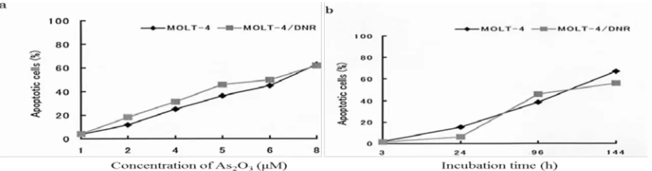
As2O3 inhibited the growth and survival of MOLT-4 and MOLT-4/DNR cells
in a time- and dose dependent manner. As2O3 induced apoptotic morphology in
dose-dependent. As2O3 did not change the percentage of
P-glycoprotein-expressing cells or the efflux ability of MOLT-4/DNR cells.
Thus, the data in this Chapter showed that As2O3 inhibited growth and induced
apoptosis equally in MOLT-4 and MOLT-4/DNR cells, and this suppressive effect
Fig. 1 Induction of apoptosis by As2O3 in M OLT-4 and MOLT-4/ DNR cells. a; Percent apoptotic cells as a function of As2O3 concentration after culture for 4 days, b; Percent
apoptotic cells as a function of incubation time in the presence of 5μM As2O3. Values are the
means of three independent experiments.
was not influenced by P-glycoprotein expression or function in MOLT-4/DNR cells.
Chapter 2. Arsenic trioxide induces apoptosis in cells of MOLT-4 and its daunorubicin-resistant cell line via depletion of intracellular glutathione, disruption of mitochondrial membrane potential, and activation of caspase-3
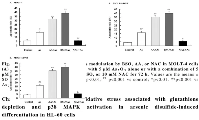
MOLT-4 cells and MOLT-4/DNR cells were similarly sensitive to the apoptosis-inducing effect of As2O3. Buthionine sulfoxide (BSO), a selective
inhibitor of γ-glutamylcysteine synthetase, and ascorbic acid (AA), having pro-oxidant properties, rendered these cells more sensitive to As2O3, whereas
N-acetylcysteine (NAC), an antioxidant since it donates a cysteine to the de novo synthesis of glutathione, reduced this sensitivity. BSO and AA decreased, but NAC increased, the intracellular glutathione contents of both MOLT-4 and MOLT-4/DNR cells. Decreasing glutathione with BSO potentiated As2O3-mediated growth inhibition, disruption of mitochondrial membrane
potential, activation of caspase-3, and apoptosis of cells. Clinically relevant doses of AA enhanced the anticancer effects of As2O3 via the disruption of
mitochondrial membrane potential, activation of caspase-3, and induction of apoptosis. In contrast, increase in glutathione levels with NAC attenuated all of these As2O3-mediated actions.
Thus, MOLT-4 and MOLT-4/DNR cell sensitivity to As2O3 was associated
and MOLT-4/DNR cells expressing functional P-glycoprotein via depletion of intracellular glutathione, and subsequent disruption of mitochondrial membrane potential and activation of caspase-3.
Fig. 2 As2O3-induced apoptosis and its modulation by BSO, AA, or NAC in MOLT-4 cells
(A) and M OLT-4/DNR cells (B) treated with 5 μM As2O3 alone or with a co mbination of 5
μM As2O3 with 125 μM AA, 100 μM BSO, or 10 mM NAC for 72 h. Values are the means ± SD of three independent experiments. # p<0.01, # # p<0.001 vs control; *p<0.01, **p<0.001 vs As2O3 alone.
Chapter 3. Involvement of oxidative stress associated with glutathione depletion and p38 MAPK activation in arsenic disulfide-induced differentiation in HL-60 cells
As2S2 induced cell differentiation based on the increment in expression of
CD11b, antibody specific for the myeloid differentiation marker, nitroblue tetrazolium-positive cells, and cell size. A transient increase in generation of reactive oxygen species level along with intracellular glutathione level was also observed. p38 MAPK activation gradually increased after generation of reactive oxygen species and sustained during the cell differentiation. Decreased CD11b expression was accompanied by p38 MAPK activation, and p38 MAPK inhibitor restored the CD11b expression.
Thus, the data in this Chapter showed that As2S2 induced differentiation in
HL-60 cells, and moderate levels of oxidative stress induced by As2S2 positively
contribute to HL-60 cell differentiation. The activation of p38 MAPK resulted from oxidative stress seems to be implicated in the negative regulation of the differentiation.
Fig. 3 The modified effects of inhibitor (SB203580) of
p38 MAPK on As2S2-induced differentiation in HL-60
cells. Cells were incubated with 10μM SB203580 (SB)
and 8 μM As2S2 (A8) alone or in combination (SB +
stained
for CD11b and then analyzed with flow cytometer. Data are the mean ± SD of 3 independent experiments. Means were compared by 1-way ANOVA. *p< 0.05 as compared with 8 μM As2S2 (A8) alone.
Chapter 4. Arsenic disulfide induced apoptosis and concurrently promoted erythroid differentiation in cytokine-dependent MDS-progressed leukemia cell line F-36p with complex karyotype including monosomy 7.
As2S2 inhibited the proliferation of F-36p cells. The apoptotic cells
significantly increased and were in a dose-dependent manner. The cell viabilities were significantly inhibited by As2S2 and were in dose-dependent. Significant
increases of expression of CD235a, antibody specific for the erythroid differentiation marker glycophorin A (Gly A), were concurrently observed and were also in a dose-dependent manner.
In this Chapter, I showed that F-36p cell line might provide a desirable cell model for the study of effective mechanisms of As2S2 in the treatment of MDS or
MDS/AML. As2S2 could inhibit proliferation and viability, induce apoptosis, and
concurrently promote erythroid differentiation in F-36p cells, which were in a dose-dependent manner.
Fig. 4 Comparison of levels of CD235a-positive
cells. Cells were treated with As2S2 at
concentrations of 0, 2, 8 and 16 μM for 72 h. The data are the mean ± SD of three independent experiments. *p<0.05 as compared with control group, and in dose-dependent by the multiple comparisons.
Conclusions
Effects of As2O3 are not confined to APL cells but can also be observed in
various other cell lines and in drug-resistant sublines. As2O3 induced apoptosis
in parent MOLT-4 cells and MOLT-4/DNR cells expressing functional P via depletion of intracellular glutathione, and subsequent disruption of mitochondrial membrane potential and activation of caspase-3. As2S2 was a candidate for its
good therapeutic reputation and perceived low toxicity in traditional medicines. The data showed that moderate levels of oxidative stress induced by As2S2
regulation of the differentiation. Furthermore, it is the first description that As2S2 can inhibit proliferation and viability, induce apoptosis, and concurrently
promote erythroid differentiation in cytokine-dependent MDS-progressed human leukemia cell line F-36p with complex karyotype including karyotype -7. The precise action mechanisms of As2S2 demonstrated in this study in human
malignant cells might imply the rationale and future directions of As2S2 as a
potential anticancer drug candidate.
PUBLICATIONS
1. Hu XM, Oka K, Hirano T. Cancer Chemother Pharmacol 2003;51:119-126. 2. Hu XM, Oka K, Hirano T. Cancer Chemother Pharmacol 2003;52:47-58.
3. Hu XM, Yuan B, Tanaka S, Onda K, Toyoda H, and Hirano T. Leuk Lymphoma DOI: 10.3109/10428194.2013.802779