九州大学学術情報リポジトリ
Kyushu University Institutional Repository
ヒトDNA ポリメラーゼεと結合したCTF18-RFCによる PCNAローディングはそのDNA合成の継続に寄与する
藤澤, 遼
https://doi.org/10.15017/1931744
出版情報:Kyushu University, 2017, 博士(理学), 課程博士 バージョン:
権利関係:
Human CTF18-RFC complexed with DNA polymerase ɛ loads PCNA efficiently and maintains the DNA synthesis
ヒト DNA ポリメラーゼ ɛ と結合した CTF18-RFC による PCNA ローディングはその DNA 合成の継続に寄与する
九州大学大学院 システム生命科学府 分子生命科学 染色体機能学講座 (釣本 敏樹 教授) 平成 25 年度入学 藤澤 遼 平成 30 年 1 月 22 日提出
CONTENTS
Abbreviations ………3
Abstract ………4
Introduction ………6
Materials and methods………12
Results………21
Discussion………29
References………34
Figure legends……… 53
Acknowledgement………60
Abbreviations
APB: 4-azidophenacyl bromide, ATP: adenosine triphosphate,ATPγS:
adenosine-5′-O-(3-thiotriphosphate), ATR: Ataxia telangiectasia and Rad3 related protein, BSA:
bovine serum albumine, CBB: coomasie brilliant blue, Cdc: Cell division cycle, CDK, Cell cycle-dependent kinase, Cdt1: Cdc10-dependent transcript 1, CHAPS: 3-[(3-cholamidopropyl) dimethyl ammonio] propane sulfonate, CMG: Cdc45-Mcm2–7-GINS, CTF: Chromosome Transmission Fidelity, CV: column volume, DCC1: Defective in sister Chromatid Cohesion 1, DDK, Dbf4-Dependent Kinase, dpb: DNA Polymerase B subunit, DEAE: diethylaminoethyl, dsDNA: double strand DNA, DTT: dithiothreitol, EDTA: ethylenediamine tetra acetic acid, Elg:
Enhanced Level of Genomic instability, EMSA: electrophoretic mobility shift assay, exo–:
exonuclease deficient, FenI: Flap end nuclease I, GINS: Go-ichi-ni-san, HBS: Hepes Buffered Saline, Hepes: 4-(2-hydroxyethyl)-1-piperazineethanesulfonic acid, HU: hydroxyl urea, Mcm:
Mini chromosomal maintenance, Mrc1: Mediator of the replication checkpoint 1, NP-40: Nonidet P-40, nt: nucleotide, ORC: Origin recognition complex, PBS: Phosphate buffered saline, PCNA:
Proliferating cell nuclear antigen, PMSF: phenylmethylsulfonyl fluoride, Pol: DNA polymerase, pre-RC: pre-replicative complex, Psf1: Partner of sld five, Rad: Radiation sensitive, PVDF:
polyvinylidene difluoride, RFC: Replication factor C, RPA: Replication protein A, SDS: sodium dodecyl sulfate, SDS-PAGE: sodium dodecyl sulfate-polyacrylamide gel electrophoresis, sld:
synthetic lethal with dpb11-1, ss: single strand, Tris: tris hydroxymethyl aminomethane, WT: wild type, 3′ primer-template junction: 3′-end recessed primer-template junction
ABSTRACT
At initiation of DNA replication in eukaryotic cells, “replisome”, a high-ordered protein assembly is formed to unwind double-stranded DNA and synthesize leading and lagging strands coordinately. The replicative DNA helicase complex, CDC45-MCM2–7-GINS (CMG) complex recruits three distinct DNA polymerases (Pol) α, δ, and ε, which are specialized to synthesize RNA/DNA hybrid primers, lagging- and leading-strand DNAs, respectively. A toroidal protein complex, proliferating cell nuclear antigen (PCNA) functions as a DNA sliding clamp, which encircles double-stranded DNA to provide an assembly platform for various replisome proteins including Pols.
Polε is required for the leading-strand synthesis, specifically forming a stable complex with an alternative PCNA loader complex CTF18-RFC via the N-terminal half of the catalytic p261 subunit (p261N). Unlike the canonical PCNA loader RFC, CTF18-RFC alone has a limited clamp-loading activity under physiological conditions. I found that CTF18-RFC complexed with Polε restored the activity and became an alternative of RFC. This suggests that CTF18-RFC functionally loads PCNA especially when it forms complex with Polε. A 3′-end recessed primer-template junction is the target DNA structure for efficient PCNA loading of the CTF18-RFC–Polε complex. Proteins directly interacting with the primer-template junction DNA were analyzed in the presence of CTF18-RFC, PCNA, and p261N by site-specific photo-crosslinking. p261N bound to the target site most of the time and binding of CTF18-RFC was limited. However, in the presence of ATPγS, the binding of CTF18 increased. Thus, CTF18-RFC complexed with Polε would access to the primer terminus for PCNA loading transiently. Polε could be placed in DNA synthesis mode using a deoxidized 3′ primer end. I demonstrated that CTF18-RFC complexed with synthesizing mode of Polε exhibited less efficient PCNA loading, indicating that DNA synthesis and PCNA loading of the CTF18-RFC–
Polε complex are mutually exclusive at the 3′-end of a primer-template junction. In a DNA synthesis mode, PCNA and the CTF18-RFC–Polε complex engaged in stable trimeric assembly on the template DNA and actively synthesized DNA. Therefore, the CTF18-RFC–Polε complex will be an active configuration of the leading-strand DNA polymerase, in which CTF18-RFC will monitor and, if necessary, restore DNA synthesis by de novo loading of PCNA.
In summary, two sets of PCNA loader and DNA polymerase in eukaryotes have distinct roles for leading- and lagging-strand DNA synthesis. RFC and Polδ respectively target free 3′
primer ends and will be most applicable for discontinuous lagging-strand synthesis, whereas,
CTF18-RFC and Polε form a stable complex along with the loaded PCNA at synthesizing 3′ DNA ends and will be specialized to maintain continuous leading-strand synthesis.
Introduction
Eukaryotic DNA replication
DNA replication is a fundamental process for all living organisms. Its dysregulation leads to spontaneous mutations, DNA damage and gross chromosomal rearrangements. These genomic instabilitys cause cell senescence and cancer cell development (Weinert et al., 2009, Truong and Wu, 2011, Lopez et al., 2014, Gaillard et al., 2015, Tomasetti and Vogelstein, 2015).
Unlike prokaryotes’ genomes, eukaryotes’ genomes have multiple replication origins (Leonard and Méchali, 2013). To maintain the gene dosage constant through cell divisions, the activation of replication origins must occur only once per cell-cycle, and strict regulation for the loading and activation of replicative helicase ensures the mechanism (Bell and Labib, 2016). Recent studies with purified proteins of S. cerevisiae have demonstrated the reconstitution of origin dependent DNA replication (Yeeles et al., 2015, 2017, Devbhandari et al., 2016, Lõoke et al., 2017).
The mechanism of replication initiation in S. cerevisiae will be described as follows; The six subunits complex ORC (Origin Recognition Complex) binds to the replication origin in the presence of ATP (Bell and Stillman, 1992, Diffley and Cocker, 1992, Li and Stillman, 2012).
Using the ORC as the assembly platform, pre-RC (pre-replication complex) is formed with two Mcm2–7 complexes, the hexametric AAA+ motor core of the replicative helicase in collaboration with Cdc6 and Cdt1 from telophase to G1 phase when CDK/cyclin levels are low (Evrin et al., 2009, Remus et al., 2009, Riera et al., 2014). In S phase when the activities of two essential kinases, DDK and S-CDK increase, sequential protein recruitments including Sld3-Sld7-Cdc45, Dpb11, Sld2, GINS, Mcm10, and Polɛ occur on the phosphorylated Mcm2–7 in a head to head configuration, resulting in formation of the active replicative DNA helicase CMG consisted of Cdc45, Mcm2–7 and GINS (Kamimura et al., 2001, Tanaka et al., 2007, Zegerman and Diffley, 2007, Muramatsu et al., 2010, Yardimci et al., 2010, Heller et al., 2011, Deegan et al., 2015, Riera et al., 2017). Upon completion of the assembly, two CMG helicases bidirectionaly
translocate on the leading-strand template DNA in a 3′-5′ direction to unwind duplex DNA (Fu et al., 2011, Yu et al., 2014).
Eukaryotic DNA polymerases and the elongation process
To date 16 DNA-dependent DNA polymerases are identified in human cell (Vaisman and Woodgate, 2017). Among them, Polα, Polδ, and Polɛ are replicative DNA polymerases for genomic DNA replication in nucleus, unlike that only one replicative DNA polymeraseIII is responsible for replication of eubacteria genome (Mclnerney et al., 2007, Johansson and Dixon,
2013). Polα, Polδ, and Polɛ are composed of one catalytic subunit and other two or three subunits and in division of labour in the replisome. The CMG helicase associates with Polɛ via the interaction between Dpb2/p59 second subunit of Polɛ and Psf1 subunit of GINS (Sengupta et al., 2013) and synthesizes leading-strand DNA processively (Pursell et al., 2007, Georgescu et al., 2014, Daigaku et al., 2015). The other replicative DNA polymerase Polα complexed with the primase is also connected physically with CMG helicase via Ctf4, which functions as a hub in replisome (Gambus et al., 2009, Tanaka et al., 2009, Villa et al., 2016). Polα synthesizes short RNA/DNA primer on unwound ssDNA of the lagging-strand DNA template (Pellegrini, 2012, Georgescu et al., 2015a). The third DNA polymerase Polδ synthesizes lagging-strand DNA discontinuously (Nick McHelinny et al., 2008, Daigaku et al., 2015) without direct connection with CMG helicase (Sun et al., 2015, Schauer and O’Donnell, 2017). The discontinuous
lagging-strand DNAs are processed by FenI and Dna2 nucleases and ligated by Lig1 (Bae et al., 2001, Stith et al., 2008). Since most of yeast replication proteins are conserved in metazoan (Bell and Labib, 2016), the basic principles of DNA replication among eukaryotes are suggested to be identical.
Polδ and Polɛ possess a 3′-5′ exonuclease activity and highly accurate fidelity for DNA polymerization activity (Kunkel et al., 1987, Morrison et al., 1990, 1991). For efficient, processive DNA synthesis, however, they require additional factors (Masuda et al., 2007, Chilkova et al., 2007) such as RPA (Replication Protein A), PCNA (Proliferating Cell Nuclear Antigen), and RFC (Replication Factor C). RPA is a single-stranded DNA binding protein, resolving secondary structure of ssDNA (Fanning et al., 2006). PCNA is a ring-shaped sliding clamp protein, clamping DNA polymerase onto DNA for their stable DNA synthesis, and its loader, RFC loads PCNA at a 3′-end recessed primer-template junction (3′ primer-template junction) DNA with ATP-hydrolysis actions (see below sections). Thus, these factors are also essential components of the replisome.
PCNA forms a homo-trimeric ring complex embracing double-strand DNA in its central hole (Krishna et al., 1995), and slides along DNA freely at 100-150 bp/0.2 microseconds (De March et al., 2017). This sliding clamp interacts not only with replicative DNA polymerases, but also with various proteins involved in translesion DNA synthesis, Okazaki fragment processing, chromatin assembly, DNA methylation, DNA damage response and sister chromatid cohesion (Moldovan et al., 2007, Choe and Moldovan, 2017). Thus, PCNA provides a reaction-platform on replicating DNA not only for DNA synthesis but also for diverse range of reactions (Bell and Labib, 2016).
The replicative leading-strand DNA polymerase, Polɛ
Polɛ consists of four subunits, Pol2, Dpb2, Dpb3, and Dpb4 in S. cerevisiae (Chilkova et al., 2003), and p261/POLE1, p59/POLE2, p17/POLE3, p12/POLE4 in H. sapiens (Li et al., 1997).
Pol2 (p261 in human) encoding the catalytic subunit and Dpb2 (p59 in human) are essential, whereas Dpb3 and Dpb4 (p17 and p12 in human) are non-essential in yeast (Araki et al., 1991a, Araki et al., 1991b, Ohya et al., 2000). A unique property of Polɛ is that its catalytic subunit consists of two tandem exonuclease-polymerase modules, but N-terminal one is only catalytically functional (Tahirov et al., 2009). In S. cerevisiae and S. pombe, its N-terminal polymerase domain is dispensable, while the inactive C-terminal one is necessary for cell viability (Kesti et al., 1999, Feng and D’Urso, 2001). Thus, the essential Polδ will complement Polɛ to synthesize leading-strand DNA when its catalytic activity is nonfunctional (Miyabe et al., 2015, Yeeles et al., 2017). The C-terminal module along with Dpb2 plays a role for assembly of CMG helicase by recruiting GINS complex to pre-RC, and keeps the coupling with CMG helicase during replisome progression (Muramatsu et al., 2010, Handa et al., 2012, Kang et al., 2012, Yeeles et al., 2015, 2017). Dpb3 and Dpb4 form a hetero-dimer via their histone-fold like structures, bind to dsDNA (Tsubota et al., 2006), and are important for the processivity of both DNA synthesis and exonucleolytic degradation (Aksenova et al., 2010).
In addition to its direct roles in DNA replication, Polɛ has diverse roles, such as in DNA repair (Moser et al., 2007, Ogi et al., 2010), epigenetic information inheritance (Iida and Araki, 2002, Tsubota et al., 2006, Li et al., 2011), cell senescence (Deshpande et al., 2011, Saka et al., 2016) and S-phase checkpoint response (Navas et al., 1995, 1996, Dmowski et al., 2017). The replisome inevitably encounters stochastic disassembly or stalling by various obstacles as DNA damage on template DNA and by deficiency of dNTP. Under such circumstances, checkpoint kinases Mec1-Rad53 in S. cerevisiae, and ATR-Chk1 in vertebrate are activated and regulate the ribonucleotide reductase activity, the temporal program of replication origin firing, and cell cycle progression. (Giannattasio and Branzei, 2017, Saldivar et al., 2017). Because Polɛ is the primary DNA polymerase for leading-strand DNA, Polɛ might monitor its own DNA synthesis state in leading-strand DNA and transduce signals to checkpoint pathway (Puddu et al., 2011).
Indeed, mutations in the C-terminus of Pol2 affect the S-phase checkpoint function (Navas et al., 1995), and the presence of the catalytically functional Polɛ in replisome is required to activate Rad53 (García-Rodríguez et al., 2015). Polɛ also interacts with the checkpoint mediator protein Mrc1/CLASPIN, and one of the alternative PCNA loader Ctf18-RFC as described below, both of which are implicated in the checkpoint response (Lou et al., 2008, García-Rodríguez et al., 2015).
Even with tight relevance between replisome and the checkpoint response, the actual mechanism in checkpoint response mediated thorough Polɛ is still poorly understood.
The alternative PCNA loader, CTF18-RFC
Clamps and their loader complexes are the essential components for DNA replication in all organisms. Loader complexes consist of five similar or same AAA+ ATPase family proteins.
As exemplified above, the eukaryotic canonical loader for PCNA clamp is RFC, consisting of one large subunit (RFC1) and four small subunits (RFC2–5) (Bloom, 2009, Hedglin et al., 2013, Kelch, 2016). An ATP-bound form of RFC binds to PCNA (Shiomi et al., 2000, Gomes and Burgers, 2001), opens PCNA ring structure and further binds to the 3′ primer-template junction (Tsurimoto and Stillman, 1991, Hingorani and Coman, 2002, Shiomi et al., 2004). Upon binding, RFC hydrolyses ATP and leaves from PCNA and DNA (Sakato et al., 2012, Marzahn et al., 2015), consequently the loaded PCNA staying on DNA.
Three RFC1 paralogues, Ctf18, Rad17 (Rad24 in S. cerevisiae) and Elg1, exist in eukaryotes and associate with RFC2–5 to form alternative clamp loader complexes, Ctf18-RFC, Rad17-RFC and Elg1-RFC. Ctf18-RFC and Elg1-RFC target PCNA (Berumudez et al., 2003, Kanellis et al., 2003), whereas the checkpoint loader Rad17-RFC targets the checkpoint clamp Rad9–Hus1–Rad1 (9-1-1 complex; Ddc1–Rad17–Mec3 in S. cerevisiae; Ellison and Stillman, 2003, Navadgi-Patil and Burgers, 2009) for the activation of checkpoint kinases (Saldivar et al., 2017). Elg1-RFC functions as a PCNA unloader (Kubota et al., 2013a, Shiomi and Nishitani, 2013, Yu et al., 2014). As compared to other clamp loader complexes, actual functions of Ctf18-RFC as a clamp loader are less understood.
Ctf18-RFC has two additional subunits, Dcc1 and Ctf8, and forms a heptameric-subunit complex (Mayer et al., 2001, Merkle et al., 2003). CTF18, DCC1, and CTF8 were originally identified as genes whose defects resulted in mitotic chromosome stability (Spencer et al., 1990, Kouprina et al., 1993). Ctf18, Dcc1 and Ctf8 function together, and are involved in the same epistasis group. Their defects affect a variety of DNA replication-coupled events such as double strand break-recombination repair, chromatin regeneration, triplet repeats stability and proper telomere maintenance (Hiraga et al., 2006, Ogiwara et al., 2007, Khair et al., 2010, Gellon et al., 2011, Foltman et al., 2013, Gao et al., 2014). Important feature of Ctf18-RFC is its requirement for sister chromatid cohesion, which ensures faithful segregation of replicated sister chromatids (Mayer et al., 2001, Hanna et al., 2001, Xu et al., 2007, Takahashi et al., 2010). This explains why mutations of CTF18 affect mitotic chromosome stability. The ring-shaped SMC complex,
cohesin entraps replicated sister chromosomes until anaphase (Michaelis et al., 1997, Losada et al., 1998, Peters and Nishiyama, 2012, Uhlmann, 2016). One of the key steps in the establishment of sister chromatid cohesion is acetylation of Smc3 cohesin subunit mediated by cohesin acetyltransferase Eco1 (xEco1/xEco2 in Xenopus and Esco1/Esco2 in human; Skibbens et al., 1999, Hou and Zou, 2005, Zhang et al., 2008), leading the cohesin to be resistant to its unloading factors Wapl-Pds5 (Rolef Ben-Shahar et al., 2008, Sutani et al., 2009). Eco1 interacts with PCNA for its efficient acetylation of Smc3 (Moldovan et al., 2006, Higashi et al., 2012, Song et al., 2012). Ctf18-RFC is suggested to contribute for the proper function of cohesin acetyltransferase at the replication fork via its loading of PCNA (Lengronne et al., 2006, Terret et al., 2009, Borges et al., 2013). Ctf18-RFC also functions in S-phase checkpoint response redundantly with the Ddc1–Rad17–Mec3 complex and Rad24-RFC in S. cerevisiae (Naiki et al., 2001, Ansbach et al., 2008, Crabbé et al., 2010, Kubota et al., 2011, García-Rodríguez et al., 2015). Its deficiency partially reduces the activation of Rad53/Cds1 checkpoint kinase, and leads to the firing of dormant replication origins under replication stress condition.
Ctf18 localizes at the replication fork and recruits a subpopulation of PCNA in S.
cerevisiae (Lengronne et al., 2006, Kubota et al., 2011). In metazoan, such as worm, xenopus, and human, Ctf18 (CTF18) is also enriched at the replication fork (Sirbu et al., 2013, Alabert et al., 2014, Dewar et al., 2017, Sonneville et al., 2017). In human cell, CTF18-RFC contributes for PCNA-dependent Cdt1 degradation during S-phase, differentially from RFC (Shiomi et al., 2012).
Knockout of DCC1 in human cell severely affects cell proliferation, and the cells showed hypersensitivity to HU and aphidicolin, leading to cell senescence, indicating that the functional CTF18-RFC complex might be required for normal replication-fork progression, DNA-damage response and proper cohesion establishment (Terret et al., 2009). Knockout mice of CTF18 (CHTF18) are viable, although they show slow embryonic development, and have a defect in meiotic recombination (Berkowitz et al., 2012).
In contrast to these genetic and cell-biological studies, there are less biochemical studies on CTF18-RFC. CTF18-RFC loads PCNA at the 3′ primer-template junction with ATP hydrolysis and supports DNA synthesis by Polδ (Bermudez et al., 2003, Shiomi et al., 2004). It has been reported that in the presence of RPA, Ctf18-RFC directed unloading PCNA (Bylund and Bergers, 2005), though a contradictory result exists that the unloading would not be a major role of Ctf18-RFC in vivo (Kubota et al., 2011). One of Y-family DNA polymerases, human Polη is identified as a DNA polymerase that is specifically stimulated by CTF18-RFC (Shiomi et al.,
2007). However, these in vitro studies also reported that the presence of DCC1 and CTF8 subunits did not affect significantly in their assay being unable to explain significances of Dcc1 and Ctf8 in vivo. Thus, the actual functions of CTF18-RFC in replication fork and variety of events remain to be elucidated.
Ctf18, Dcc1, and Ctf8 function together in the direct interaction of CTF18-RFC with the leading-strand DNA polymerase Polε (Murakami et al., 2010, García-Rodríguez et al., 2015, Okimoto et al., 2016). Their interaction was reported with buddying yeast and human proteins, suggesting that the interaction mechanism is highly conserved. The interaction occurs between the trimeric assembly consisting of CTF18, DCC1, CTF8 and the N-terminal region of the catalytic subunit Pol2/p261 of Polε (Murakami et al., 2010, García-Rodríguez et al., 2015). In S.
cerevisiae, the interaction in DNA replication fork is required for activation of the Mec1-Rad53-dependent checkpoint pathway (García-Rodríguez et al., 2015) and maintenance of genome stability (Okimoto et al., 2016). Because either defect of ctf18, dcc1 or ctf8 leads to loss of the interaction with Polε and also diverse defects in cellular activities, understanding of the biochemical significance of their interaction is necessary.
In this study, I demonstrated functional significances of their interaction by variety of biochemical analyses. Though CTF18-RFC alone has a limited PCNA loading activity in near-physiological conditions, it restores the activity when it forms a complex with Polε and can load PCNA at a 3′ primer-template junction. Furthermore, I examined how the complex binds to 3′ primer-template junctions during PCNA loading. Polε occupies the target structure most of the time, but CTF18-RFC transiently accesses it for PCNA loading. CTF18-RFC loads PCNA efficiently when Polε is not in DNA synthesizing mode. In addition to the efficient PCNA loading, Polε, CTF18-RFC and the loaded PCNA form a stable complex at the 3′ primer end and synthesize DNA more efficiently than Polε with PCNA loaded by RFC. These results indicate that CTF18-RFC will be involved in the replisome through interaction with Polε and load PCNA by monitoring the DNA synthesis mode of Polε to maintain efficient synthesis of the leading-strand DNA.
MATERIALS AND METHODS Buffers
PBS: 140 mM NaCl, 2.8 mM KCl, 6.1 mM Na2HPO4, and 1.7 mM NaH2PO4. (TaKaRa)
Buffer H: 25 mM Hepes [4-(2-hydroxyethyl)-1-piperazineethanesulfonic acid]-NaOH (pH7.8), 1 mM EDTA (ethylenediamine-tetra-acetic acid), 0.01% (v/v) NP-40 (Nonidet P-40) and 10% (v/v) glycerol.
Buffer B: 50 mM Tris [tris(hydroxymethyl)aminomethane]-HCl (pH8.0), 1 mM EDTA and 10%
(v/v) glycerol.
PC Buffer: 50 mM KPO4 (pH7.5), 1 mM EDTA, 1 mM CHAPS (3-[(3-Cholamidopropyl) dimethyl ammonio] propane sulfonate), 10% (v/v) glycerol
Buffer AK: 0.01% (v/v) NP40, 10% (v/v) glycerol and indicated concentration of KPO4 (pH7.5) 1×HBS: 10 mM Hepes-NaOH (pH7.8), 3.2 mM EDTA, 0.05% (v/v) Tween20 and 0.15 M NaCl loading buffer: 10 mM Hepes-NaOH (pH7.8), 0.05% (v/v) Tween20, 10 mM MgCl2, 0.2 mM EDTA, 0.01% (w/v) BSA and 0.5 mM DTT
2×SDS sample buffer: 100 mM Tris-HCl (pH6.8), 200 mM DTT, 4% (w/v) SDS, 0.1% (w/v) bromophenol blue and 20% (v/v) glycerol.
Running buffer for SDS-PAGE (polyacrylamide gel electrophoresis): 25 mM Tris, 20 mM glycine and 0.1% (w/v) SDS.
Transfer buffer for immunoblotting: 25 mM Tris and 20 mM glycine.
TBS-T: 30 mM Tris-HCl (pH7.5), 200 mM NaCl and 0.1% (v/v) Tween20.
TE: 10 mM Tris-HCl (pH8.0) and 1 mM EDTA BW Buffer: 1 M NaCl, 10 mM Tris-HCl (pH8.0) SDS-PAGE and Immunoblotting
Protein samples were mixed with SDS sample buffer, heated at 95˚C for 3 min, and applied to SDS-PAGE in 10, 12.5 or 15% (w/v) polyacrylamide gel. Proteins in polyacrylamide were transferred to Hybond-P PVDF membrane (GE Healthcare) with semi-dry blotting apparatus with transfer buffer at 1.5 mA/cm2 for 1 h. The membrane was masked with 2.5% (w/v) skim milk in TBS-T (TBS-T milk) for 30 min, washed for 5 min with TBS-T 3 times, and incubated with the primary antibody in TBS-T milk or CANGETSIGNAL solution1 (TOYOBO) at RT for 1 h.
Subsequently, the membrane was washed for 5 min 3 times with TBS-T, incubated with the secondary antibody in TBS-T milk or CANGETSIGNAL solution2 (TOYOBO) at RT for 1 h and soaked in ECL Detection Reagent (GE Healthcare) after 3 washes with TBS-T for 5 min. The
reacted protein bands were visualized with LAS-3000mini (FUJIFILM). For CBB staining of separated proteins in the gels, Rapid stain CBB Kit (nacalai tesque) was used.
Antibodies
Anti-CTF18 mouse monoclonal antibody (ABNOVA M01 886-975: 1/2000), a culture sup of the mouse hybridoma producing anti-Polε p261 monoclonal antibody (ATCC CRL-2284: 1/1), anti-Polδ p125 goat polyclonal antibody (Santa Cruz C-20, sc-8769: 1/3000), anti-PCNA rabbit polyclonal antiserum (Laboratory preparation C-2: 1/3000), anti-RPA polyclonal antibody (Laboratory preparation: 1/2000), anti-mouse IgG goat antibody conjugated with HRP (Bio-Rad, 170-6516: 1/2,000), anti-rabbit IgG goat antibody conjugated with HRP (Bio-Rad, 170-6515:
1/2,000), were used at indicated dilution rates.
Construction of the baculovirus for expression of His-p261Nexo–
A recombinant baculovirus (bv) to express His-p261Nexo–, a mutant of human p261N, which harbors from 1 to 1342 residues of p261 with the substitution of D275, the highly conserved residue in the exonuclease domain (Henninger and Pursell, 2014) to alanine was prepared as follows: 261Nter cassette containing a stop-codon substitution at the 1342 residue was prepared by annealing of p260NFw and p260EcoRV, and inserted between FseI and EcoRI ends of pBacPAKHisp261D275A (This plasmid has the full-length coding sequence of p261 with the mutation of D275A; a kind gift from Dr. Kouji Hirota, Tokyo Metropolitan University). The resulted plasmid, pBacPAKHisp261ND275A-6 was then treated with SalI. The 4 kb DNA fragment carrying p261ND275A coding sequence was separated and inserted into pFastbac1 vector using its SalI end and blunted XbaI ends, and pFastBac p261ND275A-1 was then modified to pFastBac His p261ND275A-3 by in-frame insertion of HisSalI cassette prepared by annealing of HisSalIFw and HisSalIRv at the SalI site, locating in front of the start codon of p261ND275A. The baculovirus genome DNA for expression of His-p261Nexo– was obtained by transposition of the Tn7 unit of the donor plasmid into the bacmid genome in E.coli DH10Bac (Invitrogen) by 72 h culturing after transformation. The resulted recombinant bacmid clones appeared as white colonies on an X-gal/IPTG plate were further selected by PCR confirming transposed DNA fragment. The high molecular weight plasmid DNA from a selected colony was prepared and transferred into Sf9 insect cells with Cellfectin (Invitrogen). The culture supernatant was recovered 72 h post-transfection and a genuine virus clone expressing His-p261Nexo– was isolated after single-plaque isolation from the supernatant as bvHis-p261Nexo–.
Amplification of baculoviruses
1.0×10⁷ Sf9 insect cells, which is cultured at 0.5-2.0×10⁶ cells/ml in a spinner flask, were plated onto a 150 mm culture dish with 20 ml of Grace medium (GIBCO) supplemented with 10%
(v/v) FBS (Fetal Bovine Serum), 100 units/ml penicillin, and 100 µg/ml streptomycin. After adhesion of the cells to a dish, the medium was removed and an appropriate volume (typically 0.1-0.5 ml) of culture sup containing bv for expression of an aimed protein was added to the dish by mixing with the same medium up to 3 ml, and left at RT for infection. After 1 h, the same medium was added up to 20 ml and the cells were incubated at 27˚C for 72 h. The culture sup was collected by centrifugation at 800 g for 5 min and stored at 4˚C.
Expression of recombinant proteins with baculoviruses
bv for expression of human proteins were amplified from laboratory stocks (Shiomi et al., 2007, Narita et al., 2010, Murakami et al., 2010) or newly prepared as bvHis-p261Nexo–. All epitope-tags were inserted at the N-termini of target proteins. 1.5×10⁷ High5 insect cells, maintained at 0.5-2.0×10⁶ cells/ml in a spinner flask, were plated onto a 150 mm culture dish with Express five medium (GIBCO) supplemented with L-glutamine, 100 units/ml penicillin, and 100 µg/ml streptomycin. After adhesion of the cells to dish, medium was removed and Sf9-culture sups containing bv were added to the dish by mixing the same medium up to ~4 ml.
RFC and Polδ complexes were expressed by mixed infections of bvFLAG-RFC1, bvp40, bvp38, bvp37 and bvp36 or of bvp125, bvp66, bvHis-p50, and bvp12 in ratios optimized by pilot experiment. CTF18-RFC or CTF18-RFC(5) complexes were prepared by mixed infections of byFLAG-CTF18, bvp40, bvp38, bvp37 and bvp36 with or without bvDCC1 and bvCTF8. Polε and Polεexo– were prepared with bvHis-p261 or bvHis-p261exo– along with bvFLAG-p59, bvp17 and bvp12. p261N and p261Nexo– proteins were prepared by infection of bvHis-p261NWT and bvHis-p261Nexo– respectively. After 1 h infection at RT, fresh Express five medium was added up to 20 ml and the cells were incubated at 27˚C for 48 h, except for CTF18-RFC and CTF18-RFC(5) for 72 h. After expression, cells in 150 mm dishes were washed with 5 ml PBS, and suspended with 0.8 ml of Buffer B per dishes containing 0.15 M NaCl, 2 µg/ml leupeptin (Peptide Institute) and 0.1 mM PMSF (nacalai tescue). The cells were lysed by addition with final 0.5% (v/v) NP-40 and vigorous mixing with vortex for 30 sec. The samples was left on ice for 10 min and further added with final 0.5 M NaCl followed by incubation on ice for 20 min and applied to centrifugation at 66,000 g for 30 min at 4˚C. The cell lysate was obtained as the supernatant, frozen with liquid nitrogen and stored at –80˚C.
Purified recombinant human proteins
in Figure 1. Their protein concentrations were quantified with these CBB bands of the largest subunit mostly, by ImageJ using BSA as a standard.
Purification of RFC, CTF18-RFC, CTF18-RFC(5)
All purification procedures were done at 4˚C unless otherwise noted. A cell lysate from 7.5 x 10⁷ High5 cells expressing FLAG-tagged RFC, or CTF18-RFC or CTF18-RFC(5) was applied onto a DEAE sepharose column (2 ml, GE Healthcare) in Buffer B with 0.5 M NaCl. The flow-through fractions were pooled and loaded onto an anti-FLAG antibody sepharose column (1 ml, SIGMA) and washed successively with 5 CV (column volume) of PC buffer with 0.5 M NaCl, 5 CV of PC buffer containing 0.5 M NaCl, 10 mM MgSO4, 5 mM ATP at RT and with 5 CV of PC buffer with 0.5 M NaCl. The target proteins was eluted from the column with 250 µl of PC buffer containing 0.5 M NaCl, 100 µg/ml 1xFLAG peptide (SIGMA), 2 µg/ml leupeptin and 0.1 mM PMSF at 16 times each 4 minutes. The peak fraction (200 µl) was applied to a 5 ml 15-35% (v/v) glycerol gradient sedimentation in PC buffer with 0.1 M NaCl and centrifuged at 220,000 g at 4˚C for 14 h with SW50.1 rotor. After dropwise fractionation of the samples from the bottom of the centrifugation tube, their protein-peak fractions were frozen in liquid N2 and stored at -80 ˚C.
Purification of p261NWT and p261Nexo–
A cell lysate expressing His-tagged p261NWT or His-tagged p261Nexo– was prepared as above. The lysates were passed through a DEAE sepharose column (2 ml) in Buffer B with 0.5 M NaCl and the unbound fractions were loaded onto a Ni-sepharose High Performance column (1 ml, GE Healthcare), and eluted successively with 10 CV each of Buffer H containing 0.5 M NaCl and 20 mM or 50 mM or 100 mM imidazole. The 100 mM imidazole-eluted fractions containing p261N were pooled and loaded onto a Heparin sepharose 6 Fast Flow column (0.5 ml, GE Healthcare) after 2.5-fold dilution with Buffer H without NaCl. The bound proteins were eluted with 10 CV of a 0.2-1.0 M NaCl gradient in Buffer H. DNA polymerase activities in eluted fractions were monitored with dA/dT [poly dA (ave. 250 nt) and oligo dT (11 nt) at 25 : 1 nucleotides ratio; Pharmacia] as a template (see following sections) and the active p261N fractions around 0.5 M NaCl were pooled, concentrated 5 fold with YM-10000 (Millipore), and centrifuged with a 15-35% (v/v) glycerol gradient in Buffer H with 0.1 M NaCl at 220,000 g at 4˚C for 15 h in SW50.1 rotor. The protein-peaks were collected and stored at -80 ˚C as above.
Purification of PolɛWT and Polɛexo–
Cell lysates and the DEAE sepharose unbound fractions for PolɛWT or Polɛexo– were prepared as above. The obtained samples were loaded onto an anti-FLAG antibody column (1
ml) and washed with 10 CV of Buffer H with 0.5 M NaCl. The bound proteins were eluted with 4 CV of Buffer H with 0.5 M NaCl and 100 µg/ml 1xFLAG peptide. The eluates were then loaded onto a Ni-sepharose High Performance column (0.3 ml), and the bound proteins were eluted by 5 CV of Buffer H containing 100 mM imidazole and 0.5 M NaCl after a wash with 30 CV of Buffer H with 0.5 M NaCl and 20 mM imidazole. The eluate pool with 100 mM imidazole (200 µl) was purified by glycerol gradient sedimentation, and stored as p261NWT/exo–, except for the centrifugation for 11 h.
Purification of Polδ
The DEAE sepharose unbound fractions from a lysate for Polδ was prepared as above and loaded onto a Ni-sepharose High Performance column (1 ml) in Buffer H with 20 mM imidazole and 0.5 M NaCl. The bound proteins were eluted by a 50-250 mM imidazole gradient in 10 CV Buffer H with 0.5 M NaCl after a wash of 5 CV of Buffer H with 50 mM imidazole and 0.5 M NaCl. Fractions containing Polδ bands were pooled and loaded onto MonoQ (1 ml, GE Healthcare) after dilution with Buffer H with 0 M NaCl to 0.1 M NaCl condition. The bound proteins were eluted by a 0.1-0.6 M NaCl gradient in 6 CV of Buffer H. Four-subunit Polδ complex was obtained in fractions eluted with 0.3 M NaCl condition. Their pool were diluted with Buffer H with 0 M NaCl to 0.1M NaCl and loaded onto SOURCE15S (0.25 ml, GE Healthcare).
The bound proteins were eluted by a 0.1-0.75 M NaCl gradient in 8 CV Buffer H. The peak fractions (200 µl) around 0.28 M NaCl containing the Polδ complex were pooled, further purified by glycerol gradient sedimentation, and stored as p261NWT/exo–, except for the centrifugation at 10˚C for 12 h.
Purification of PCNA
9.6 g of the frozen BL21 (DE3) expressing human PCNA from pT7 PCNA plasmid (Fukuda et al., 1995) was thawed and suspended in 20 ml of Buffer H containing 0.15 M NaCl, 2 µg/ml leupeptin and 0.1 mM PMSF. The cells were lysed by sonication and the supernatant was recovered as the lysate by centrifugation at 66,000 g for 30 min. The lysate was loaded onto a DEAE sepharose column (20 ml) with Buffer H with 0.15 M NaCl, followed by a wash with 5 CV Buffer H with 0.15 M NaCl. The bound proteins were eluted with 5 CV Buffer H with 0.4 M NaCl, and loaded onto a Q-sepharose column (10 ml) after dilution with Buffer H with 0 M NaCl to 0.1 M NaCl, and eluted with an 8 CV gradient of 0.1-0.6 M NaCl in Buffer H. Fractions containing PCNA band around 0.4 M NaCl were pooled and loaded onto Superdex200 (1.5 cm x 50 cm, GE Healthcare) in Buffer H with 0.1 M NaCl. Eluted fractions containing PCNA were pooled and
loaded onto MonoQ, and the bound PCNA was eluted with an 8 CV gradient of 0.1 M-0.6 M NaCl in Buffer H. The peak fractions (200 µl) were pooled and further purified by a glycerol gradient sedimentation and stored as p261NWT/exo–, except for the centrifugation at 10 ˚C for 20 h.
Purification of RPA
Rosetta2 carrying p11dt RPA plasmid for expression for human RPA (Henricksen et al., 1994) was cultured in 3.2 L of L-Broth at 37˚C for 16 h and suspended in 30 ml of Buffer H containing 0.5 M NaCl 2 µg/ml leupeptin and 0.1 mM PMSF. The lysate was prepared as PCNA, diluted to 0.1 M NaCl with Buffe H without NaCl and loaded onto a Q-sepharose column (10 ml) equilibrated with Buffer H with 0.1 M NaCl. The bound proteins were eluted with a 9.6 CV gradient of 0.1-0.6 M NaCl gradient in Buffer H. Fractions containing RPA were pooled and loaded onto an ssDNA agarose column (10 ml) equilibrated Buffer H with 0.15 M NaCl. The column was washed successively with 3 CV each of Buffer H with 0.1 M and 0.75 M NaCl, and RPA was eluted with 1.5 CV Buffer H containing 1.5 M NaCl, 50% (v/v) ethylene glycol. The eluate was diluted four-fold with Buffer H with 0 M NaCl and loaded onto a hydroxylapatite column (1.6 ml; Type I, Bio-Rad). The column was washed successively with 5 CV of Buffer H with 0.1 M NaCl, and 10 CV of Buffer AK with 20 mM KPO4, and RPA was eluted with 5 CV of Buffer AK with 100 mM KPO4. The peak fractions (200µl) were pooled and purified by a glycerol gradient sedimentation and stored as PCNA.
Preparation of gapped-DNA beads
Cfr10I-digested ends of 30 µg of 2.7 kb pUC19GAP1 (Sun et al., 2014) were biotinylated by incubation with 33 µM biotin-dCTP (PromoKine), 33 µM dGTP and 2 units of Klenow fragment (Clontech) in a 75 µl Klenow reaction mixture [10 mM Tris-HCl (pH 7.5), 7 mM MgCl2, 0.1 mM DTT] at 37°C for 1 h. A gapped structure was introduced to this DNA using two Nt.BbvCI nicking endonuclease sites flanking 38 nt distance. The linearized DNA was treated with 10 units of Nt.BbvCI (NEB) at 37°C for 1 h, then heated at 80°C for 1 min followed by sepharose CL-4B chromatography (1.6 ml, GE Healthcare) in TE containing 0.1 M NaCl. The gapped DNA was eluted in the excluded fractions and the short ssDNA from the gapped region was in the included fraction. The prepared gapped DNA (3 µg) was mixed with 500 µg of Dynabeads M-280 streptavidin (Life Technologies) in 100 µl of BW buffer at RT for 1 h, and gapped-DNA beads bound with ~2 µg of DNA were obtained.
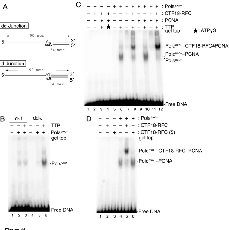
Incorporation of ddAMP at the 3′ end of the gap (Figure 11) was achieved by incubation of 3.6 µg of gapped DNA in a 70 µl Klenow reaction mixture with 4 units of Klenow fragment, 140
µM each of TTP, dCTP and ddATP (GE Healthcare) on ice for 2 h, followed by addition of 5 µl of 0.5 M EDTA. The DNA was conjugated with Dynabeads M-280 streptavidin as above.
Preparation of oligo-DNA beads
Biotinylated 90 mer-oligonucleotide BTN3 was obtained from Sigma Genosys. BTN3 was annealed with BTN30 or BTN28 at a 1:2 ratio as described previously (Waga and Stillman, 1998a), resulting in 3′ and 5′ recessed (3′/5′) or 3′ recessed (3′) primer-template DNAs. Similarly, BTN5 was annealed with BTN32, resulting in 5′ recessed primer-template DNA (5′) (see Table 1 for primer sequences). Oligo-DNA beads were prepared by binding 100 pmol of biotinylated ssDNA (ss) or primer-template DNAs with 50 µl of streptavidin agarose ultra-performance beads (Solulink) in BW buffer (Figure 6A).
PCNA loading
Gapped-plasmid-DNA beads containing 15 ng of DNA were suspended in 10 µl of reaction mixture [10 mM Hepes-NaOH (pH 7.8), 0.05% (v/v) Tween 20, 10 mM MgCl2, 0.2 mM EDTA, 0.01% (w/v) BSA and 0.5 mM DTT] containing 2 mM ATP, 6.2 pmol of PCNA and additional components as indicated. Assays in Figure 3B and C included additionally 40 mM creatine phosphate and 250 ng of creatine-phosphate kinase. The DNA beads were incubated at 32°C for 30 min after addition of the indicated components for loading, and free proteins were removed by four washes with 100 µl of 1xHBS [10 mM Hepes-NaOH (pH 7.8), 3.2 mM EDTA, 0.05% (v/v) Tween20 and 0.15 M NaCl]. The assay with oligo-DNA beads was carried out similarly, except for using of DNA beads with 500 fmol of oligonucleotides and 4.2 pmol of RPA for loading and five washes of 500 µl of 1xHBS after the reaction.
DNA polymerase assay with dA/dT
DNA synthesis reaction was performed in 5 µl of a mixture containing 30 mM Hepes-NaOH (pH7.8), 7 mM MgCl2, 0.01% BSA, 50 µM [α-32P] TTP (Perkin Elmer), 0.5 mM DTT and 200 pmol dA/dT and indicated amounts of DNA polymerases at 37˚C for 15 min.
Incorporated TMP into was determined by adsorption of the products to DE81 paper (Whatman) followed by Cherenkov counting with liquid scintillation counter (Beckman).
Labeling of primer DNA
10 pmol of ssDNA primer was labeled with [γ-32P]ATP (Perkin Elmer) using T4 polynucleotide kinase (TAKARA) in 10 µl reaction mixture at 37˚C for 1 h and purified by phenol-chloroform extraction and ethanol precipitation. The labeling efficiency of primer was measured by adsorption to DE81 paper as above.
Exonuclease assay
5 µl of loading buffer containing 80 fmol 32P-TEMP60, 30 mM NaCl, and indicated amounts of p261N was incubated at 37˚C for 10 min. The reaction was stopped by addition of 1 µl of 10 x sample buffer (TAKARA) containing SDS and the product was electrophoresed in 10%
polyacrylamide gel with TAE. The gel was fixed in 40% (v/v) methanol, 20% (v/v) acetic acid for 5 min followed by washing with water for 5 min and gel-drying on 3MM paper (Whatman) under vacuum, and the radiolabeled DNA was visualized by autoradiography with FLA-7000 (GE Healthcare).
Site-specific DNA photo-crosslinking
To prepare 3′ end-labeled oligonucleotide substrates for photo-crosslinking, 45 pmol of RF-30 primer was annealed with 30 pmol of TEMP90-R to create AP-Junction or with 30 pmol of TEMP90-Rneo to create AP-End, and incubated with 3.1 µM [α-32P]TTP (PerkinElmer Life Sciences), 20 µM α-S-dCTP (ChemCyte) and 4 units of Klenow fragment at 10°C for 30 min in a 20 µl Klenow reaction mixture, followed by a chase with 50 µM TTP at 10°C for 30 min. After phenol–chloroform (1:1) extraction, the product DNA was incubated with 2.1 nmol of azidophenacyl bromide (APB; Sigma) in a 50 µl volume for 3 h at room temperature in the dark.
After removal of unreacted reagent by ethanol precipitation with ethachinmate (NIPPON GENE), the product DNA was dissolved in TE at 50 nM (calculated from the estimated recovery from incorporated TMP), and stored at 4°C in the dark.
Photo-crosslinking oligonucleotide substrate labeled at a fixed position on the template strand was prepared by extension of RF64 on BTN3; 300 pmol of RF64 primer was annealed with 150 pmol of BTN3 and bound to 5 µl of streptavidin agarose ultra-performance beads. The DNA beads were incubated in a 25 µl of Klenow reaction mixture with 4 µM [α-32P]TTP, 24 µM α-S-dCTP and 2 units of Klenow fragment at 10°C for 30 min. After three washes with BW buffer and one wash with TE, the attached RF64 was completely elongated with 250 µM dNTP and 2 units of Klenow fragment at 10°C for 30 min in a 20 µl Klenow reaction mixture. Free nucleotides and proteins were removed by two washes with BW buffer; then the elongated RF64 strand was eluted twice with 50 µl of 0.1 M NaOH. The eluted sample was neutralized by addition of 10 µl of 1 M Tris-HCl (pH 8.0) and 10 µl of 10-fold diluted HCl and reacted with 2.1 nmol of APB in a 130 µl reaction mixture, as above. Aliquots of the product were annealed at 1:1.5 ratio with RF21–
RF56 oligonucleotides (Table 1) in TE containing 0.1 M NaCl, and stored at 4°C in the dark.
A 10 µl reaction mixture in a 1.5 ml test tube containing 25 fmol of labeled
oligonucleotide substrate, 60 mM NaCl and additional components as indicated was incubated at RT for 10 min and irradiated with 8 W ultraviolet (UV) C for 5 min at a 10 cm distance from the light source in a UVC 500 Ultraviolet crosslinker (GE Healthcare). The crosslinked DNA–protein complexes were further treated with 5 units of TurboNuclease (Accelagen) for 1 h at RT, and proteins conjugated with labeled nucleotides were separated by SDS-PAGE and autoradiographed as above.
Electrophoretic-mobility-shift assay (EMSA) after glutaraldehyde fixation
As the assay substrates, AP-Junction DNA obtained as described above was further extended at their 3′ end with ddAMP or dAMP, and a dd-Junction and a d-Junction were prepared, respectively (Figure 12A). The 25 fmol substrate was incubated in 5 µl of reaction mixture containing 60 mM NaCl and proteins at RT for 10 min and treated with 0.5 µl of 10% (v/v) glutaraldehyde for 5 min. The crosslinked sample was supplemented with 0.5 µl of electrophoretic-mobility-shift assay (EMSA) loading solution [100 mM Tris-HCl (pH 8.0), 20%
(w/v) sucrose, 1 mg/ml bromophenol blue] and separated on a 5% polyacrylamide gel in TAE buffer [20 mM Tris-acetate (pH 7.8), 1.25 mM EDTA] at RT. The shifted DNA was visualized by autoradiography as above.
Holoenzyme assay with Polδ and Polε
A singly-primed template DNA was prepared by annealing M13mp18 ssDNA (TAKARA) to 3-fold molar excess amount of TEMP90-R. The template DNA (30 ng) was then incubated in a 10 µl reaction mixture containing 25 mM Hepes-NaOH (pH 7.8), 10 mM Mg-acetate, 60 mM NaCl, 2 mM ATP, 0.01% (w/v) BSA, 0.5 mM DTT, 10 µM dNTP, [α-32P]TTP and indicated amounts of proteins at 32°C for 30 min. The product DNA was precipitated with ethanol, dissolved in 5 µl alkaline electrophoresis solution [0.3 M NaOH, 2 mM EDTA, 5% (w/v) Ficoll]
followed by electrophoresis in a 0.8% alkaline agarose gel at 40 V for 5 hours, and the products were visualized by autoradiography as above.
RESULTS
PCNA loading with CTF18-RFC and its stimulation by addition of Polε
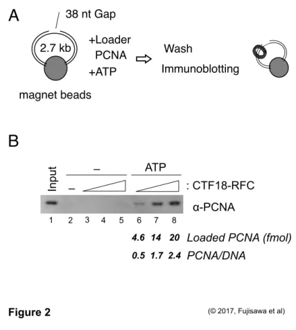
To examine quantitatively PCNA-loading activity by CTF18-RFC, magnetic beads were conjugated with 2.7 kb dsDNA to prepare the substrate DNA in pseudo-circular situation as described in Preparation of gapped DNA beads (Figure 2A, Sun et al., 2014). This DNA harbored a 38 nt-gapped region to generate 3′ primer-template junction where CTF18-RFC would target to load PCNA. Indeed, DNA beads-retained PCNA was observed according to the amounts (20-60 fmol) of CTF18-RFC only in the presence of ATP (Figure 2B, lanes 2-8). The ATP-dependent manner represented that the retained PCNA was the result of PCNA loading by CTF18-RFC. I quantified the PCNA band intensities and estimated that 2.4 PCNA trimer molecules were loaded to a substrate DNA molecule with 60 fmol of CTF18-RFC.
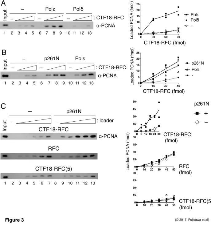
Next, in order to investigate the functional significance of the interaction between CTF18-RFC and Polε, I examined the effect of Polε for PCNA loading by CTF18-RFC along with another replicative DNA polymerase Polδ as a negative control, which does not interact with CTF18-RFC. Neither Polε nor Polδ alone exhibited any PCNA loading (Figure 3A, lanes 6, 10).
Addition of Polε to CTF18-RFC resulted in 3-5 fold augmentation of the PCNA loading (lanes 2–
9). The augmentation was not observed with Polδ (lanes 10-13), indicating that Polε specifically augments PCNA loading reaction by CTF18-RFC. The N-terminal half of Polε p261 (p261N), carrying the region necessary for interaction with CTF18-RFC (Murakami et al., 2010), showed similar stimulation (Figure 3B, lanes 7–9). PCNA loading was also examined with two other loader complexes, the canonical PCNA loader RFC and CTF18-RFC(5), the pentameric derivative of CTF18-RFC lacking DCC1 and CTF8. Neither RFC nor CTF18-RFC(5) interacts stably with Polε (Murakami et al., 2010). In the absence of p261N, PCNA loading activity was nearly the same with RFC, CTF18-RFC and slightly lower with CTF18-RFC(5) at 30 mM NaCl (Figure 3C, lanes 2–7). In the presence of p261N, only CTF18-RFC exhibited enhanced PCNA loading (Figure 3C, lanes 8–13), indicating that the specific interaction of CTF18-RFC with Polε is important to augment PCNA loading by CTF18-RFC.
Polε stimulates loading of PCNA by CTF18-RFC under near-physiological conditions CTF18-RFC has been reported to be less active for PCNA loading than RFC, reflecting higher salt sensitivity of ssDNA-stimulated ATPase and binding to 3′ primer-template junction DNA in CTF18-RFC than in RFC (Bermudez et al., 2003). Therefore, the effects of salt concentration on PCNA loading were examined. RFC and CTF18-RFC loaded similar amounts
of PCNA at 30 mM NaCl condition. PCNA loading by RFC increased as the NaCl concentration increased, whereas PCNA loading by CTF18-RFC decreased with increasing NaCl and was very low at near-physiological salt concentrations over 100 mM NaCl (Figure 4A). However, in the presence of p261N, significant PCNA loading was observed even at 100 mM NaCl (Figure 4B).
Active PCNA loading by CTF18-RFC at physiological salt concentrations requires interaction with Polε.
ssDNA binding protein RPA is involved in various DNA metabolic reactions including DNA replication and repair (Fanning et al., 2006). It is expected that presence of RPA in the PCNA loading reaction would make it more physiological. When I added 42.5 and 85 fmol of RPA in PCNA loading reaction with 16 fmol of the gapped DNA by CTF18-RFC alone at 60 mM NaCl, less PCNA was loaded than without RPA (Figure 4C, lanes 3-5). This result is consistent with a previous report with S. cerevisiae Ctf18-RFC that saturating amounts of RPA inhibit the PCNA loading (Bylund and Bergers, 2005). In the presence of Polε, however, PCNA loading by CTF18-RFC was maintained or slightly increased even with RPA (Figure 4C, lanes 6-8).
Collectively, these data indicate that at near-physiological conditions. i.e., in the presence of RPA and a higher salt, CTF18-RFC can load PCNA after it has formed a complex with Polε.
The CTF18-RFC–p261N complex loads PCNA efficiently through cooperative DNA binding
Polε has intrinsically high affinity for various DNA structures, while Polδ uses its high affinity to PCNA to access to a template DNA (Chilkova et al., 2007). This led me to hypothesize that CTF18-RFC might be recruited to its target site through its interaction with DNA-bound Polε.
Therefore, protein binding to gapped DNA beads was analyzed during PCNA loading (Figure 5).
p261N bound to the gapped DNA in the absence of other proteins, and addition of PCNA did not affect this binding (Figure 5A, lanes 2, 3). Similar DNA binding was observed with Polε, but not Polδ, even with PCNA (Figure 5B and C, lanes 2, 3). CTF18-RFC alone bound to DNA at a very low level, and a limited PCNA loading occurred (Figure 5A-C, lanes 4, 5). When p261N was present, >10% of the input CTF18-RFC was retained on the DNA with or without PCNA, and 3-fold more PCNA was loaded than in the absence of p261N (Figure 5A, lanes 6, 7). Similarly, increased CTF18-RFC retention and PCNA loading were observed with Polε, but not Polδ (Figure 5B, C lanes 6, 7). Notably, about 2-fold greater binding of p261N and Polε to DNA occurred in the presence of CTF18-RFC than in its absence (Figure 5A, B lanes 2, 3, 6, 7).
These results indicated that CTF18-RFC and p261N/Polε bound to DNA cooperatively. Thus,
CTF18-RFC could access target DNA through the cooperative binding, which would further lead to enhanced PCNA loading at near physiological salt concentrations.
PCNA loading by the CTF18-RFC–p261N complex occurs at 3′ primer-template junctions Because CTF18-RFC is able to support PCNA-dependent DNA synthesis by Polδ (Bermudez et al., 2003, Shiomi et al., 2004), 3′ primer-template junctions are considered as a target structure for PCNA loading by CTF18-RFC. However, PCNA loading by the CTF18-RFC–
p261N/Polε complex would be possibly a novel mechanism, I studied the target DNA structures for PCNA loading. Prior to the experiments, I prepared p261Nexo–, in which the highly conserved Asp275 of the exonuclease motif of B-family polymerases (Henninger and Pursell, 2014) was substituted with alanine to avoid degradation of substrate DNAs during experiments. Purified p261Nexo– did not exhibit any detectable nuclease activity (Figure 6A), and augmented PCNA loading by CTF18-RFC slightly more effectively than p261N (Figure 6B), indicating that the substitution did not affect the stimulation.
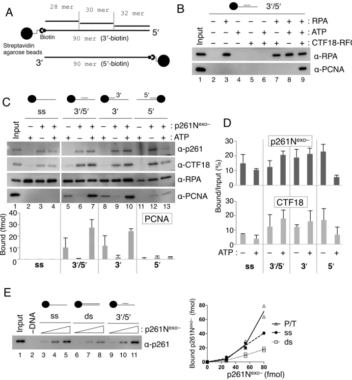
RFC targets 3′ primer-template junctions to load PCNA (Ellison and Stillman, 2003). The target DNA of PCNA loading by CTF18-RFC and the CTF18-RFC–p261N/Polε complex were analyzed with various DNA structures, which were attached to agarose beads (Figure 7A).
PCNA loading by CTF18-RFC was observed with 3′ and 5′ recessed primer-template DNA (3′/5′) only in the presence of RPA, which prevents PCNA from sliding off the DNA ends (Figure 7B).
Among ssDNA (ss), 3′ and 5′ recessed (3′/5′), 3′ recessed (3′), and 5′ recessed primer-template DNAs, CTF18-RFC alone in the presence of ATP loaded PCNA onto 3′/5′ and 3′ DNAs (Figure 7C, lanes 5, 8). Thus, CTF18-RFC, like RFC, specifically loads PCNA at 3′ primer-template junctions. In the presence of p261Nexo– and ATP, increased PCNA loading was observed specifically with 3′/5′ and 3′ DNAs (Figure 7C, lanes 7, 10), but not with ss (lane 4). Small amount of PCNA were detected with 5′ DNA (lanes 12, 13) in the presence of p261Nexo– and even in the absence of ATP. This could be due to p261Nexo–-mediated binding of PCNA to DNA not to direct loading of PCNA onto DNA. In the absence of ATP, both CTF18-RFC and p261Nexo– bound non-specifically to all the DNA structures that were tested (lanes 3, 6, 9, 12, Figure 7D). Notably, in the absence of other proteins, p261Nexo– showed dose-dependent, and high affinity binding to several DNA structures (Figure 7E), implying that p261Nexo– could be the dominant cause of this non-specific DNA binding. In the presence of PCNA and ATP, bindings of CTF18-RFC and p261Nexo– to DNA with 3′ primer-template junctions increased (Figure 7C, lanes 7, 10), whereas binding to other structures decreased (Figure 7C, lanes 4, 13 and Figure 7D), compared with the
absence of ATP. Thus, the cooperative action of CTF18-RFC and p261Nexo– could increase specificity for 3′ primer-template junctions during PCNA loading.
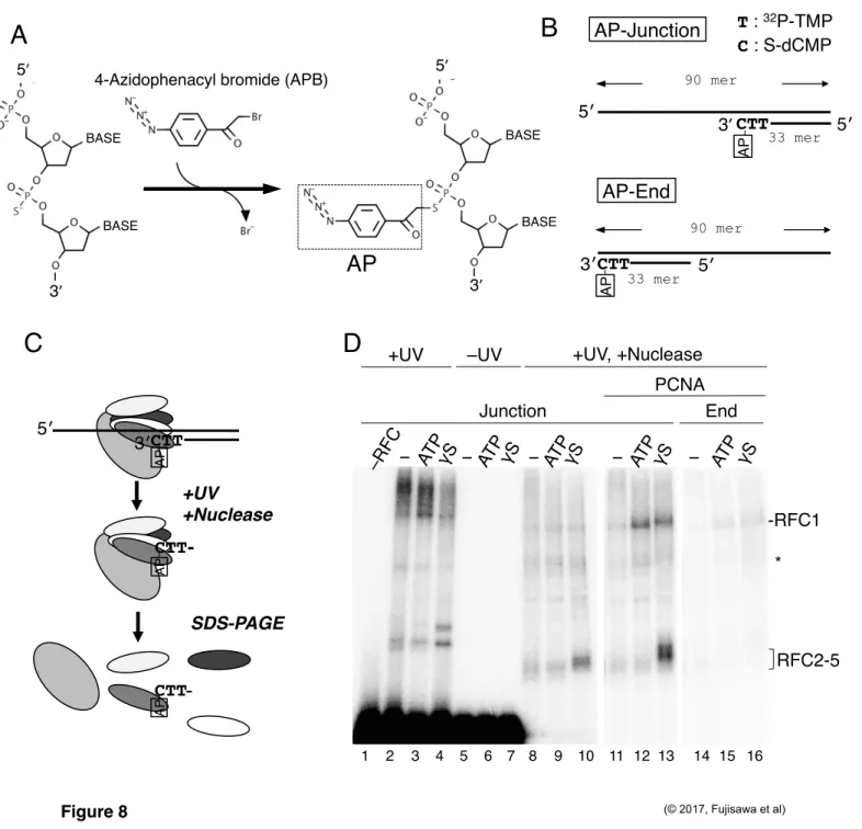
Analysis of binding modes of RFC to a 3′ primer end by photo-crosslinking
It has been demonstrated that DNA-bound Polε recruits CTF18-RFC to DNA, and they bind to cooperatively on the DNA and load PCNA at the site. DNA polymerases bind to 3′
primer-template junctions to elongate primer DNA. Thus, both CTF18-RFC and Polε can recognize 3′ primer-template junctions during the PCNA loading. How does they share the same binding DNA when they are in a complex? To identify which protein subunit directly interacts with the target DNA during PCNA loading, site-specific DNA photo-crosslinking analysis with APB was employed (Yang and Nash, 1994, Lagrange et al., 1996, Lee and Bell, 1997). This reagent couples with the thiol group of S-dNMP in a substrate DNA (Figure 8A). The substrate
“AP-Junction” had AP coupled to a 3′ primer-template junction along with the next 32P-labeled nucleotide (Figure 8B). After UV-irradiation and nuclease treatment, proteins most proximal to the AP-labeled nucleotides during a defined period of UV irradiation can be detected as
32P-labeled bands by electrophoresis (Figure 8C). Therefore, a band of higher intensity will have a higher probability of being attached to the target site than a band of lower intensity.
When I examined RFC and AP-Junction at 60 mM NaCl without nuclease treatment, a high-molecular-mass smear and one or two bands migrating slower than the raw RFC1 and RFC2–5 peptides were detected only after UV-irradiation (Figure 8D, lanes 2–7). After nuclease digestion, signal intensities were decreased in general and bands corresponding to RFC2–5 became prominent in the presence of ATPγS (Figure 8D, lanes 8–10). Thus, RFC2–5 are the major docking protein when ATP-bound RFC is associated with the 3′ primer-template junction.
Addition of PCNA in the presence of ATP enhanced the RFC1 signal (Figure 8D, lane 12), and the RFC2–5 signals increased in the presence of ATPγS (Figure 8D, lane 13). These results represent two binding modes of RFC to the 3′ primer-template junction. One is via RFC2–5 bound by ATP (an ATPγS plus condition). The other is via RFC1 in the presence of PCNA on the DNA. These signals were specific for the 3′ primer-template junction, because moving the target 3′ primer end to a double-stranded end decreased most of the signals (“AP-End”; Figures 8B and D, lanes 14–16). The molecular mass of labeled protein could not be determined precisely with this assay, so the specific subunits of RFC2–5 that bound to 3′ primer end could not be distinguished.
Analysis of binding modes of CTF18-RFC and p261Nexo– to 3′ primer ends
As representing weak PCNA loading activity of CTF18-RFC, its binding to a 3′ primer end at 60 mM NaCl was hardly detectable even with PCNA and ATPγS (Figure 9A, lanes 1–3).
At 10 mM NaCl in the presence of ATPγS and PCNA (lane 9), however, a similar binding profile was observed for CTF18 and RFC2–5 as was seen with RFC, though only limited bindings occurred with ATP and PCNA or without PCNA (lanes 4–8). This result indicated that binding of CTF18-RFC to a 3′ primer end only occurred in the intermediate state of PCNA loading, and that binding in the ATP-bound state without PCNA, or after PCNA loading and ATP hydrolysis was less stable than the binding seen with RFC, demonstrating the weak intrinsic DNA binding of CTF18-RFC.
When p261Nexo– was added to the experiments, it showed a much greater level of binding to a 3′ primer end than CTF18-RFC (Figure 9B, lanes 2, 3). Addition of p261Nexo– and CTF18-RFC together in the presence of PCNA and ATPγS, representing a condition of the PCNA-loading intermediate, increased bindings of both p261Nexo– and CTF18-RFC to DNA.
About 3-fold more p261Nexo– and 5-fold more CTF18 bound to a 3′ primer end than when they were included individually (Figure 9C), demonstrating their cooperative binding as in the DNA pull-down assay (Figure 5). Again, this stimulation of the DNA binding depended on their specific interaction, as CTF18-RFC(5) did not exhibit any significant increase (Figure 9B, lane 6).
Even when p261Nexo– and CTF18-RFC bound to DNA cooperatively in the presence of PCNA and ATPγS, the signal of CTF18 binding was <5% of the p261Nexo– signal, indicating that the 3′ primer end was mostly occupied by p261N (Figure 9B, lane 4, Figure 9D, lane 3). In the presence of ATP or the absence of nucleotides to minimize the level of the intermediate state of PCNA loading, the ratio of binding signal of CTF18 to p261N was decreased to a half (Figure 9D).
Thus, p261N attaches mostly with a 3′ primer end and CTF18-RFC is tethered by the p261N.
Then, temporal access of CTF18-RFC to a 3′ primer end will occur for loading of PCNA.
Analyses of the binding modes of CTF18-RFC and p261Nexo– on the template strand at primer-template junctions
The binding of CTF18-RFC and p261Nexo– on the template strand at primer-template junctions were investigated with a 90-mer ssDNA labeled with 32P-TMP and AP-crosslinker through S-dCMP at positions 25 nt and 26 nt from the 3′ end, respectively (AP-Template, Figure 10A). Six primers, from 21 nt to 41 nt, were annealed at the 3′ end side of the 90 mer to make six constructs in which the position of the AP-crosslinker relative to the 3′ primer end (single–
double-strand junction) varied from −5 (ssDNA region) to +20 (double-stranded-DNA region)
(Figure 10B).
Binding profiles of p261Nexo– and CTF18-RFC in the presence of PCNA and ATPγS were studied with the six AP-Template substrates. p261Nexo– was crosslinked to the AP obviously when it was in positions −5 to +15, whereas CTF18-RFC crosslinked to AP only in positions −5 and ±0 (Figure 10C, lanes 2–7 and 9–14). In assays containing both p261Nexo– and CTF18-RFC (Figure 10C, lanes 16–21), increased bindings of both proteins were observed, corresponding to 3′ primer-template junctions-specific binding of CTF18, and cooperative DNA binding of the CTF18-RFC–Polε complex. The binding of CTF18 at position −5 corresponded to ~25% of the signal for binding of p261Nexo–, indicating that the association of CTF18-RFC with the template strand at this point (relative to that of p261Nexo–) was greater than the association with the 3′
primer end (Figure 9B). The prominent binding of CTF18 at −5 and ±0 in the presence of ATPγS was not observed in the presence of ATP (Figure 10D), where the level of the intermediate state of PCNA loading should be low as described above. Thus, in the presence of ATPγS, the greater association of CTF18 with the region from the single–double-strand junction to the ssDNA template strand suggests a temporal association of CTF18-RFC to the region during PCNA loading.
PCNA loading by CTF18-RFC–Polε is blocked if Polε is in the DNA-synthesis mode All the preceding experiments were carried out without dNTPs, so demonstrated with Polε in a DNA non-synthesizing mode. To investigate the interactions with Polε in a DNA-synthesis mode, I prepared a primer-template junction substrate with dideoxynucleotide (ddNMP) at the 3′ primer end (deoxidized 3′ primer end, Figure 11A). In the presence of this substrate and the next-incoming dNTP, Polε would be trapped in the act of extending DNA with deoxynucleotides (a DNA-synthesis mode), as demonstrated by a structural study of yeast Polε (Hogg et al., 2014). Indeed, discrete bindings of Polα and Polδ to a similar substrate DNA was observed only in the presence of the incoming dNTP (Tsurimoto and Stillman, 1991). To test the DNA-synthesis mode of Polε at the deoxidized 3′ primer end, a 32P-labelled primer-template DNA substrates with incorporated ddAMP or dAMP at its 3′ end were prepared (“dd-Junction”
“d-Junction”, Figure 11A). When this substrate was incubated with Polεexo– in the presence of TTP, the next incoming nucleotide, stronger shifted bands were observed than without TTP (Figure 11B, lanes 5, 6) or with a substrate lacking the deoxidized 3′ primer end (lanes 2, 3). This result demonstrated a strategy to produce Polεexo– in synthesizing mode on a substrate DNA.
On the basis of Figure 11 that addition of next incoming nucleotide can control DNA
binding modes of Polε at the deoxidized 3′ primer end, gapped-DNA beads with ddAMP at the 3′
primer end of gap were prepared (Figure 12A), to examine the effect of dGTP, the incoming nucleotide, on PCNA loading by CTF18-RFC in the presence of Polεexo–. dGTP did not affect PCNA loading by CTF18-RFC alone in the presence of ATP (Figure 12B, lanes 3, 8), but it suppressed the stimulation of PCNA loading by the addition of Polεexo– (Figure 12B, lanes 4–6, 9–11). This result indicates that Polεexo– in the DNA synthesizing mode does not augment PCNA loading as well as Polεexo– in the non-DNA synthesizing mode.
Assembly of the CTF18-RFC and PCNA with Polε in DNA-synthesis mode
By the same EMSA assay, binding of RFC to a 3′ primer-template junction was observed in the presence of PCNA and ATPγS (Tsurimoto and Stillman, 1991). However, DNA binding of CTF18-RFC by the same method was not detected even with PCNA and ATPγS (Figure 11C, lanes 2–4). This difference between two loaders demonstrates again the relatively low affinity of CTF18-RFC for DNA. I examined the effects of PCNA and CTF18-RFC on the mobility of the Polεexo–-DNA band appeared in synthesizing mode. The band was slightly supershifted by the addition of PCNA (Figure 11C, lane 10), representing the formation of a Polεexo––PCNA complex on the DNA. In this case, PCNA was spontaneously entrapped from the free end of DNA in the absence of clamp loaders (Burgers and Yoder, 1993). Addition of CTF18-RFC to Polεexo–
supershifted the Polεexo– band (Figure 11C, lane 8), indicating that CTF18-RFC is tethered to Polε in synthesizing mode at the 3′ primer end. Addition of PCNA to CTF18-RFC and Polεexo–
produced a 2-fold to 3-fold increase in intensity of the supershifted band, compared with no PCNA (Figure 11C, lane 12), suggesting the presence of a stable assembly of Polεexo–– CTF18-RFC including PCNA on the dd-Junction substrate. By addition of CTF18-RFC(5) instead of CTF18-RFC, the supershifted band could not be observed, indicating that the complex formed via the specific interaction of CTF18-RFC and Polε (Figure 11D). Note that all shifted bands were observed in the presence of TTP, indicating that these complexes formed on Polεexo– in synthesizing mode.
DNA synthesis by CTF18-RFC–Polε–PCNA
The trimeric assembly of CTF18-RFC–Polε–PCNA forms at the 3′ primer-template junction when Polε is synthesis mode. To study DNA synthesis by this assembly, I performed a holoenzyme assay with CTF18-RFC, Polε, PCNA and RPA using a singly primed M13mp18 as template DNA. Efficient DNA synthesis was observed with them (Figure 13A, lane 5). Omission of one of the components resulted in the severe or total loss of DNA synthesis except for when
RPA was omitted (lanes 1–4); in this case, DNA products with a size of about 1.5 kb accumulated. Thus, efficient initiation of DNA synthesis occurred with the trimeric complex, and RPA was further required for DNA elongation, probably by its ability to resolve secondary structures on the template DNA. A decrease in the amount of CTF18-RFC resulted in reduced DNA synthesis, but the mean-product-lengths were not affected significantly (lanes 5–7), suggesting that CTF18-RFC might be required for the efficient initiation of DNA synthesis by Polε.
To determine whether the specific assembly of CTF18-RFC–Polε–PCNA mediates the efficient DNA synthesis, I compared DNA syntheses with Polδ and Polε in the presence of RPA in reactions where PCNA was loaded by either RFC or CTF18-RFC. Similar to previously published results (Bermudez et al., 2003, Shiomi et al., 2004), Polδ synthesized DNA efficiently with PCNA loaded by RFC, but less efficiently with PCNA loaded by CTF18-RFC (Figure 13B, lanes 10–15). This difference might reflect the difference in the efficiency of PCNA loading by these two loaders. PCNA loaded by RFC also stimulated the DNA synthesis by Polε via the previously reported interaction between Polε and PCNA (Chilkova et al., 2007, Bermudez et al., 2011, lanes 3–5). Interestingly DNA synthesis in the presence of CTF18-RFC was more efficient and produced longer DNA than in the presence of RFC (lanes 6–8). Collectively, these results suggest that CTF18-RFC is more adapted as a PCNA loader for Polε than RFC and vice versa for Polδ. The CTF18-RFC–Polε–PCNA complex synthesized DNA more processively than Polε in the presence of PCNA loaded by RFC, suggesting that it functions as a genuine functional DNA polymerase holoenzyme.