Title
脳動静脈奇形摘出術の基本戦略( 本文(Fulltext) )
Author(s)
岩間, 亨; 吉村, 紳一; 矢野, 大仁; 大江, 直行; 榎本, 由貴子; 山
田, 清文; 高木, 俊範
Citation
[脳卒中の外科 = Surgery for cerebral stroke] vol.[37] no.[5]
p.[390]-[394]
Issue Date
2009-09-30
Rights
The Japanese Society on Surgery for Cerebral Stroke (日本脳卒
中の外科学会)
Version
出版社版 (publisher version) postprint
URL
http://hdl.handle.net/20.500.12099/38513
は じ め に
脳動静脈奇形(arteriovenous malformation: 以下 AVM) に対する治療は,外科的摘出,定位的放射線治療,血管内 治療によって,あるいはこれらのモダリティを組み合わせ て複合的に行われている.AVM の部位や大きさ,症候や 出血の有無などによって個々に治療方針が検討されるが, 現時点での治療指針として Spetzler-Martin 分類3)の grade 1–3 に対しては外科的摘出術が推奨され,外科手術の危険 性が高く病巣が小さい場合には定位的放射線治療が勧めら れている2).しかし,実際に AVM 手術の経験を有する脳 神経外科医は必ずしも多くなく,摘出術を比較的安全に行 うことができる場合であっても定位的放射線治療が選択さ れることも多いと思われる.AVM 摘出術の難易度は高い が最も確実な治療手段であることから,多くの脳神経外科 医が AVM 手術の要点を理解し,手術が適応となる症例に 対しては適切に摘出術が行われるべきであると考えられ る.本稿では AVM 手術の基本戦略と基本手技についてわ れわれの考えを述べる. AVM 摘出術の基本戦略 AVM 摘出術の基本戦略をきわめて単純に表現するなら 脳卒中の外科 37: 390 ∼ 394,2009
脳動静脈奇形摘出術の基本戦略
岩間 亨,吉村 紳一,矢野 大仁,大江 直行
榎本由貴子,山田 清文,高木 俊範
Basic Surgical Strategy for Cerebral Arteriovenous
Malformations
Toru IWAMA, M.D., Shin-ichi, YOSHIMURA, M.D., Hirohito YANO, M.D., Naoyuki OHE, M.D., Yukiko ENOMOTO, M.D., Kiyofumi YAMADA, M.D., and Toshinori TAKAGI, M.D.
Department of Neurosurgery, Gifu University Graduate School of Medicine, Gifu, Japan
Summary: We describe basic surgical strategy and techniques for resection of cerebral arteriove-nous malformations (AVMs). Understanding of the vascular structure of AVMs is important to plan surgical strategies for AVMs. The craniotomy should be large enough as to expose all the structures of the AVMs, such as nidus, feeders and drainers. In the first step of surgery, feeders are secured to control bleeding and to reduce tension of the nidus and drainers. Preoperative occlusion of the hidden feeders by endovascular surgery is a useful option. For an approach to the buried nidus, ret-rograde dissection of the main drainer is effective. When bleeding is difficult to control during dis-section of the nidus, the disdis-section plane has got into the nidus and should be reset outward. Feeding arteries are coagulated and cut after they are confirmed to contribute to the AVM. The main drain-er should be presdrain-erved up to the final step of the resection. Intraopdrain-erative angiography is useful to identify the feeders and to confirm the residuals of the AVM.
Key words: ・ arteriovenous malforma-tion ・ resection ・ surgical strategy ・ basic technique
Surg Cereb Stroke (Jpn) 37: 390–394, 2009
岐阜大学大学院医学系研究科 脳神経外科学分野(受稿日 2008. 12. 22)(脱稿日 2009. 3. 25)〔連絡先:〒501–1194 岐阜市柳戸 1–1 岐阜大 学大学院医学系研究科 脳神経外科学分野 岩間 亨〕[Address correspondence: Toru IWAMA, M.D., Department of Neurosurgery, Gifu University Graduate School of Medicine, 1–1 Yanagito, Gifu 501–1194, Japan]
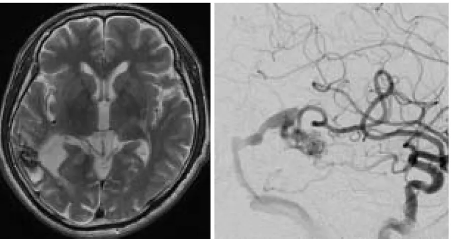
ば,「主な feeder を確保し,AVM への血流をコントロー ルしつつ drainer と nidus を周囲脳から 離し,順次 feed-er と従たる drainfeed-er を切断し,最後に main drainfeed-er を切断 して AVM を摘出する」こととなる. 実例を呈示し,具体的な手順とともに,各ステップでの 留意点,対策を述べる. 症 例 57 歳の男性でてんかん発作にて発症した.MRI にて右 側頭葉に陳旧性の血腫を伴う flow-void が認められた(Fig.
1A).脳血管撮影(DSA)にて中大脳動脈(MCA)と後大脳 動 脈(PCA)の 側 頭 枝 を feeder と し 横 静 脈 洞 へ 流 出 す る drainer を有する AVM が確認された(Fig. 1B).
AVM 摘出術 1.手術準備 術中の feeder の同定や遮断の確認,残存 AVM の確認 に術中 DSA はきわめて有用であり1),原則として必須と 考えるべきである.術中 DSA 対応のカーボン製 Mayfield を用いて頭部を固定しておく. 2.開頭と硬膜切開 AVM の 摘 出 に あ た っ て は 構 成 要 素 で あ る feeder, nidus,drainer の全体像を把握することが重要である.ま た,AVM 摘出術では術中になんらかの問題が生じた場合 には著しい脳腫脹をきたす危険性がある.そのため,すべ ての症例で可能ではないが,できる限り術野に feeder, nidus,drainer のすべてが観察でき,かつ十分に大きめの 開頭を行っておくことが望ましい.Fig. 2 に本例の皮切, 開頭範囲を示す. 流出路となる静脈洞の圧は上昇しており,開頭時の損傷 に注意する.もし,静脈洞上の硬膜面から出血する場合に は,fibrin glue 付きの Surgicel により十分時間をかけて圧 迫止血する.脳表の AVM ではしばしば硬膜との癒着がみ られる.癒着が強い場合には無理に 離せず癒着の周囲 で硬膜を切断し AVM 側に癒着硬膜を残す. 3.Feeder の確保と drainer の 離 出血のコントロールと nidus,drainer の 離操作を容 易にするために,最初に feeder の確保を行う.術野内で 確保が困難な feeder は,血管内治療により予め塞栓して おくとよい.
Surgery for Cerebral Stroke 37: 2009 391
Fig. 1 A: Preoperative T2 weighted MR image demonstrates a flow-void with a high-intensity area at the right temporal cortex.
B: Lateral view of the preoperative right carotid angiogram shows a small arte-riovenous malformation (AVM). The AVM is fed by temporal branches of the middle and posterior cerebral arteries, and drained to the transverse sinus via 2 drainers.
A B
Fig. 2 Intraoperative view.
U-shape skin incision and craniotomy is drawn on the scalp.
Fig. 3 に本例の脳表所見と,術野に方向を一致させた
DSA を示す.本例は nidus も小さく構造も比較的単純で, 前上方と前下方からそれぞれ MCA,PCA からの feeder が流入し,後方と下方に drainer が存在していた.nidus 前上方の sulcus を開放して MCA からの main feeder を確 保したのち,脳表の drainer 周囲のくも膜を全周性に浅 く 離した.main feeder と PCA からの feeder と思われ た細血管をクリップにて遮断したのち(Fig. 4A),nidus
の 離を開始した.feeder の遮断により,わずかながら drainer の色が暗く変化している. 本例のように AVM の位置や範囲がわかりやすい例では 問題ないが,症例によっては nidus が脳溝内に存在して脳 表から観察されなかったり,逆に脳表に多数の異常血管が 観察され,nidus 本体の範囲がわかりにくい場合には, main drainer を逆行性に 離していくと確実に nidus 本体 に到達することができる4).通常,drainer の壁は肥厚し
Fig. 3 A: Preoperative angiogram matched with intraoperative view.
B: Intraoperative view after dural opening. The main drainer is confirmed through
whitened arachnoid membrane. A B
Fig. 4 Intraoperative views.
A: The main feeder and 2 small feeders are clipped to reduce the tension of the AVM. Color of the main drainer is partly turned to dark.
B: The nidus is almost totally dissected. Color of the main drainer is turned to dark more.
C: The main feeder is coagulated at the final stage of the dissecion. D: The coagulated main feeder is cut.
A B C D
ており 離操作による損傷の危険性は少ないが,tension が低いほうがより安全であり,可能な範囲で feeder を遮 断したのちに 離したほうがよい.もしも損傷した場合 には,凝固せずに Surgicel と fibrin glue による圧迫止血 を行う. AVM 周囲の白濁して肥厚したくも膜や drainer の 離 には血管吻合用の鑷子,剪刀が有用である. 4.Nidus の 離 確実な 離操作と出血のコントロールのため,nidus の 離は全周性に浅いところから順次深部に進めるよう に心がけ,一部分だけ深く 離を進めることは避ける4).
Surgery for Cerebral Stroke 37: 2009 393
Fig. 5 Intraoperative views.
A: The nidus is lifted up from the cortex.
B: The nidus is completely dissected with connection to the brain by the main drainer.
C: Intraoperative control DSA demonstrating the AVM.
D: Intraoperative DSA obtained after resection of the AVM. Disappearance of the AVM is confirmed.
Fig. 6 A: Postoperative CT shows neither bleedings nor infarctions.
B: Lateral view of the postoperative right carotid angiogram demonstrates extirpa-tion of the AVM.
A B C D
feeder の中には passing artery として AVM とともに正 常脳へも灌流している場合があり,最終的に nidus に入る ことが確実となった時点で凝固切断し,それまではクリッ プによる遮断を行っておく1).drainer もすべてを温存し ながら nidus を 離することはできない.main drainer は 必ず最後まで温存し,それ以外の drainer は細いものから 順次凝固・切断しつつ 離を進める. nidus の辺縁は多数の異常血管のループにより形成され ていることをイメージし,これらの血管を feeder や drain-er と誤って凝固・切断しないように留意する1). 離中 に nidus に切り込んでしまった場合は,出血のコントロー ルが困難となる.コントロールが困難な出血を生じたら 「nidus に切り込んだ」との認識を持ち,出血点でむやみ に操作を続けることなく,やや外側の正しい境界で 離, 止血を行う.nidus 辺縁のいわゆる“赤虫”と呼ばれる fragile な血管はバイポーラーによる凝固のみでは弾けて 出血するため,ミニクリップをかけて血管の圧を下げたの ち,バイポーラーの出力を下げて凝固する.
本例では main feeder と main drainer を残してほぼ全周 性に 離を行ったのち(Fig. 4B),main feeder が nidus に 入る直前で凝固・切断した(Fig. 4C, D).
5.Main drainer の切断と AVM の摘出
すべての feeder が切断されると,nidus の tension が下
が り red vein を 呈 し て い た drainer の 色 が 暗 く な る . drainer を十分に凝固して切断し,AVM を摘出する.
本例でも最終的に AVM が main drainer のみで脳と繋 がった状態とし(Fig. 5A, B),main drainer を切断して摘 出した.術後出血の原因となりうる残存 AVM のないこと を術中 DSA にて確認し手術を終了した(Fig. 5C, D).Fig.
6 に術後の CT,DSA を示す. ま と め 単純な血管構築をもつ AVM を実例にあげ,AVM 摘出 術の基本的な戦略と手技,各ステップにおける留意点を述 べた. 文 献
1) Hashimoto N, Nozaki K, Takagi Y, et al: Surgery of cere-bral arteriovenous malformations. Neurosurgery 61(SHC Suppl 1): SHC-375–SHC-389, 2007
2) 篠原幸人,吉本高志,福内靖男,ほか:脳動静脈奇形.脳 卒 中 治 療 ガ イ ド ラ イ ン 2004,協 和 企 画 ,東 京 ,2004, pp119–122
3) Spetzler RF, Martin NA: A proposed grading system for arteriovenous malformations. J Neurosurg 65: 476–483, 1986
4) Yamada S, Brauer FS, Knierim DS: Direct approach to arteriovenous malformations in functional areas of the cerebral hemisphere. J Neurosurg 72: 418–425, 1990