Constitutive and inducible defensive metabolites in Hordeum species and wheat
(オオムギ属植物とコムギにおける
構成的および誘導性防御物質に関する研究)
Naoki Ube 宇部尚樹
2020
CONTENTS
Contents ………..………..1
Chapter 1 General introduction ··· 2
Chapter 2 Evolutionary changes in defensive specialized metabolism in the genus Hordeum 2.1. Introduction ··· 7
2.2. Results ··· 8
2.3. Discussion ··· 19
2.4. Experimental ··· 24
Chapter 3 Identification of phenylamide phytoalexins and characterization of inducible phenylamide metabolism in wheat 3.1. Introduction ··· 31
3.2. Results ··· 33
3.3. Discussion ··· 46
3.4. Experimental ··· 49
Chapter 4Biosynthesis of phenylamide phytoalexins in pathogen-infected barley 4.1. Introduction ··· 54
4.2. Results ··· 56
4.3. Discussion ··· 73
4.4. Experimental ··· 77
Chapter 5 Conclusion ··· 86
References ··· 88
Acknowledgments ··· 100
Summary ··· 101
摘要 ··· 104
List of publications ··· 107
Appendix··· 108
Chapter 1
General introduction
Plants possess chemical defense systems to protect themselves against biotic stress.
Antifungal compounds in plants, such as phytoanticipins and phytoalexins, contribute to the rejection of pathogen infections. Phytoanticipins are defined as low molecular weight, antimicrobial compounds that are present in plants before challenge by microorganisms or are produced after infection solely from preexisting constituents (VanEtten et al., 1994). On the other hand, phytoalexins are defined as low molecular weight, antimicrobial compounds produced by plants in response to pathogen infection, and provide a chemical barrier against pathogen attacks.
The significance of phytoanticipin and phytoalexin accumulation in the interaction between plants and pathogenic microorganisms has been demonstrated experimentally. For example, a wild oat species (Avena longigluminis) lacking the oat phytoanticipins, avenacins, demonstrated susceptibility to Gaeumannomyces graminis var. tritici, the causal agent of take-all disease of wheat, while oat (Avena sativa) that accumulates avenacins showed resistance to the disease (Osbourn et al., 1994).
Incompatible combinations of oat cultivars and crown rust fungus (Puccinia coronata f.
sp. avenae) races were found to induce a rapid accumulation of phytoalexins at high concentrations (Mayama et al., 1982). In Arabidopsis thaliana, plants without the ability to produce phytoanticipins and phytoalexins were shown to be more susceptible to pathogenic infection compared with phytoanticipin- and phytoalexin-producing wild- type plants (Schlaeppi et al., 2010; Thomma et al., 1999). The genetically modified tobacco (Nicotiana tabacum) plant that accumulates a foreign phytoalexin, resveratrol, showed enhanced resistance to pathogens (Hain et al., 1993). Moreover, tolerance to phytoanticipins and phytoalexins, due to their degradation and exclusion, has been identified as a factor contributing to virulence of pathogenic microorganisms (Sandrock
In species belonging to the family Poaceae, benzoxazinones (Bxs) are representative phytoanticipins. Bxs are present in wheat (Triticum aestivum), rye (Secale cereale), maize (Zea mays), and other Poaceae species (Niemeyer, 1988; 2009;
Sicker et al., 2000; Kokubo et al., 2017). The major Bxs, 2,4-dihydroxy-1,4-
benzoxazin-3-one (DIBOA) and its 7-methoxy derivative (DIMBOA), are constitutively stored in vacuoles in the form of glucosides (DIBOA-Glc and DIMBOA-Glc). Due to their anti-aphid, antimicrobial, and phytotoxic activities, Bxs have been considered to function as defense mechanisms against various biological stresses (Argandoña et al., 1980; Barnes and Putman, 1987; Niemeyer, 1988; 2009; Sicker et al., 2000; Meihls et al., 2013). Gramine (N,N-dimethyl-3-aminomethylindole) is another class of defensive metabolites found in barley (Hordeum vulgare ssp. vulgare) and several poaceous species (Kokubo et al., 2017). Similar to Bxs, gramine has been implicated in resistance against aphids and pathogenic fungi (Zúñiga et al., 1986; Matsuo et al., 2001). Both Bxs and gramine are derived from the tryptophan biosynthetic pathway (Frey et al., 1997;
Gross et al., 1974).
In contrast to the general accumulation of Bxs among Poaceae species, hordatines are specialized metabolites characteristic to barley. These compounds possess strong antifungal activity and accumulate at high concentrations in young seedlings (Stoessl, 1967; Stoessl and Unwin, 1970). Hordatine A (HA) is a dimer of p- coumaroylagmatine (CouAgm), whereas hordatine B (HB) is a heterodimer of CouAgm and feruloylagmatine (FerAgm) (Stoessl, 1966). Hordatines are also present in the form of glycosides (Kageyama et al., 2011; Kohyama and Ono, 2013), and other minor hordatines have been detected in seeds and seedlings of barley by mass spectroscopic analysis (Gorzolka et al., 2014; Pihlava, 2014). Hordatines are biosynthesized via two reactions from agmatine and hydroxycinnamoyl-CoA esters (Fig. 1.1). The first reaction comprises the condensation of agmatine and hydroxycinnamoyl-CoA esters by agmatine coumaroyltransferase to form hydroxycinnamic acid amides (HCAAs) of agmatine (CouAgm and FerAgm) (Bird and Smith, 1981, 1983; Burhenne et al., 2003). The
second reaction involves the dimerization of the HCAAs. The enzyme that catalyzes the second reaction has not been identified to date (Nomura et al., 2007), although a radical coupling reaction catalyzed by peroxidase has been shown to generate hordatines in vitro (Stoessl 1966, 1967; Negrel and Smith, 1984).
Poaceous species accumulate phytoalexins with diverse chemical structures, including phenylamides, flavonoids, and terpenoids (Fig. 1.2). For example, rice (Oryza sativa) plants accumulate 19 phytoalexins — 15 labdane-related diterpenes
(momilactones, oryzalexins, and phytocassanes), 1 casbene-type diterpene (ent-10- oxodepressin), 1 flavanone (sakuranetin), and 2 phenylamides (N-benzoyltryptamine and N-cinnamoyltryptamine) (Kodama et al., 1992a,b; Park et al., 2013; Yamane et al., 2013; Horie et al., 2016). In maize (Zea mays), the diterpenoids, kauralexins and dolabralexins, (Schmelz et al. 2011; Mafu et al., 2018) and the sesquiterpenoid, zealexin, were identified as phytoalexins (Huffaker et al., 2011). In oats,
avenanthramide phytoalexins, which are hydroxycinnamic acid amides with
anthranilates, have been extensively studied in the interaction with crown rust fungus (Mayama et al., 1982; Miyagawa et al., 1995). It has been shown that the dimers of serotonin, 5′-dihydroxy-2,4′-bitryptamine, and 3-(2-aminoethyl)-3-hydroxyindolin-2- one are induced in barley leaves infected by Bipolaris sorokiniana, the causal agent of brown spot in the Poaceae plants. They were considered to serve the function of
phytoalexins in barley (Ishihara et al., 2017). It may be reasonable to assume that barley possesses additional phytoalexins because other Poaceae species accumulate various kinds of specialized metabolites as phytoalexins. To the best of our knowledge, the presence of phytoalexins has not been reported in wheat to date, but it seems somewhat strange considering the dense distribution of phytoalexins in the poaceous species.
In many poaceous species, Bxs are representative phytoanticipins, but barley possesses characteristic specialized metabolites, hordatines. These differences in defensive metabolites between barley and the other Poaceae species suggest that
addition, inducible defensive metabolites in barley and wheat remain to be elucidated while various kinds of phytoalexins have been identified in other poaceous species. In this study, we focus on the constitutive and inducible defensive system using specialized metabolites in Hordeum species and wheat.
Fig. 1.1. Biosynthetic pathways for defensive specialized metabolites found in poaceous species.
Gramine
Hordatine A (HA, R: H) Hordatine B (HB, R: OCH
3)
p-Coumaroylagmatine (CouAgm, R : H ) Feruloylagmatine (FerAgm, R: OCH3)
Chorismic acid L-Tryptophan
L-Phenylalanine
p-Coumaroyl-CoA Agmatine
3-Amino- methylindole Indole-3-
glycerol phosphate
DIBOA-Glc
Fig. 1.2. Inducible defensive metabolites found in poaceous species.
HN
HN O
BenTry (Rice) Sakuranetin (Rice)
Kauralexin A1 (Maize)
Terpenoid
Hydroxycinnamic
acid amide Flavonoid
OH O
Momilactone A (Rice) 3-(2-aminoethyl)-
3-hydroxyin- idolin-2-one
NH
HO NH2 O
NH NH2 HO
H2N NH
HO NH
NH2
HO
Serotonin
Dimer of serotonin
Indole amine
O
O H3CO
OH
OH
O H
O O
H
Chapter 2
Evolutionary changes in defensive specialized metabolism in the genus Hordeum
2.1 Introduction
Over the course of evolution, plants have acquired chemical defense systems using specialized metabolites to protect themselves against biotic stresses. In general, closely related species accumulate similar specialized metabolites. However, in the genus Hordeum (Poaceae), which includes cultivated barley (Hordeum vulgare L. ssp.
vulgare), the occurrence of multiple defensive metabolites such as hordatines, benzoxazinones (Bxs), and gramine has been reported (Stoessl, 1966; Barria et al., 1992; Grün et al., 2005; Smith and Best, 1978).
Hordatines are characteristic specialized metabolites present in barley and the accumulation was limited. On the other hand, a few wild Hordeum species have been shown to accumulate Bxs (Barria et al., 1992; Grün et al., 2005) as well as other Poaceae species. Bxs are present in wheat (Triticum aestivum), rye (Secale cereale), maize (Zea mays), and other Poaceae species (Niemeyer, 1988; 2009; Sicker et al., 2000; Kokubo et al., 2017). Gramine (N,N-dimethyl-3-aminomethylindole) is another class of defensive metabolites found in barley. Both Bxs and gramine are derived from the tryptophan biosynthetic pathway (Frey et al., 1997; Gross et al., 1974).
The genus Hordeum, which belongs to the Triticeae tribe in the Poaceae family, consists of 31 species. The genus has been classified into four clades H, Xu, Xa, and I, among which H and Xu comprise a monophyletic group, and I and Xa form a separate monophyletic group (Taketa et al., 1999, 2005; Blattner, 2009). Cultivated barley belongs to the H clade, together with the closely related wild species H. vulgare ssp.
spontaneum (C. Koch.) Thell. Phylogenetic relationships among the 31 species of the genus Hordeum have been analyzed in detail; thus, it is the ideal genus to track
metabolic evolution. The presence or absence of defensive specialized metabolites in Hordeum has been analyzed in several species (Smith and Best, 1978; Barria et al., 1992; Grün et al., 2005). However, the correlation between the phylogenetic
relationships among species and the distribution of defensive metabolites remains to be elucidated because of the lack of information from some of the phylogenetically
important species.
In the present study, we analyzed the presence or absence of hordatines, Bxs, and gramine in representative Hordeum species, including all of the four clades.
Furthermore, we focused on the metabolites in H. murinum ssp. glaucum (Steudel.) Tzvelev., as this species belongs to the Xu clade, which is the major branch of the lineages leading to cultivated barley in the phylogenetic tree of the genus Hordeum. We detected two compounds that differed from these known metabolites in H. murinum ssp.
glaucum. These compounds were also detected in H. murinum spp. leporinum (Link.) Arcang. and H. bulbosum L. We purified these two compounds, determined their chemical structures by spectroscopic analysis, and characterized their biological activity.
2.2 Results
2.2.1. Distribution of defensive specialized metabolites in Hordeum species We analyzed the contents of hordatines, Bxs, gramine, and HCAAs (CouAgm and FerAgm) in Hordeum species at the seedling stage by HPLC and LC-MS/MS. The species analyzed were H. vulgare ssp. vulgare, H. vulgare ssp. spontaneum, and H.
bulbosum in the H clade; H. murinum ssp. glaucum and H. murinum ssp. leporinum in the Xu clade; H. marinum ssp. marinum Hudson. in the Xa clade; and H. bogdanii Wil., H. brachyantherum Nevski. ssp. californicum (Cov. & Steb.) Both. & et al., H. chilense Roem. & Schult., H. flexuosum Steud., H. pusillum Nutt., and H. jubatum L. in the I
DIBOA-Glc and DIMBOA-Glc.
Hordeum species accumulated different varieties of defensive metabolites, depending on the clade (Fig. 2.1 and Table 2.S1). H. vulgare ssp. vulgare and H. vulgare ssp. spontaneum contained CouAgm, HA, and HB at high concentrations. H. bulbosum did not accumulate hordatines, but it accumulated low levels of CouAgm and FerAgm.
Gramine content varied in the H clade: H. vulgare ssp. vulgare cv. Shunrai and H.
vulgare ssp. spontaneum accumulated gramine at high concentrations in the shoots, whereas gramine concentrations in the shoots and roots of H. vulgare ssp. vulgare cv.
Sachiho Golden were below the detection limit, and those in H. bulbosum were also low. Bxs were not detected in the H clade species studied. In addition, the HPLC analysis for hordatines in the extracts from H. bulbosum showed two peaks (1 and 2), which were absent in the extracts from H. vulgare.
Xu clade species did not accumulate hordatines or Bxs, but they contained CouAgm and FerAgm. The high concentration of FerAgm was characteristic of Xu clade species. H. murinum ssp. glaucum did not contain gramine, but H. murinum ssp.
leporinum accumulated gramine at a level similar to that in H. vulgare cv. Shunrai.
Importantly, compounds 1 and 2 were detected in Xu clade species, as well as in H.
bulbosum (an H clade species).
All the Xa and I clade species accumulated DIBOA-Glc. H. bogdanii, H.
jubatum, H. brachyantherum, H. chilense, and H. pusillum also contained gramine in the roots, although its concentrations were far lower than those in H. vulgare ssp.
vulgare cv. Shunrai, ranging from 51.8 nmol/gFW (H. brachyantherum) to 10.2
nmol/gFW (H. jubatum). Among these species, only H. jubatum accumulated detectable amounts of gramine (19.9 nmol/gFW) in the shoots. In the I and Xa clades, species other than H. pusillum accumulated CouAgm to some extent, but hordatines were below the detection limits. DIMBOA-Glc was not detected in any of the Hordeum species analyzed.
Fig. 2.1. Accumulation of p-coumaroylagmatine, hordatine A, hordatine B, gramine, and DIBOA-Glc in Hordeum species. Shoots (A) and roots (B) of young seedlings of Hordeum species were extracted with 80% methanol. The bars in the graphs show the amounts of p-coumaroylagmatine (light blue), feruloylagmatine (light green), hordatine A (dark blue), hordatine B (dark green), gramine (yellow), and DIBOA-Glc (red). Data are means with standard deviation from three independent experiments.
2.2.2 Identification of hydroxycinnamic acid amide dimers in H. bulbosum, H. murinum ssp. glaucum, and H. murinum ssp. leporinum
Compounds 1 and 2 were purified from 96-h-old seedlings of H. murinum ssp. glaucum by ODS column chromatography and preparative HPLC. Compound 1 had the
molecular formula C30H43N8O6, as determined by HRMS (m/z 611.3301, [M+H]+), which suggested that 1 was a dehydrodimer of FerAgm. 1H-NMR spectra of 1 (Table
proton (d 7.27 ppm), two coupled methines (d 3.75 and 4.32 ppm), and two methoxy groups (d 3.79 and 3.88 ppm) (Fig. 2.2). Although HRMS indicated the presence of 30 carbons in the structure, the 13C-NMR spectra showed only 29 carbon signals. The carbon signal that was not observed in the 13C-NMR spectra overlapped with the solvent signal at dC 49.0 ppm because the HMQC spectra showed a cross peak of the coupled methine proton at d 4.32 ppm and a carbon signal overlapping with the signal of the solvent. All protonated carbons were assigned using the HMQC spectra.
Because the aromatic protons at d 6.43 and 6.93 ppm did not show coupling, ring A was a 1,3,4,6-tetrasubstituted benzene ring. The coupling pattern of proton signals at d 6.51, 6.69, and 6.76 ppm indicated that ring B was a 1,3,4-trisubstituted benzene ring. COSY and HMQC spectra indicated the presence of two -CH2-CH2-CH2-CH2- units and two carbon signals at dc 158.57 and 158.64 ppm, suggesting the presence of guanidino carbons. Thus, 1 was deduced to contain two agmatine chains derived from FerAgm.
Assuming that 1 was a dehydrodimer of FerAgm, the major differences in the comparison of the spectra of 1 with those of FerAgm were (1) the disappearance of two double-bond protons and appearance of two coupled methines at d 3.75 and 4.32 ppm, (2) the disappearance of another double-bond proton, and (3) the disappearance of an aromatic proton at C-6 in ring A. Thus, the carbons involved in the linkages were two coupled methine carbons, one of double-bond carbons, and C-6 in ring A.
According to HMBC spectra, ring A and the double-bond proton consisted of one FerAgm unit, while ring B and two coupled methines consisted of the other
FerAgm unit (Fig. 2.2). The signals of C-1, 2, and 6 in ring A showed cross-peaks with the double-bond proton (H-7) at d 7.27 ppm, indicating a connection between C-1 in ring A and C-7. The signals of C-1′, 2′, and 6′ in ring B showed cross-peaks with a coupled methine proton (H-7′) at d 4.32 ppm, indicating a connection between ring B and the coupled methine.
The HMBC spectrum also displayed correlations between one of the coupled methine protons at d 3.75 ppm (H-8′) and the double-bond carbons (C-7 and C-8),
indicating a linkage of C-8 and C-8′ (Fig. 2.2). In addition, the signal of C-1, C-5, and C-6 showed cross-peaks with the coupled methine proton H-7′ at d 4.32 ppm, indicating a linkage between C-6 and C-7′. Therefore, we concluded that 1 was a dehydrodimer of FerAgm with two FerAgm units connected at 8-8′ and 6-7′.
The relative stereochemistry between the coupled methines at d 3.75 and 4.32 ppm were assigned by a combination of NOESY and analysis of coupling constants.
The NOESY spectrum displayed a correlation between the coupled methines, and the coupling constant between the coupled methines was 7.1 Hz. Thus, 1 had a cis configuration of the coupled methines.
Compound 2 had the same molecular formula as 1, C30H43N8O6, as revealed by HRMS (m/z 611.3301, [M+H]+), suggesting that 2 was also a dehydrodimer of FerAgm.
The 13C-NMR spectra showed 30 carbon signals. 1H-NMR spectra of 2 (Table 2.1) revealed the presence of two aromatic rings (C and D), a double-bond proton (d 7.48 ppm), two coupled methines (d 3.99 and 4.61 ppm), and two methoxy groups (Fig. 2.2).
Rings C and D were 1,3,4-trisubstituted rings with resonance for ortho- and meta-coupled protons at d 6.80, 6.99, and 7.01 ppm for ring C, and at d 6.73, 6.79, and 6.82 ppm for ring D. The presence of two agmatine units was confirmed by COSY, HMQC, and 13C-NMR.
The comparison of the spectra of 2 with those of FerAgm showed the
following differences: (1) the disappearance of two double-bond protons of FerAgm and the appearance of two coupled methines at d 3.99 and 4.61 ppm and (2) the absence of another double-bond proton of FerAgm. Thus, the carbons involved in the linkages were two coupled methine carbons and one double-bond carbon. In addition, because all the other carbons seemed to be intact, a nitrogen or oxygen atom was considered to be involved in the connection.
In the HMBC spectrum, the signals of C-1, 2, and 6 in ring C showed cross- peaks with the double-bond proton (H-7), indicating the connection between ring C and
cross-peaks with H-7′, indicating a connection between ring D and C-7′. Thus, ring C and the double-bond comprised one FerAgm unit, whereas ring D and the coupled methines comprised the other FerAgm unit.
HMBC spectrum displayed correlations between H-8′ and C-8, indicating the connection between C-8 and C-8′. In addition, the signal of C-9 showed cross-peaks with H-7′, indicating a connection between C-7′ and N in the amide bond. Therefore, 2 was an FerAgm dehydrodimer with the 8-8′ and 9-N-7′ linkages.
The relative stereochemistry between H-7′ and H-8′ was assigned as trans configuration by a combination of the NOESY spectrum and analysis of coupling
constants. The J7′-8′ was 2.8 Hz, indicating that the dihedral angle between H-7′ and H-8′
was about 60° or 120° on the basis of the Karplus rule. In addition, the NOESY spectrum did not display a correlation between H-7′ and H-8′.
According to the chemical shift of H-7 at d 7.48 ppm, the configuration of the double-bond between C-7 and C-8 was suggested to be of E geometry. The alkene protons in alkylidene lactone and lactams with E geometry tend to appear at 7.5–7.7 ppm, whereas the protons of the Z isomer appear upfield by 0.5–0.6 ppm (Younai et al., 2010). Bisavenanthramide (dehydrodimer of N-feruloyl-5-hydroxyanthranilate) and heliotropamide (dehydrodimer of N-caffeoyltyramine) have similar alkylidene lactams with E geometry. In these compounds, the chemical shifts of the corresponding alkene protons were d 7.69 ppm and d 7.47 ppm, respectively (Okazaki et al., 2004; Guntern et al., 2003), which are close to d 7.48 ppm of H-7 in 2.
Because 1 and 2 are compounds that have not been reported before, we designated them as murinamides A (MA) and B (MB) (derived from “murinum”) (Fig.
2.3).
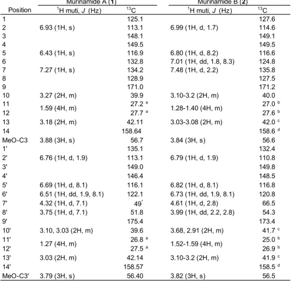
Table 2.1. 1H (600 MHz) and 13C (150 MHz) NMR spectral data for murinamides A and B in methanol-d4
Position 1H muti, J (Hz) 13C 1H muti, J (Hz) 13C
1 125.1 127.6
2 6.93 (1H, s) 113.1 6.99 (1H, d, 1.7) 114.6
3 148.1 149.1
4 149.5 149.5
5 6.43 (1H, s) 116.9 6.80 (1H, d, 8.2) 116.6
6 132.8 7.01 (1H, dd, 1.8, 8.3) 124.8
7 7.27 (1H, s) 134.2 7.48 (1H, d, 2.2) 135.8
8 128.9 127.5
9 171.0 171.2
10 3.27 (2H, m) 39.9 3.10-3.2 (2H, m) 40.0
11 27.2a 27.0b
12 27.7a 27.6b
13 3.18 (2H, m) 42.11 3.03-3.08 (2H, m) 42.0c
14 158.64 158.6d
MeO-C3 3.88 (3H, s) 56.7 3.84 (3H, s) 56.6
1' 135.1 132.4
2' 6.76 (1H, d, 1.9) 113.1 6.79 (1H, d, 1.9) 110.8
3' 149.0 149.8
4' 146.4 148.5
5' 6.69 (1H, d, 8.1) 116.1 6.82 (1H, d, 8.1) 116.8
6' 6.51 (1H, dd, 1.9, 8.1) 122.1 6.73 (1H, dd, 1.9, 8.1) 120.8
7' 4.32 (1H, d, 7.1) 49* 4.61 (1H, d, 2.8) 66.5
8' 3.75 (1H, d, 7.1) 51.8 3.99 (1H, dd, 2.2, 2.8) 54.3
9' 175.4 173.4
10' 3.10, 3.03 (2H, m) 39.6 3.68, 2.91 (2H, m) 41.7c
11' 26.8a 25.0b
12' 27.5a 26.9b
13' 3.03 (2H, m) 42.14 3.10-3.2 (2H, m) 41.9c
14' 158.57 158.5d
MeO-C3' 3.79 (3H, s) 56.40 3.82 (3H, s) 56.5
* Overrapping with solvents.
a-d Assignments may be interchanged.
Murinamide A (1) Murinamide B (2)
1.59 (4H, m) 1.28-1.40 (4H, m)
1.27 (4H, m) 1.52-1.59 (4H, m)
Fig. 2.2. Key HMBC (indicated by arrows from 13C to 1H) and COSY (indicated by bold lines) for 1 and 2.
Fig. 2.3. Chemical structures of murinamides A and B.
2.2.3. Dimerization of feruloylagmatine by peroxidase
To verify that 1 was a dehydrodimer of FerAgm, we performed a peroxidase reaction using FerAgm as a substrate under acidic conditions. The reaction resulted in the formation of 1, with a 22% yield, thereby confirming the identity of 1 (Fig. 2.S1A).
Compound 2 was not formed by the peroxidase reaction with FerAgm in the presence of H2O2 under acidic conditions. Therefore, we performed the reaction under basic
conditions (Fig. 2.S1B). The reaction resulted in the formation of 2 with 8.0% yield.
Hordatine C (HC, Fig. 2.S2) was generated as the major product in the peroxidase reactions for the syntheses of 1 and 2 under acidic and basic conditions (Fig. 2.S1). The determination of the structure is described in the supplementary material.
2.2.4. Optical resolution of murinamides A and B
Synthesized MA was separated into two peaks with almost the same peak areas by chiral HPLC. The CD spectra of the compounds obtained from the two peaks were mirror images of each other, indicating that synthesized MA was a racemic mixture (Fig. 2.S3). MB also separated into two peaks with the same peak area by chiral HPLC, and their CD spectra indicated that the synthesized MB was also a racemic mixture (Fig.
2.S4).
MA and MB purified from H. murinum ssp. glaucum were subjected to chiral HPLC, which separated MA into two peaks corresponding to the enantiomers. The percentages of the peak areas of the earlier and later peaks were 20% and 80%,
respectively. On the other hand, MB eluted as a single peak, corresponding to the later peak in the analysis of synthesized MB.
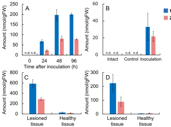
2.2.5. Determination of the concentrations of murinamides and feruloylagmatine in H.
bulbosum and H. murinum
We determined the amounts of MA, MB, and HC in the shoots and roots of H.
bulbosum accumulated MA and MB in the shoots and MA in the roots. H. murinum ssp.
glaucum accumulated MA in the shoots and roots and MB in the shoots at high
concentrations, whereas H. murinum ssp. leporinum accumulated MA and MB in both the shoots and roots. HC was detected in the shoots and roots of H. murinum spp.
leporinum (27.0 nmol/gFW and 15.8 nmol/gFW, respectively), although its
concentrations were far lower than those of MA and MB. HC was not detected in H.
bulbosum or H. murinum ssp. glaucum. HC was present in the shoots of H. vulgare ssp.
vulgare at a low concentration (11.9 nmol/gFW). In other Hordeum species, MA, MB, and HC were below the detection limits.
2.2.6. Antimicrobial activities of murinamides A and B
The antifungal activities of MA and MB against pathogenic fungi Bipolaris sorokiniana and Fusarium asiaticum were evaluated by the inhibition of conidia
germination and germ tube elongation. Murinamides showed a marked inhibitory effect in both assays, but FerAgm showed no activity (Fig. 2.4). MA and MB showed similar activity, completely inhibiting the spore germination of B. sorokiniana at 1 mM (Fig.
2.4A). In an assay for the inhibition of conidia germination of F. asiaticum, MB exhibited stronger activity than MA did; MB completely inhibited conidia germination
Table 2.2. Accumulation of murinamides A and B in H. bulbosum and H. murinum.
Plant Extracted part Murinamide A (1) Murinamide B (2) (nmol/gFW) (nmol/gFW)
H. bulbosum Shoota 3530 ± 315 102 ± 19.4
Roota 273 ± 97.6 n.d.c
H. murinum ssp. glaucum Shootb 2000 ± 129 1570 ± 108
Rootb 2280 ± 162 n.d.
H. murinum ssp. leporinum Shootb 83.0 ± 0.62 890 ± 14.0
Rootb 385 ± 120 223 ± 65.7
bShoots and roots of 3-day-old seedlings.
cn.d.: not detected.
aYoung shoots and roots emerged from bulbs of H. bulbosum.
at 0.3 mM, whereas MA inhibited conidia germination by only 50% even at 1 mM (Fig.
2.4B). MB almost completely inhibited the germ tube elongation of B. sorokiniana at 0.3 mM, while FerAgm and MA inhibited it only by 35% and 66%, respectively, even at 1 mM (Fig. 2.4C). On the other hand, MA and MB showed similar dose-response
curves for the inhibition of germ tube elongation of F. asiaticum, with almost complete inhibition at 0.3 mM (Fig. 2.4D). The activity against Pseudomonas syringae pv.
japonica was assayed by growth inhibition in liquid culture. Murinamides did not show inhibitory effects on the growth of P. syringae pv. japonica even at 1 mM.
Fig. 2.4. Inhibition of conidia germination (A and B) and germ tube elongation (C and D) of Bipolaris sorokiniana (A and C) and Fusarium asiaticum (B and D) by
murinamides A (circles) and B (diamonds), and feruloylagmatine (triangles). Data are means with standard deviation from three independent experiments.
2.3. Discussion
In the present study, we analyzed the presence or absence of three groups of defensive specialized metabolites in Hordeum species. The analysis provides an overall picture of the distribution of these metabolites in Hordeum species for the first time. First, species in the I and Xa clades accumulated the Bx glucoside DIBOA-Glc but not hordatines.
Second, in H clade species, H. vulgare ssp. vulgare and H. vulgare ssp. spontaneum accumulated hordatines but not Bxs. Third, H. bulbosum in the H clade and H. murinum in the Xu clade accumulated MA and MB but not Bxs. Fourth, species that accumulated gramine at high concentrations were found only in the H and Xu clades. This biased distribution indicates that specialized metabolism has changed dynamically during the evolution of Hordeum species.
On the basis of these findings, Hordeum species were classified into two groups: (1) I and Xa clade species, which accumulate the Bx glucoside DIBOA-Glc and (2) H and Xu clade species, which accumulate dehydrodimers of HCAAs with
agmatine, although their chemical structures showed interspecific variation among H and Xu clade species. Bxs are present in a wide range of Poaceae species (Niemeyer, 1988; Kokubo et al., 2017); thus, the common ancestor of Hordeum species is
considered to have accumulated Bxs. In the evolution of the genus Hordeum, the defensive specialized metabolism has shifted from Bxs to dehydrodimers of HCAAs of agmatine in H and Xu clade species (Fig. 2.5). Moreover, H. bulbosum, a wild H- genome species closest to barley, together with Xu clade species, accumulated
murinamides. The plausible scenario is that murinamide biosynthesis was acquired in the common ancestor of the H and Xu clade species, and that murinamides were subsequently replaced by the other type of HCAA dimers—hordatines—during speciation between H. vulgare and H. bulbosum.
Grün et al. (2005) suggested that the presence of gramine and Bxs was mutually exclusive on the basis of the results obtained by the analysis of these
compounds in two subspecies of H. vulgare in the H clade and four species in the I clade. This seems reasonable because gramine and Bxs share a common biosynthetic pathway. Our analysis in the genus Hordeum supported this notion. We analyzed seven additional species in the genus and found that most species accumulated detectable amounts of gramine, but the concentrations of gramine in the I and Xa clade species accumulating DIBOA-Glc were much lower than the concentrations of gramine in H.
vulgare. Because Bxs and gramine were detected outside of the Hordeum species (Niemeyer et al., 1988; Kokubo et al., 2017), their origin predated the genus formation.
Thus, the common ancestor of Hordeum species should have contained both metabolites. After the split of the H/Xu clades from the I/Xa clades, the gramine
biosynthetic pathway might have been activated in ancestral species of the H/Xu clades that had lost the Bx pathway, and thus had large capacity for the supply of gramine precursors.
Among H and Xu clade species, H. vulgare ssp. vulgare cv. Sachiho Golden and H. murinum ssp. glaucum did not accumulate gramine. H. murinum ssp. leporinum is a tetraploid derived from H. murinum ssp. glaucum and an extinct species with an Xv genome (Cuadrado et al., 2013). Gramine accumulation in H. murinum ssp. leporinum might be a trait introduced from the extinct Xv species. In cultivated barley, a lack of gramine accumulation in some cultivars has been reported (Lovett et al., 1994). The deletion of the gene encoding the methyltransferase catalyzing the conversion of AMI to gramine was indicated to be responsible for the lack of gramine (Larsson et al., 2006).
Murinamides are hitherto undescribed dehydrodimers of FerAgm in H.
bulbosum and H. murinum instead of hordatines (dehydrodimers of CouAgm).
Hordatines have been detected in H. vulgare ssp. vulgare and H. vulgare ssp.
spontaneum by Smith and Best (1978), who also reported that H. murinum and H.
bulbosum contained hordatines. They separated the compounds by TLC, and detected spots by spraying with Sakaguchi reagent, which visualizes compounds bearing
bulbosum and H. murinum extracts were somewhat smaller than those of HA and HB.
Thus, the spots detected in the analyses of extracts from H. bulbosum and H. murinum probably corresponded to MA and MB.
On the basis of their chemical structures, MA and MB can be classified into the lignanamides, which are lignans possessing at least one amide group (McCredie et al., 1969). MA and MB had common 8′-8′ linkages in the hydroxycinnamic acid moieties.
This type of linkage has also been found in cannabisins (Sakakibara et al., 1995), jacpaniculines (Henrici et al., 1994), hyoscyamide (Ma et al., 2002), lignanamides from Porcelia macrocarpa (Chaves and Roque, 1997), and bisavenanthramides (Okazaki et al., 2004, 2007). The 8′-8′ linkages can be formed by a radical coupling reaction catalyzed by peroxidase. We found that reactions of FerAgm with peroxidase led to the generation of MA and MB. Hordatines are also generated by a peroxidase reaction in vitro (Stoessl, 1966, 1967; Negrel and Smith, 1984). It is likely that similar enzymes are involved in the generation of hordatines and murinamides. The accumulation of HCAAs with agmatine is not limited to the H and Xu clades; some I clade species accumulated CouAgm at low concentrations. Thus, species in the H and Xu clades might have acquired an enzyme that catalyzes this coupling reaction between two HCAA units.
It is noteworthy that the products of the peroxidase reaction with FerAgm differed depending on the reaction conditions. In an acidic reaction mixture, the generation of MA was predominant and MB was hardly detectable, whereas MB was the main product under basic conditions. The reaction scheme leading to MA and MB is shown in Fig. 2.S5. The preferred generation of MB in alkaline conditions is reasonable because these conditions enhance the nucleophilicity of the amide nitrogen. The
nucleophilic attack of the amide nitrogen to the 7′ position of the other FerAgm unit leads to the formation of a C-9-N-C-7′ linkage. In the biosynthesis of MA and MB in plant cells, some factor enabling the simultaneous production of MA and MB might be present at moderate pH in plant cells.
The analysis of the stereochemistry of murinamides extracted from H. murinum ssp. glaucum indicated that MA is a 1:4 mixture of enantiomers, whereas MB is
optically pure. If murinamides are biosynthesized by a peroxidase reaction in plant cells, a factor controlling the configuration of intermediate species formed with two FerAgm units linking 8′ positions might exist (Fig. 2.S5). Hordatines have also been shown to be optically pure (Wakimoto et al., 2009). In lignin biosynthesis, a protein has been shown to control the stereochemistry of the radical coupling reaction (Davin et al., 1997; Pickel et al., 2007). In this context, it is of interest that a factor might be involved in the control of cyclization after the radical coupling between two FerAgm molecules leading to murinamides.
MA and MB showed inhibitory effects against fungal pathogens at
concentrations roughly higher than 100 µM, although the activities depended on the fungal species and bioassay method. The total concentrations of MA and MB ranged from 270 nmol/gFW (root of H. bulbosum) to 3600 nmol/gFW (shoot of H. bulbosum) in murinamide-accumulating species. Thus, it is reasonable to assume that murinamides play defensive roles in these species. The inhibitory activities of MA and MB were similar in bioassays for conidia germination in B. sorokiniana and germ tube elongation in F. asiaticum, whereas MB had a higher inhibitory activity towards conidia
germination of F. asiaticum and germ tube elongation of B. sorokiniana than MA, suggesting that MA and MB may play distinct roles in plant defense against pathogens.
Murinamides showed strong inhibitory activity against pathogens compared with FerAgm. HA is also a strong antifungal compound (Stoessl and Unwin, 1970), whereas the activity of CouAgm is weak. The viniferins of grapevines are phytoalexins comprising a dimer and oligomers of resveratrol, and they demonstrate stronger
antifungal activities than those of resveratrol (Langcake, 1981). Thus, dimerization and oligomerization are considered to be efficient mechanisms that increase the diversity of plant metabolites by building new carbon skeletons, thereby creating compounds with
Fig. 2.5. Schematic representation of evolutionary changes in defensive specialized metabolism in the genus Hordeum.
In the present study, we found that dramatic metabolic changes have occurred after the separation of the H/Xu claddes from the I/Xa clades in the genus Hordeum.
The changes were the replacement of Bxs by gramine and the dehydrodimers of agmatine HCAAs such as hordatines and murinamides in the H/Xu clades (Fig. 2.5).
The accumulation of dehydrodimers of agmatine HCAAs appears to have occurred by the activation of precursor supply, expression of coupling enzyme for HCAAs, and acquisition of factors that control the stereo- and regioselectivity of cyclization reactions after the coupling reaction.
2.4. Experimental
2.4.1. General
1H- NMR, 13C-NMR, and 2-D NMR spectra (COSY, HMQC, HMBC, and NOESY) were recorded using an Avance II 600 spectrometer (Bruker, Billerica, MA, USA).
HRMS analyses were performed using an Exactive mass spectrometer (Thermo Fisher Scientific, Waltham, MA, USA), and ESI-MS mesurements were performed using a Quattro Micro API mass spectrometer (Waters, Milford, MA, USA) connected to an Acquity UPLC system (Waters). HPLC was performed using a 10Avp HPLC system (Shimadzu, Kyoto, Japan). CD spectra were recorded using a J-820 CD
spectrophotometer (JASCO, Tokyo, Japan).
2.4.2. Plant materials
H. vulgare L. ssp. vulgare ʻSachiho Golden’, H. vulgare L. ssp. spontaneum (C. Koch.) Thell., H. bulbosum L., H. murinum ssp. glaucum (Steudel.) Tzvelev., H. marinum ssp.
marinum Hudson., H. chilense Roem. & Schult., H. brachyantherum Nevski. ssp.
californicum (Cov. & Steb.) Both. & et al., H. bogdanii Wil., H. flexuosum Steud., H.
pusillum Nutt. (these nine species are all diploids), H. murinum ssp. leporinum (Link.) Arcang. and H. jubatum L. (both species are tetraploids) were stocks from the Institute of Plant Science and Resources, Okayama University, Japan. Taxonomy of the Hordeum species follows Bothmer et al. (1995). The details of Hordeum species that we used in this study are summarized in Table 2.S2. H. vulgare L. ssp. vulgare ‘Shunrai’ was purchased from JA Tottori Chu-Oh (Tottori, Japan). Seeds were sown on a layer of filter paper in a petri dish, kept at 4°C in the dark for 2 days to stimulate germination, and incubated at 15°C in the light for several days. For the self-incompatible species H.
bulbosum, shoots and roots were obtained from a clone at the tillering stage.
The causal agent of spot blotch of barley, B. sorokiniana (OB-25-1), was a stock from the Laboratory of Natural Product Chemistry, Faculty of Agriculture, Tottori University.
The causal agent of Fusarium head blight, F. asiaticum (MAFF 305134), was obtained from the National Institute of Agrobiological Sciences (NIAS) Genebank
(https://www.gene.affrc.go.jp/about.php). B. sorokiniana and F. asiaticum were inoculated to V8 agar plates. B. sorokiniana was cultured for 4 weeks at 25°C under black light bulb (BLB: FL15BL-B, Hitachi, Tokyo) radiation to obtain conidia. F.
asiaticum was cultured at 25°C in the dark for 1 week, and under BLB for several days after aerial hyphae were removed for simultaneous conidia formation.
The causal agent of bacterial black node, P. syringae pv. japonica (MAFF 301072), was obtained from the NIAS Genebank. P. syringae was cultured on potato semi-synthetic medium (300 g potato, 2 g Na2PO4·12H2O, 15 g sucrose, 5 g peptone, 0.5 g Ca(NO3)2·4H2O, and 1 L distilled water) at 25°C with shaking at 200 rpm for 24 h.
2.4.4. Chemicals
HA and HB were purified from 96-h-old seedlings of barley (H. vulgare cv. Shunrai) according to a previously described method (Nomura et al., 2007) with slight
modifications. Whole seedlings (304 g) were extracted with 3 L of 80% methanol containing 2% acetic acid for 24 h. After concentrating the extract to 200 mL by evaporation, we added 80% methanol (200 mL) to the concentrate, and applied it to an ODS column (Cosmosil 75C18-OPN, Nacalai Tesque, Kyoto, Japan) equilibrated with 80% methanol to remove hydrophobic materials. The column was eluted with 80% and 100% methanol. The flow-through fraction and 80% methanol fraction were combined and concentrated to a small volume by evaporation, and the solution was applied to an ODS column equilibrated with water containing 1% acetic acid. The column was eluted with 0%, 5%, 10%, 20%, and 30% methanol containing 1% acetic acid. HA and HB eluted in the 20% methanol fraction, so this fraction was concentrated and subjected to
preparative HPLC. The HPLC conditions were as follows: column, Cosmosil 5C18-AR- II 20 I.D. × 200 mm (Nacalai Tesque); solvents, 22% B/(A+B), A: 0.1% trifluoroacetic acid aq., B: acetonitrile; flow rate, 6.0 mL/min; column temperature, 40°C; detection, 280 nm. HA and HB eluted at 18.0 min and 16.4 min, respectively. The peaks
corresponding to HA and HB were collected and concentrated to dryness to yield HA (29.8 mg) and HB (0.8 mg). The identities of the compounds were confirmed by comparison of 1H-NMR spectra with reported data (Wakimoto et al., 2009; Stoessl, 1967).
p-Coumaroyl- and feruloyl-N-hydroxysuccinimide esters were synthesized from p-coumaric acid and ferulic acid, respectively, according to the methods described by Stöckigt and Zenk (1975). CouAgm and FerAgm were synthesized from the
corresponding N-hydroxysuccinimide esters and agmatine sulfate (Sigma Aldrich) in line with the method described by Negrel and Smith (1984). The authenticity of the compounds was confirmed by comparing their 1H-NMR spectra with previously reported spectra (Ogura et al., 2001; Jin et al., 2003).
DIBOA-Glc and DIMBOA-Glc were prepared from the seedlings of rye and maize, respectively, according to Sue et al. (2000). Gramine was purchased from Wako Pure Chemical Industries (Osaka, Japan).
2.4.5. Analysis of defensive specialized metabolites
Shoots and roots of Hordeum species at the seedling stage were extracted with 80%
methanol for 24 h. The extracts were subjected to HPLC for the analysis of CouAgm, FerAgm, HA, and HB, and to LC-MS/MS analysis in multiple-reaction monitoring mode for Bxs and gramine. HPLC conditions were as follows: column, Cosmosil 5C18- AR-II 4.6 I.D. × 150 mm (Nacalai Tesque); gradient, 10%–20% B/(A+B) within 30 min, A: 0.1% trifluoroacetic acid aq., B: acetonitrile; flow rate, 0.8 mL/min; column temperature, 40°C; detection, 280 nm. HC, MA, and MB were analyzed under the same
HB, HC, MA, and MB were 11.8, 14.3, 25.3, 24.8, 26.4, 13.2, and 17.4 min, and 2.5, 5.5, 6.0, 8.5, 7.5, 14.0, and 10.6 nmol/g FW, respectively. The trans isomers of the compounds were analyzed because the peak areas of cis isomers were negligible in freshly prepared plant extracts. The LC conditions for LC-MS/MS analysis were as follows: column, Acquity UPLC BEH C18 column 2.1 × 50 mm (1.7 µm) (Waters);
gradient, 5%–40% B/(A+B) within 5 min, A: 0.1% formic acid aq., B: 0.1% formic acid in acetonitrile; flow rate: 0.2 mL/min; column temperature, 40°C. Monitored mass transitions were from m/z 175 to m/z 130 for gramine, m/z 344 to m/z 182 for DIBOA- Glc, and m/z 374 to m/z 212 for DIMBOA-Glc in positive ESI mode. Cone voltage and collision energy were optimized by using authentic compounds. Detection limits for gramine, DIBOA-Glc, and DIMBOA-Glc were 0.08, 10, and 2.0 nmol/g FW, respectively.
2.4.6. Isolation of unidentified compounds from the seedlings of H. murinum ssp.
glaucum
We detected two hitherto unidentified compounds, 1 and 2, in the seedlings of H.
bulbosum, H. murinum ssp. glaucum, and H. murinum ssp. leporinum. Compounds 1 and 2 were purified from 96-h-old seedlings of H. murinum ssp. glaucum. Seedlings (37 g) were extracted with 80% methanol (370 mL) for 24 h. The extract was applied to an ODS column (Cosmosil 75C18-OPN) equilibrated with 80% methanol to remove hydrophobic materials. The column was eluted with 80% and 100% methanol. The flow-through and 80% methanol fractions were combined and concentrated by
evaporation, and the solution was subjected to preparative HPLC. The HPLC conditions were as follows: column, Cosmosil 5C18-AR-II 10 I.D. × 200 mm; gradient, 11%–18%
B/(A+B) within 35 min, A: 0.1% trifluoroacetic acid aq., B: acetonitrile; flow rate, 3.0 mL/min; column temperature, 40°C; detection, 280 nm. Compounds 1 and 2 were eluted at 15.5 min and 18.6 min, respectively.
1 [murinamide A (MA), 7.1 mg], HRMS (ESI): m/z 611.3301 [M+H]+ (calcd
for C30H43O6N8, m/z611.3300); UV-Vis (acetonitrile-water containing 0.1% formic acid): lmax 238, 277 (sh), 328 nm; 1H-NMR and 13C-NMR (Table 2.1).
2 [murinamide B (MB), 3.7 mg], HRMS (ESI): m/z 611.3301 [M+H]+ (calcd for C30H43O6N8, m/z 611.3300); UV-Vis (acetonitrile-water containing 0.1% formic acid): lmax 234, 284 (sh), 324 nm; 1H-NMR and 13C-NMR (Table 2.1).
2.4.7. Enzymatic preparation of murinamides A and B
MA and MB were synthesized from FerAgm using an enzymatic reaction with horse radish peroxidase (HRP; Wako Pure Chemical Industries) according to Zoia et al.
(2008).
FerAgm (200 mg, 0.65 mmol) was dissolved in DMSO-ethanol-McIlvaine buffer (pH 3.5) (9:91:10, v/v, 10 mL) and cooled to 0°C. A mixture of 0.8 M H2O2 aq.
(0.5 mL, 0.4 mmol) and 9.0 mg/mL HRP aq. (0.5 mL, 450 U) was added to the solution.
The mixture was stirred at 0°C for 6 h, and 2 M HCl (0.2 mL) was added to stop the reaction. The mixture was concentrated by evaporation to remove ethanol, and was applied to an ODS column (Cosmosil 75C18-OPN) equilibrated with water. The column was eluted with 0%, 5%, 10%, 20%, and 30% methanol in water (each solvent was divided into three fractions). The 10%-3 and 20%-1 fractions, which contained MA and MB, were combined, concentrated by evaporation, and subjected to preparative HPLC.
The HPLC conditions were as follows: column, Cosmosil 5C18-MS-II 10 I.D. × 200 mm; solvent, 13% B/(A+B), A: 0.1% trifluoroacetic acid aq., B: acetonitrile; flow rate, 3.0 mL/min; column temperature, 40°C; detection, 280 nm. MA was eluted at 16.4 min.
The identity of synthetic MA (44.6 mg, 0.073 mmol, yield 22%) was confirmed by 1H- NMR spectra.
MB was also synthesized using a peroxidase reaction under basic conditions.
FerAgm (100 mg, 0.33 mmol) was dissolved in 9.11 mL of DMSO-ethanol-water (90:810:11, v/v, 9.11 mL) and heated to 40°C. A mixture of 0.8 M H2O2 aq. (0.25 mL,
30 s, 1 M NaOH aq. (0.09 mL) was added to the mixture, followed by a 2-h incubation at 40°C. The reaction was stopped by the addition of 2 M HCl (0.06 mL). The reaction mixture was concentrated and applied to an ODS column (Cosmosil 75C18-OPN) equilibrated with water. The column was eluted with 0%, 5%, 10%, 20%, and 30%
methanol in water. The 5% and 10% fractions were concentrated by evaporation and subjected to preparative HPLC. The HPLC conditions were as follows: column,
Cosmosil 5C18-AR-II 20 I.D. × 200 mm; solvent, 16% B/(A+B), A: 0.1% trifluoroacetic acid aq., B: acetonitrile; flow rate, 7.0 mL/min; column temperature, 40°C; detection, 280 nm. MB was eluted at 21.2 min. The identity of synthetic MB (8.1 mg, 0.013 mmol, yield 8.0%) was confirmed by 1H-NMR spectra.
2.4.8. Optical resolution of murinamides A and B
The HPLC conditions for the optical resolution of MA were as follows: column,
CHIRAL PAK IA 4.6 I.D. × 150 mm (Daicel, Osaka, Japan); solvent, 10% B/(A+B), A:
0.1% trifluoroacetic acid aq., B: acetonitrile; flow rate, 0.6 mL/min; column
temperature, 30°C; detection, 280 nm. MA synthesized by peroxidase reaction eluted as two peaks at 21.2 min and 23.5 min. The fractions corresponding to the peaks were collected, and the CD spectra of MA enantiomers were recorded (Fig. 2.S3). The HPLC conditions for the optical resolution of MB were the same for MA, except that the column was CHIRAL PAK IB 4.6 I.D. × 150 mm (Daicel) and the solvent was changed to 9% B/(A+B). MB synthesized by the peroxidase reaction eluted as two peaks at 35.8 min and 37.9 min. The fractions corresponding to the peaks from synthetic MB were collected, and their CD spectra were recorded (Fig. 2.S4).
2.4.9. Antimicrobial activity
In the assay for the inhibition of conidia germination, conidia of pathogenic fungi were suspended (1.5 × 10⁵ conidia/mL) in 10, 30, 100, 300, and 1000 µM solutions of FerAgm, MA, and MB containing 0.5% DMSO, and droplets of the suspensions were
placed on a slide glass and incubated at 25°C in the dark. The number of germinated conidia was counted after 18-h (for B. sorokiniana) and 10-h incubation (for F.
asiaticum) using a microscope (BX43, Olympus, Tokyo, Japan).
In the assay for inhibition of germ tube elongation, the conidial suspension (1.5 x 10⁵ conidia/mL) was incubated for 2 h, and a photomicrograph of germinated conidia was recorded. After adding the same volumes of solutions of compounds at 20, 60, 200, 600, and 2,000 mM in 1% DMSO, the suspension was incubated for an additional 4 h, and another photomicrograph of the conidia was recorded. The length of the germ tube was measured using the public domain image analysis software ImageJ
(http://rsb.info.nih.gov/ij/).
P. syringae pv. japonica was inoculated into a potato semi-synthetic medium (5 mL) and cultured at 25°C with shaking at 200 rpm for 24 h. Two microliters of cultured medium was inoculated into 198 µL of medium containing FerAgm, MA, and MB at 1 mM containing 1% DMSO in a well of a microplate. After incubation for 24 h at 25°C in the dark, the OD600 of the medium was determined using a microplate reader (Infinite M200PRO, Tecan Group Ltd., Männedorf, Switzerland).
Chapter 3
Identification of phenylamide phytoalexins and characterization of inducible phenylamide metabolism in wheat
3.1. Introduction
Phenylamides are amide compounds of hydroxycinnamic or benzoic acids with biogenic amines, which function in plant defense responses. Representative
hydroxycinnamic acids composing phenylamides are cinnamic (Cin), p-coumaric (Cou), caffeic (Caf), ferulic (Fer), and benzoic (Ben) acids, while amines parts were derived from tryptamine (Try), serotonin (Ser), tyramine (Tyr), agmatine (Agm), and putrescine (Put). Herein, we refer to specific phenylamides using combined abbreviations of the acid parts and amines, e.g. CinTry (cinnamoyltryptamine). Induced phenylamide
accumulation has been reported in a wide range of plant species. In rice, infection by the brown spot fungus (Cochliobolus miyabeanus) induced the accumulation of FerTry, CouSer, and FerSer, together with Ser (Ishihara et al., 2008). In addition, rice leaves irradiated with ultraviolet (UV) light accumulate CinTry, CinTyr, CouSer, and BenTry (Park et al., 2013, 2014). In barley, Lee et al. (1997) reported the induced accumulation of CouAgm in methyl jasmonate (MeJA)-treated leaves. Similarly, Ogura et al. (2001) reported the accumulation of CouPut and Cou-3-hydroxy-Put, as well as CouAgm, in barley leaves treated with jasmonic acid (JA). In oats, avenanthramides, which are phytoalexins, are hydroxycinnamic acid amides with anthranilic acids (Miyagawa et al.
1995).
In wheat, benzoxazinoids (Bxs) are the representative defensive specialized metabolites (Niemeyer, 1988; 2009; Sicker et al., 2000). Because of their anti-aphid, antimicrobial, and phytotoxic activities, Bxs are thought to function in the defense against various biological stresses (Argandoña et al., 1980; Barnes and Putnam, 1987;
Niemeyer, 1988, 2009; Sicker et al., 2000; Meihls et al., 2013). Conversely, reports on
inducible specialized metabolites in wheat are limited. From a metabolic analysis, Gunnaiah et al. (2012) found the differential accumulation of metabolites in rachis and spikelets of near isogenic wheat lines with resistant or susceptible alleles of Fhb1 upon Fusarium graminearum infection. Phenolics, lignans, hydroxycinnamic acid amides (phenylamides), flavonoids, compounds related to signaling molecules, terpenoids, indole alkaloids, and compounds related to methionine biosynthesis were suggested to be involved in Fhb1 mediated resistance against F. graminearum. Those authors suggested that cell-wall thickening due to the deposition of hydroxycinnamic acid amides and flavonoids was the potential resistance mechanism.
Plants often activate multiple specialized metabolic pathways upon pathogen infection. Rice plants accumulate diterpenoid phytoalexins and a flavonoid phytoalexin in addition to serotonin and hydroxycinnamic amides, which are biosynthesized by different pathways. Similarly, maize plants accumulate Bxs as constitutive defensive compounds, and produce kauralexins, dolabralexins, and zealexins as phytoalexins in response to pathogen attack. This variety of defense compounds may be important for forming multiple layers of chemical barriers, which are effective against different classes of pathogens with different sensitivity to each compound. In addition, the multiplicity of defense metabolites may be important for preventing acquisition of tolerance against a specific compound. However, in wheat, the compounds involved in inducible defense mechanisms have not been identified, although the accumulation and function of Bxs have been widely studied. Since wheat is likely possess a phytoalexin defense mechanism, the leaves infected with B. sorokiniana were metabolically profiled, and undescribed inducible phenylamides were identified. In addition, we analyzed the accumulation of other phenylamides in wheat leaves to characterize the activated metabolic pathways. This study provides a frame work of multiple chemical barriers endowed by phytoalexins in wheat that protect against pathogen attack.
3.2. Results
3.2.1. Detection of inducible metabolites in wheat leaves infected by B. sorokiniana B. sorokiniana conidia were inoculated to the third leaves of 3-week-old wheat seedlings. We extracted metabolites from the leaves with 80% methanol 48 h after inoculation, and subjected the extracts to HPLC analysis. As shown in Fig. 3.1, the accumulation of compounds 1–6 was induced in leaves inoculated with B. sorokiniana.
Compounds 3–6 were collected by preparative HPLC and subjected to LC-MS analysis.
3–6 showed [M+H]+ ions at m/z 251, 293, 291, and 277, respectively. Comparison of the detected ions and retention time by HPLC analysis with those of authentic
compounds revealed 6 was CouAgm. LC-MS/MS analysis of 3–6 showed different fragment ions at m/z 88 in 3, m/z 130 in 4, m/z 128 in 5, and m/z 114 in 6 in addition to a commonly detected fragment ion at m/z 147 corresponding to coumaroyl moiety (Fig.
3.S1). The fragment ion at m/z 114 in 6 corresponded to an agmatine moiety.
Differences in fragment ions among 3–5 were due to the conversion of a guanidine group to amine (-42), the addition of a hydroxy group (+16), and dehydrogenation (-2) in an agmatine moiety, respectively. Accordingly, we predicted 3–5 as p-coumaric acid amides of hydroxyputrescine, hydroxyagmatine, and hydroxydehydroagmatine,
respectively. The induction of these compounds was previously shown in wheat and barley. p-Coumaroyl-3-hydroxyputrescine accumulated in wheat leaves inoculated with rust fungus (Stoessl et al., 1969). p-Coumaroyl-3-hydroxyagmatine and CouAgm accumulation was induced under snow cover in wheat (Jin et al., 2003). p-Coumaroyl-3- hydroxydehydroagmatine accumulation was induced in response to powdery mildew in barley leaves (von Röpenack et al., 1998), as well as CouAgm and p-coumaroyl-3- hydroxyagmatine. Compounds 1 and 2 in the upper left inset showed [M+H]+ ions at m/z 321 and m/z 305, respectively. These ions did not correspond to those of the inducible compounds previously reported in wheat.
Fig. 3.1. High-performance liquid chromatography (HPLC) analysis of extracts from B.
sorokiniana-inoculated, control, and intact leaves.
A suspension of B. sorokiniana conidia (inoculation) or distilled water containing 0.25% Tween20 (control) was placed on the third leaves of 3-week-old wheat seedlings.
The leaves were extracted 48 h after inoculation and subjected to HPLC analysis.
Untreated leaves (intact) were also extracted and analyzed by HPLC. Insets were zoom up views of the chromatograms.
3.2.2. Identification of 1 and 2
We examined the effects of CuCl2 solution on wheat leaves, because CuCl2 acts as an elicitor for other plants (Rakwal et al., 1996); we found that the accumulation of 1 and 2 was also induced by the treatment with 0.5 mM CuCl2 solutionfor 72 h. Thus, we purified the compounds from 98.4 g of wheat leaves treated with 0.5 mM CuCl2 for 72
-10 10 30 50
0 5 10 15 20 25 30
-10 10 30 50
-10 10 30 50
U V 28 0 nm (mV)
Retention time (min) Inoculation
-3 -1 1
20 25 30
-3 -1 1
20 25 30
Intact Control
Cou-3-OH- Put (3)
Cou-3-OH-Agm (4)
CouAgm (6) 1
2
-5 -3 -1
20 25 30
Cou-3-OH-dehydroAgm (5)
First, we describe the chemical structure of compound 2. Compound 2 had the molecular formula C19H16O2N2, as determined by HRMS (m/z 305.1284, [M+H]+). 1H- NMR and COSY spectrum of 2 (Table 3.1) showed two groups of aromatic proton signals at dH 7.30, 7.32, 7.59, and 8.27 ppm for a 1, 2-disubstituted benzene ring (ring A) and at dH 7.47–7.58 and 7.70 ppm for a monosubstituted benzene ring (ring B). In addition, 1H-NMR spectrum revealed the presence of two coupled double-bond protons (dH 6.96 and 7.57 ppm, J = 15.6 Hz), a methylene proton (dH 4.73 ppm) and three 1H protons (dH 8.53, 8.57, and 12.15 ppm).
Although HRMS indicated the presence of 19 carbons in the structure, the 13C- NMR spectrum showed 17 carbon signals. In the HMQC spectrum, two carbon signals at dC 127.6 and 128.9 ppm showed cross-peaks with two 2H proton signals at dH 7.70 and 7.54 ppm, respectively, indicating that these signals corresponded to the 2- and 6-, and 3- and 5-positions of ring B. Because no correlation was detected for two 1H proton signals at dH 8.53 and 12.15 ppm in the HMQC spectrum, these signals correspond to active protons. All other protonated carbons were assigned using the HMQC spectrum.
The presence of two carbonyl carbons was indicated by signals at dC 190.1 and 165.2 ppm in the 13C-NMR spectra. Taking the molecular formula into account, no oxygen atom was available for OH group, and thus, two active proton signals at dH 8.53 and 12.15 ppm were considered to correspond to NH signals. In the COSY spectrum, NH signals at dH 12.15 ppm showed a cross-peak with a 1H proton at dH 8.57 ppm.
According to the HMBC spectrum, a 1H proton at 8.57 ppm (H-2) showed cross-peaks with carbon signals at dC 136.4 (C-7a), 125.4 (C-3a), and 114.0 ppm (C-3). These results indicated that ring A, NH, C-3, and C-2 comprise an indole group (Fig. 3.3).
The carbon signal at dC 134.9 ppm (C-1’) in ring B showed a cross-peak with the double-bond protons at dH 7.57 ppm (H-7’) in the HMBC spectrum, indicating a connection between C-1’ and C-7’. Two double-bond protons (H-7’ and H-8’) were connected to the carbonyl carbons (C-9’) at dC 165.2 ppm based on HMBC correlations, indicating the presence of a cinnamoyl moiety in the structure.
The C-9’ signal showed a cross-peak with the -NH proton at dH 8.53 ppm in the HMBC spectrum, indicating an amide group (NH [NH-10] and C-9’). Furthermore, based on HMBC correlations, the linkages between this NH and the methylene group at 9-position, and between this methylene group and the carbonyl carbon at 8-position, are established.
Then, this carbonyl carbon (C-8) was considered to bind to the quaternary carbon C-3 in the indole moiety because no position can form a linkage in the molecule.
The presence of a carbonyl group at 8-position was supported by the downfield shift of the H-2 proton (dH 8.57 ppm) (Ma et al., 2009). Thus, we concluded that 2 is N- cinnamoyl-8-oxotryptamine (Fig. 3.2).
To confirm this conclusion, we attempted synthesis of compound 2 by
condensation of the N-hydroxysuccinimide ester of cinnamic acid and 8-oxotryptamine.
The reaction resulted in the formation of 2 with a 31.2% yield, thereby confirming the identity of 2.
Next, compound 1 had the molecular formula C19H16O3N2, as determined by HRMS (m/z 321.1229, [M+H]+); thus, 1 had an additional oxygen atom compared with 2. The 1H- and 13C-NMR spectra of 1 were similar to those of 2; however, there was a downfield shift in a methylene proton signal at dH 4.73 ppm (H-9) in 2 to a methine proton signal at dH 6.48 ppm in 1 (Table 3.1). This difference was attributable to the deshielding effect of the hydroxy group. Therefore, 1 was the hydroxy derivative of 2 at C-9, N-cinnamoyl-9-hydroxy-8-oxotryptamine (Fig. 3.2). These compounds were designated as triticamides A and B, respectively, because these compounds purified from wheat (Triticum aestivum). Correlations observed in the 2D NMR spectra were consistent with these structures (Fig. 3.3).
Fig. 3.2. Chemical structures of 1 and 2.
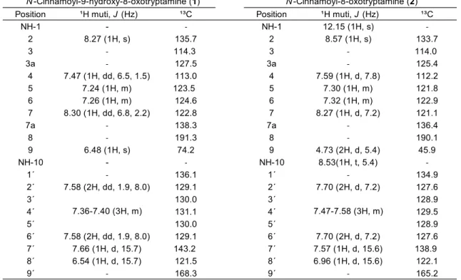
Position ¹H muti, J (Hz) ¹³C Position ¹H muti, J (Hz) ¹³C
NH-1 - - NH-1 12.15 (1H, s) -
2 8.27 (1H, s) 135.7 2 8.57 (1H, s) 133.7
3 - 114.3 3 - 114.0
3a - 127.5 3a - 125.4
4 7.47 (1H, dd, 6.5, 1.5) 113.0 4 7.59 (1H, d, 7.8) 112.2
5 7.24 (1H, m) 123.5 5 7.30 (1H, m) 121.8
6 7.26 (1H, m) 124.6 6 7.32 (1H, m) 122.9
7 8.30 (1H, dd, 6.8, 2.2) 122.8 7 8.27 (1H, d, 7.2) 121.1
7a - 138.3 7a - 136.4
8 - 191.3 8 - 190.1
9 6.48 (1H, s) 74.2 9 4.73 (2H, d, 5.4) 45.9
NH-10 - - NH-10 8.53(1H, t, 5.4) -
1´ - 136.1 1´ - 134.9
2´ 7.58 (2H, dd, 1.9, 8.0) 129.1 2´ 7.70 (2H, d, 7.2) 127.6
3´ 130.0 3´ 128.9
4´ 131.1 4´ 129.5
5´ 130.0 5´ 128.9
6´ 7.58 (2H, dd, 1.9, 8.0) 129.1 6´ 7.70 (2H, d, 7.2) 127.6
7´ 7.66 (1H, d, 15.7) 143.2 7´ 7.57 (1H, d, 15.6) 138.9
8´ 6.54 (1H, d, 15.7) 121.5 8´ 6.96 (1H, d, 15.6) 122.1
9´ - 168.3 9´ - 165.2
N-Cinnamoyl-9-hydroxy-8-oxotryptamine (1) N-Cinnamoyl-8-oxotryptamine (2)
7.36-7.40 (3H, m) 7.47-7.58 (3H, m)
Table 3.1. 1H (600 MHz) and 13C (150 MHz) NMR spectral data for compounds 1 (in CD3OD) and 2 (in DMSO-d6)