*Corresponding author: e-mail [email protected]
Rapid Screening for Cytotoxicity and Group Identification of
Secondary Metabolites in Methanol Extracts from Four Sponge
Species Found in Kapoposang Island, Spermonde Archipelago,
Indonesia
Ajuk Sapar
1,2*, Alfian Noor
1, Nunuk Hariani Soekamto
1and Ahyar Ahmad
11 Chemistry Department, Hasanuddin University, Macassar 90245 2 Chemistry Department, Tanjungpura University, Pontianak 78124
Abstract
Four sponge samples collected from Kapoposang Island, Spermonde Archipelago have been inves-tigated to estimate their cytotoxicity and existence of secondary metabolite groups in their methanol extracts at a preliminary level. The species were Niphates sp., Biemna sp., Liosina paradoxa, Haliclona (Reniera) fascigera. Rapid screening of small scale extracts for cytotoxicity was performed using a Brine Shrimp Lethality Test and the results were expressed as 50% lethal concentration (LC50). Results of the test for all extract are the followings: 391.56 ppm, 1,730 ppm, 456.04 ppm and 478.63 ppm respectively. Only the extract from Biemna sp. was inactive while the others, particularly the extract from Niphates sp. had potent cytotoxic agents. Identification of molecules using specific reagents like Lieberman-Burchard, Salkowski, Mayer, Wagner and Dragendorff showed that methanol extracts contained steroid, terpenoid and alkaloid compounds. Identification of group compounds was supported by a Fourier transform infrared spectroscopy (FTIR) instrument and reference shows that these extracts contain alkaloid, steroid and terpenoid compounds.
Key words: sponge, screening, cytotoxicity, secondary metabolite, Spermonde
Introduction
Sponges are marine animals that have been known as a source of bioactive compounds which can be used as a medicine. It is very interesting to study sponges that live as a filter feeder in competition with other species in a coral reef ecosystem. Their bioactive compounds have effects other organisms and threaten other sponges. This is a phenomenon called allelopathy mediated by allelochemicals. This phenomenon is demonstrated by Plakortis halichondroides sponge overgrowing the coral Agaricia lamarcki by forming a zone on the coral tissue by inducing necrosis of coral cells (Sebastian and Pawlik, 2000). Similarly, sponge Dysidea sp. can induce necrosis of the sponge Cacospongia sp. (Thacker et al., 1998). Secondary metabolites that cause necrosis of other species have been widely reported in Biemna triraphis Niphates sp. sponge.
Two metabolites exhibiting a moderate cytotoxicity have been isolated in Niphates sp. sponge from the New Caledonian. The metabolites were polyoxygenated
acety-lenic acids, nepheliosyne B and nepheliosyne A (Nathalie Legrave, et al., 2013). A steroid compound, 5α, 8α-Epidioxy-24(S)-ethylcholest-6-en-3β-ol was success-fully isolated from Biemna triraphis Topsent collected at Andavadoaka near Toliary (west coast of Madagascar-Indian Ocean) (Julia et al., 2008). Excretion of sec-ondary metabolites from the sponge is influenced by the environment and it is assumed that the different environ-ments induce different secondary metabolites from the same species of sponge. Based on this assumption, we have studied the secondary metabolites from four species of sponges, Niphates sp., Biemna sp., Liosina paradoxa, Haliclona (Reniera) fascigera from Kapoposang Island. This is a preliminary study to explore the bioactive sec-ondary metabolites in sponges from Kapoposang Island.
Material and methods
Tools and material
Fourier transform infrared spectroscopy (FTIR) (IRPrestige-21 Shimadzu), an analytic balance (Mettler
AE 100), and a rotary evaporator (Heidolph 4000) were used. Reagents: Spectrophotometric grade of methanol, chloroform, and sulfuric acid were used. Lieberman-Burchard reagent, Salkowski reagent, Mayer reagent, Wagner reagent, and Dragendorff reagent were pur-chased.
Sampling and identification of species
Sponges of Niphates sp., Biemna sp., Liosina paradoxa, and Haliclona (Reniera) fascigera were col-lected by hand (scuba) from various depth (6-9 m) in Kapoposang Island (Fig. 1), Spermonde Archipelago, South of Sulawesi, Indonesia on 15 August 2012. Sponge taxonomy was determined in Research Center for Oceanography LIPI Jakarta and all specimens has been deposited in Research Center for Oceanography (RCO) LIPI Jakarta, Indonesia 14430. Vouchers code for Niphates sp., Biemna sp., Liosina paradoxa, Haliclona (Reniera) fascigera were SPV 01/10/13, SPV 02/10/13, SPV 03/10/13, and SPV 04/10/13, respectively. The spe-cies data like taxonomy, morphology, consistency, skel-eton and specula are described below.
The morphology of Niphates sp. is a flabelliform with conulose surfaces at the upper side (Fig. 2A). The body colour is pale brown in a dry state. Its consistency
is spongy and the skeleton are nultispicular tracts that consist of spicules-oxeas (110- 150 μm), no microscleres (Fig. 2B and C).
The morphology of Biemna sp. is massive and cushion-shaped as shown in Fig. 3A. The body colour is brown in a dry state. Its consistency is tough and the skeleton consist of spicules Styles (310-350 μm), sigmas (60-70 μm), raphides (110-150 μm) like in Fig. 3B and C.
INDONESIA
SOUTH SULAWESI
SPERMONDE ARCHIPELAGO KAPOPOSANG ISLAND Sampling locationFig. 1. Sampling location in Kapoposang Island.
B
B
C
B
Fig. 2A. Morphology Niphates sp..
The morphology of Liosina paradoxa is massive, tube-shape with muddy surfaces as shown in Fig. 4A and its consistency is firm. The skeleton of Liosina paradoxa is loose bundles with foreign materials that consist of oxeote (600-700 μm) spicules as shown in Fig. 4A and B.
The morphology of Haliclona (Reniera) fascigera is long-tube shaped (Fig. 5A) and its consistency is sot and fragile. The skeleton Haliclona (Reniera) fascigera are unispicular tracts, isotropic and consist of oxeas (60-80 um) spicule in Fig. 5 (A) and (B), respectively.
Small scale extraction
Samples of Niphates sp., Biemna sp., Liosina para-doxa, Haliclona (Reniera) fascigera, whose amounts were 41 g, 47 g, 39 g and 45 g respectively were extracted by methanol. Extracted solutions were fil-tered and evaporated using an evaporator to obtain 52.2 mg, 46.2 mg, 67.2 mg and 45.4 mg methanol extracts respectively. All extracts were examined in a bioactivity test and group compounds were identified by specific reagents and FTIR.
Bioactivity Test
Secondary metabolites are often toxic to shrimp larvae Artemia salina. Lethality tests to shrimp Artemia salina larvae performed to get the data of toxicity LC50 of each sample. The method has been developed for the natural products to monitor cytotoxic natural compounds (Meyer et al. 1982; McLaughlin and Rogers, 1998). In this method, the sample was prepared in a concentra-tion of 1000, 100 and 10 ppm in triplets and emulsified with DMSO. The solution was tested against 10 larvae of Aremia salina. The mortality of the larvae of Aremia salina was analyzed using Probit Analysis.
Identification of secondary metabolite groups
Specific tests of each methanol extract were carried out on the sponge Agelas nakamurai Hoshino. The test using specific reagent as follows: Lieberman-Burchard (LB) reagent for steroids, Salkowski reagent for
terpe-B
C
Fig. 4A. Morphology of Liosina paradoxa.
Fig. 4B, C. Skeleton (B) and specule (C) of Liosina
para-doxa.
B C
Fig. 5A. Morphology of Haliclona (Reniera) fascigera.
Fig. 5B, C. Skeleton (B) and specule (C) of Haliclona
(Reniera) fascigera.
B C
Fig. 3A. Morphology Biemna sp..
noids, Wagner regent, Mayer reagent, and Dragendorff reagent for alkaloids. Meanwhile, analysis with FTIR was carried out to support the prediction of secondary metabolites groups in each specific test.
Result and discussion
Specific test and cytotoxicity
The LC50 value of each extract of Niphates sp., Biemna sp., Liosina paradoxa, Haliclona (Reniera) fas-cigera were 391.56 ppm, 1,730 ppm, 456.04 ppm and 478.63 ppm respectively. Only the methanol extract of Biemna sp. was not cytotoxic, while the others, were cytotoxic. Particularly the methanol extract of Niphates sp. was most toxic. Specific tests to estimate the groups of secondary metabolites in methanol extracts showed that methanol extracts contain the compounds of steroids, terpenoids and alkaloids (Table 1).
FTIR Analysis
FTIR analyses were conducted for methanol extracts of Niphates sp., Biemna sp., Liosina paradoxa, Haliclona (Reniera) fascigera. It should be noted that the current analyses can provide limited information and prediction concerning the functional groups of secondary metabolites.
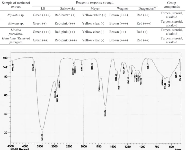
FTIR analysis of Niphates sp. methanol extract
Generally, FTIR Spectrum patterns of the methanol extract of Niphates sp., as shown in Fig. 6, were domi-nated by several broad band absorption, indicating band absorption is overlapped. Appearance of 3421.72 cm-1 was
assigned to be a typical stretching band of OH, supported by a primary or secondary bending band of OH observed at 1242.16 cm-1. A bending band of CO (medium) was
Table 1. Results of specific test group of secondary metabolites. Sample of methanol
extract LB SalkowskyReagent / response strengthMeyer Wagner Dragendorff compoundsGroup Niphates sp. Green (+++) Red-brown (+) Yellow-white (+) Brown (+++) Red (++) Terpen, steroid, alkaloid
Biemna sp. Green (+) Red-pink (++) Yellow clear (-) Brown (+++) Red (+++) Terpen, steroid, alkaloid Liosina
paradoxa, Green (+++) Red-pink (++) Yellow clear (-) Brown (++) Red (+) Terpen, steroid, alkaloid Haliclona (Reniera)
fascigera Green (++) Red-pink (+++) Yellow clear (-) Brown (+++) Red (++) Terpen, steroid, alkaloid
observed at 1037.70 cm-1, and bands absorption at
2953.02 cm-1, 2927.94 cm-1 and 2856.56 cm-1 were
esti-mated to be a C-H stretching of CH3 and CH2 groups.
A shoulder observed at about 3190 cm-1 could be
esti-mated as band absorption of =C-H olefin, supported by appearance of 1635.64 cm-1 for a C=C stretching band.
Stretching bands of CH3 and CH2 groups were supported
by their bending absorption at 1406.11 cm-1and 1452.40
cm-1, and a methyne band. Band absorption at 1735.93
cm-1 were estimated as a C=O stretching even though
medium band but supported by overtone (weak) at 3738.05 cm-1. Sharp bands absorption at 615.29 cm-1 and
659.66 cm-1 could be estimated as cis CH out of plane
olefin or stretching CBr for aliphatic bromo compounds. Broad bands observed at 3421.72 cm-1 and 1635.64 cm-1
could be assigned as overlapping band absorption of OH stretching, NH stretching, and the bending.
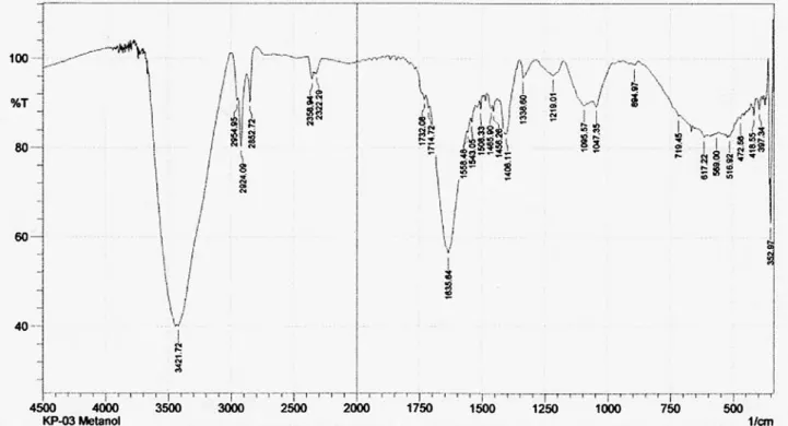
FTIR analysis of Biemna sp methanol extract
FTIR spectrum patterns of the methanol extract of Biemna sp., as shown in Fig. 7, were similar to those of Niphates sp. A broad band at 3421.72 cm-1 is assigned
as a typical stretching band of OH, supported by a weak primary or secondary bending band of OH at 1219.01 cm-1, a bending band CO (medium) at 1095.57 cm-1,
and 1047.35 cm-1. Aliphatic bands absorption of CH
stretching of CH3 and CH2 groups typically appeared at
2954.95 cm-1, 2924.09 cm-1, and 2852.72 cm-1. Bands
absorption at 1465.90 cm-1, 1456.90 cm-1, and 1406.11
cm-1 arose from typical bending absorption of CH 3 and
CH2 groups in a similar manner to bending absorption
of CH at 1336.60 cm-1. A small shoulder was observed
at about 3190 cm-1, which could be estimated to be band
absorption of =C-H olefin, supported by a stretching band of C=C at 1635.64 cm-1. Weak band absorption
at 1714.72 cm-1 could be estimated as C=O stretching.
Bands absorption overlapped at 617.22 cm-1 and 659.66
cm-1 could be estimated as cis CH out of plane olefin or
stretching C-Br for aliphatic bromo compounds. Band absorption at 719.45 cm-1 was expected to be overlapped
by several bands absorption, which is typical for rocking bending methylene groups. Bands absorption at 1558.40 cm-1 and 1635.64 cm-1 were estimated as an N-H bending. FTIR Analysis of Liosina paradoxa methanol extract
FTIR spectrum patterns of the methanol extraxt of Liosina paradoxa, as shown in Fig. 8, were similar to those of Biemna sp. It was also dominated by several broad band sorption.
FTIR Analysis of Haliclona (Reniera) fascigera methanol extract
Bands absorption patterns of the methanol extract of Haliclona (Reniera) fascigera, as shown in Fig. 9, were
simpler than those in Figs 6-8. A broad band at 3419.79 cm-1 was assigned to be a typical stretching band of OH,
supported by a primary or secondary bending band of OH at 1201.65 cm-1 and bending band CO (strong) at
1047.35 cm-1. Bands absorption at 2990.73 cm-1, 2927.94
cm-1, and 2856.56 cm-1 were estimated as a stretching
C-H of CH3 and CH2 groups, and shoulders observed at
3200 cm-1 and 3090 cm-1 could be estimated as bands
absorption of =C-H olefin and ≡C-H, supported by stretching bands at 1643.36 cm-1 and at 2067.69 cm-1 for
C=C and C≡C, respectively. Band absorption at 1739.79 cm-1 was assigned to be a stretching band of C=O,
sup-ported by overtone (weak) near 3700 cm-1. Sharp band
absorption at 611.43 cm-1 could be estimated as cis C-H
out of plane olefin or stretching C-Br for aliphatic bromo compounds. Band absorption at 740.6 cm-1 was estimated
Fig. 9. FTIR Analysis of Haliclona (Reniera) fascigera methanol extract Fig. 8. FTIR Analysis of Liosina paradoxa methanol extract
as rocking band CH2 or stretching band C-Cl. Broad
bands absorption at 3419.79 cm-1 and 1558.48 cm-1 could
be estimated by overlapping of band absorption for OH stretching and either NH or NH2 stretching and bending. Conclusion
All extracts identified contain alkaloid, steroid and terpenoid. Typical band sorption stretching CH aliphatic, CH olefin, OH, C-O, C=C, bending CH3 and CH2 appear
in all extract. Its mean in all extracts contain aliphatic, olefinic and alcohol compounds. Typical band sorption NH for alkaloid also appears in all extracts. The strength of these bands sorption varied from weak to medium, overlapping with other bands. Niphates sp., Liosina par-adoxa, Haliclona (Reniera) fascigera were cytotoxic. Acknowledgment
We gratefully acknowledge to DIKTI and Hasanuddin University for funding and facilities. And also Thanks to Tanjungpura University for all supporting and especially thanks to Mr. Tri Haryono Hadi in LON LIPI.
Reference
Julia Bensemhouna, Isabelle Bombardaa, Maurice Akninb, Jean Vaceletc and Emile M. Gaydoua, 2008. 5α, 8α-Epidioxysterol from the marine sponge Biemna triraphis Topsent. Natural Product Communications, 3: No. 5, 843.
McLaughlin, J.L, Rogers, L.L., 1998, The Use of Biological Assays to Evaluate Botanicals, Drug Information Journal, 32: 513-524.
Meyer B. N., Ferrigni N. R., Putnam J. E., Jacobsen L. B., Nichols D. E., and Mclauglin J. L. 1982. Brine shrimp: a convenient general bioassay for the active plant constituents. Planta Medica, 45: 31-34.
Nathalie Legrave, Souhir Hamrouni-Buonomo, Maeva Dufies, Vincent Guérineau, Jean Vacelet, Patrick Auberger, Philippe Amade and Mohamed Mehiri, 2013. Nepheliosyne B, a New Polyacetylenic Acid from the New Caledonian Marine Sponge Niphates sp. Mar. Drugs, 11: 2282-2292.
Sebastian Engel, Joseph R. Pawlik, 2000. Allelopathic activities of sponge extracts. Marine Ecology Progress Series, 207: 273–281.
Thacker RW, Becerro MA, Lumbang WA, Paul VJ, 1998. Allelopathic interactions between sponges on a tropical reef. Ecology, 79: 1740-1750.