Isolation and analysis of herpesviruses from wild animals
The United Graduate School of Veterinary Science,
Yamaguchi University
Keita Ishijima
March, 2019
1. GENERAL INTRODUCTION 1
1.1. History of identification of herpesviruses 2
1.2. Virus properties 3
1.3. Classification and distribution of herpesvirus 5
1.4. Replication of herpesvirus 7
1.5. Latency and persistent 8
1.6. Herpesvirus infection in animals 9
1.7. Prevention and treatment 10
1.8. Cetacean herpesviruses and bat herpesviruses 11
2. CHAPTER 1 Isolation of a novel herpesvirus from a Pacific white-sided dolphin 13
2.1. ABSTRACT 14
2.2. INTRODUCTION 15
2.3. MATERIALS AND METHODS 17
2.3.1. Primary cell culture 17
2.3.2. Identification of the putative virus by molecular analysis 17
2.3.3. Phylogenetic analysis 17
2.3.4. PCR and sequencing analysis for the complete glycoprotein B gene 18
2.4. RESULTS 20
2.4.1. Isolation of the novel virus 20
2.4.2. Identification of the novel virus 20
2.4.3. Phylogenetic analysis using a partial DNA polymerase gene 20 2.4.4. The sequence of the complete envelope glycoprotein B gene 20
2.5. DISCUSSION 22
2.6. LEGENDS FOR FIGURES 24
2.7. FIGURES 25
3. CHAPTER 2 The complete genomic sequence of Rhinolophus gammaherpesvirus 1
isolated from a greater horseshoe bat 27
3.1. ABSTRACT 28
3.2. INTRODUCTION 29
3.3. MATERIALS AND METHODS 31
3.3.1. Cell line 31
3.3.2. Primary cell culture and virus cultivation 31
3.3.3. Identification of the putative virus 32
3.3.4. Titration 32
3.3.5. Growth curve 32
3.3.6. Extraction of genomic DNA 33
3.3.7. Next generation sequencing 33
3.3.8. PCR and sequence analysis 34
3.3.9. Analysis and annotation 34
3.3.10. Phylogenetic analysis 34
3.4. RESULTS 36
3.4.1. Virus isolation 36
3.4.2. Identification of novel herpesvirus 36
3.4.3. Propagation 36
3.4.4. Determination of the complete genome 37
3.4.6. The results of phylogenetic analysis 39
3.5. DISCUSSION 40
3.6. LEGENDS FOR FIGURES 42
3.7. FIGURES AND TABLES 44
4. GENERAL CONCLUSION 53
5. ACKNOWLEDGMENTS 56
6. REFERENCE 57
1.1. History of identification of herpesviruses
The oldest description of herpesvirus infection was confirmed in A.D. 100. It was the description of herpesvirus lesion by Herodotus (Mettler, 1947). The oldest descriptions of herpesvirus infection in animals were considered to be the report of Aujeszky's disease in 1902 (Aujeszky, 1902) and the report of Marek's disease in 1907 (Marek, 1907).
The first isolation of herpesvirus is considered the report of experimental infection of human herpesvirus 1 (HHV-1) from human lesions to rabbit corneas (Loewenstein, 1919). Following this report, cultivation and titration of HHV-1 using mice was reported in 1932 (Saddington, 1932). Virus growth in cell culture was demonstrated in 1949 (Enders et al., 1949). After that, cultivation of several kinds of herpesviruses using cell cultures were reported (Sanders et al., 1953). The electron microscope was put to practical use in the 1930's. Electron microscope and cultivation of virus using cell culture system made it possible to identify a partial virus (Green, 1965). In the 1950's and 1960's, identification of several herpesviruses using immunofluorescence were reported (Biegeleisen et al., 1959; Albrecht et al., 1963; Schipper and Chow, 1968; Stevens and Watkins, 1969).
Sanger et al. (1977) and Maxam et al. (1977) developed basics of DNA sequencing methods. Since then, nucleotide sequences of viruses have been reported. DNA sequence data of herpesviral genome have been reported since 1980 (McKnight, 1980). In 1984, the complete genome sequence of human herpesvirus 4 was reported (Baer et al., 1984). Until 1990's, nucleotide sequence data of a number of herpesviruses had been accumulated. The family Herpesviridae are further classified into three subfamilies, Alpha-, Beta-, and Gammaherpesvirinae, according to the biological features. Molecular phylogenetic analysis based on nucleotide sequences of
herpesviruses indicated that the classification of herpesviruses was consist with genetic evolution of herpesviruses (McGeoch and Cook, 1994; McGeoch et al., 1995).
Van Devanter et al. (1996) designed consensus primers for amplification of DNA of various kinds of herpesviruses by PCR. This PCR method was the breakthroughs in identification of novel herpesviruses. In the assay, degenerate primers were targeted to highly conserved sequences of DNA polymerase gene of herpesviruses. Recently, several new methods for DNA sequencing were developed (Ronaghi et al, 1996; Ronaghi et al., 2000; Brenner et al., 2000; Bentley et al., 2008). These massively parallel sequencing methods are called as the next-generation sequencing (NGS) methods. These technologies made it possible to detect and identify novel herpesviruses more easily. Especially, detection and identification of herpesviruses derived from bats were reported (Wu et al., 2012; Sano et al., 2015; Shabman et al., 2016; Geldenhuys et al., 2018).
1.2. Virus properties
Herpesviruses are viruses belonging to the order Herpesvirales. The virion of herpesviruses is spherical and the diameter of HHV-1 is from 170 to 200 nm (Grünewald et al., 2003).
The virion of herpesviruses consists of nucleocapsid, tegument, and envelope. Herpesviral genome is a linear double stranded DNA. Nucleocapsid consists of an icosahedral capsid and a herpesviral genome contained in the capsid. Tegument, which is a virus structure characteristic of herpesvirus, is located in the space between the envelope and capsid. The envelope is derived from the host cell membrane. Herpesviral envelope contains viral proteins, especially glycoproteins (Pellett et al., 2011).
based on the location of repeat sequences and the number of isomers (Roizman and Pellet, 2001; Davison 2007). In type A, the genome is composed of a pair of direct repeats (Terminal Repeat; TR) and a unique region sandwiched between them. The size of TR is diverse. For example, the size of direct repeat of equine herpesvirus 5 (EHV-5) is 10 bp, and the size of direct repeat of equine herpesvirus 2 (EHV-2), belonging to the same genus as EHV-5, is more than 15k bp (Wilkie et al., 2015). In type B, the genome is composed of a pair of direct repeats and a unique region. The difference from type A is that the direct repeat consists of tandem repeat sequences, and the number of repeats is variable. In type C, the genome structure is basically similar to type B, but the repeat sequences, not related to the terminal repetitive sequence, are inserted in the unique region. In type D, there are two unique regions (UL and US) on the genome. The UL and
US are sandwiched between different inverted repeats, TRL/IRL and IRS/TRS respectively.
IRL and IRS are adjacent. In type E, the genome structure is basically similar to type D,
but the difference with type D is that the direct repeats locate at the both termini of viral genome and an inverted repeat of direct repeats insert between IRL and IRS. In type F,
the genome does t include definite direct or inverted repeats.
Herpesvirus genomes contain a lot of open reading frames (ORFs). The number of ORFs ranges from 70 to more than 200 (Pellett et al., 2011). The genomes of several herpesviruses also encode microRNA gene (Piedade and Azevedo-Pereira, 2016). In the mammalian and avian herpesvirus group, about 40 genes are commonly shared, which are termed core genes. These genes are classified into several gene blocks. They encode following proteins concerning for DNA replication machinery, peripheral enzymes, processing and packaging of DNA, egress of capsids from nucleus, capsid assembly, structure of capsid and tegument, envelope glycoproteins, multifunctional regulator of gene expression, and nuclear protein (Davison, 2007; Pellett et al., 2011).
1.3. Classification and distribution of herpesvirus
The classification of herpesviruses will be described according to virus taxonomy released by International Committee on Taxonomy of Viruses (ICTV) and information of nucleotide sequences registered in National Center for Biotechnology Information (NCBI) database.
Herpesviruses belong to the order Hepesvirales. The order Herpesvirales are classified into three families; Herpesviridae, Alloherpesviridae, and Malacoherpesviridae (Davison et al., 2009). Herpesviridae is further classified into three subfamilies: Alpha-, Beta-, and Gammaherpesvirinae.
The subfamily Alphaherpesvirinae includes mammalians, birds, and turtles herpesviruses. The subfamily Alphaherpesvirinae consists of five genera: Iltovirus, Mardivirus, Scutavirus, Simplexvirus and Varicellovirus. The type specie of the genus Simplexvirus is HHV-1. The genome organization of several simplexviruses belongs to type E. The genus Simplexvirus mainly includes primates herpesviruses. Bovine herpesvirus 2 also belongs to the genus Simplexvirus. The type specie of Varicellovirus is human herpesvirus 3 (HHV-3), varicella-zoster virus. The genome organization of several varicelloviruses belongs to type D. This genus includes herpesviruses derived from a wide range of animal species in comparison with simplexviruses. The genera Mardivirus and Iltvirus consist of viruses infecting to birds. All avian herpesviruses belong to the subfamily Alphaherpesvirinae or unassigned herpesvirus, not to Beta- or gammaherpesviruses. The type specie of the genus Mardivirus is gallid herpesvirus 2, which is the pathogen of Marek's disease. The type specie of the genus Iltovirus is gallid herpesvirus 1, which is the pathogen of infectious laryngotracheitis. The type specie of the genus Scutavirus is chelonid alphaherpesvirus 5 (ChHV-5), which is associated with
fibropapillomatosis in sea turtles. Chelonid alphaherpesvirus 6 (ChHV-6) is the alphaherpesvirus derived from turtles, but this herpesvirus was not defined as the genus Scutavirus. ChHV-6 is associated with lung-eye-trachea disease in sea turtles.
The subfamily Betaherpesvirinae consists of four genera: Cytomegalovirus, Muromegalovirus, Proboscivirus, and Roseolovirus. The type specie of the genus Cytomegalovirus is human herpesvirus 5 (HHV-5). The genus Cytomegalovirus includes herpesviruses mainly infecting to primates. The type specie of the genus Muromegalovirus is murid herpesvirus 1. The genus Muromegalovirus includes rodents herpesvirus. The type specie of the genus Roseolovirus is human herpesvirus 6A. The genus Roseolovirus includes herpesviruses in human and several non-human primates. The type specie of the genus Proboscivirus is elephantid herpesvirus 1 (ElHV-1), which is known as elephant endotheliotropic herpesvirus. The genus Proboscivirus contains several elephants herpesviruses.
The subfamily of Gammaherpesvirinae consists of four genera: Lymphocryptovirus, Macavirus, Percavirus and Rhadinovirus. The type specie of the genus Lymphocryptovirus is human herpesvirus 4 (HHV-4), known as Epstein-Barr virus. The genus Lympocryptovirus includes herpesviruses derived from human and non-human primates. The type specie of the genus Rhadinovirus is human herpesvirus 8 (HHV-8), known as Kaposi's sarcoma-associated herpesvirus. The genus Rhadinovirus include herpesviruses derived from various mammalians; human, non-human primates, rodents, ruminants, cetaceans and bats. The type specie of the genus Macavirus is alcelaphine herpesvirus 1, known as causal virus of malignant catarrhal fever virus. The genus Macavirus includes herpesviruses derived from Artiodactyla. The type specie of the genus Percavirus is EHV-2. The genus Percavirus contains herpesviruses derived from Carnivora and Perissodactyla. Some herpesviruses derived from mammals, birds
and reptiles, have not been assigned into these three subfamilies yet.
The family Alloherpesviridae consists of 4 genera, Batrachovirus, Cyprinivirus, Ictalurivirus, and Salmonivirus. Frog herpesviruses belong to the genus Batrachovirus. Fish herpesviruses belong to the genus Cyprinivirus, Ictavirus, and Salmonivirus. The members of the family Alloherpesviridae contain diverse virus species (McGeoch et al., 2006).
The family Malacoherpesviridae consists of 2 genera, Aurivirus and Ostreavirus. They infect to Oyster, Abalone and Chlamys.
1.4. Replication of herpesvirus
Generally, cell-virus adsorption and viral entry to cells are essential for establishment of virus infection of virus. In other words, binding of surface proteins of viruses to receptors on cells, followed by membrane fusion, or endocytosis are essential. Envelope glycoproteins of herpesviruses are known as binding proteins to receptor of host cells. Although the number of envelope glycoproteins varies depending on herpesvirus spiecies, at least three glycoproteins are required for entry; gB, gH, and gL (Spear and Longnecker, 2003) which are encoded in core gen. After membrane fusion, the nucleocapsid and tegument proteins of herpesvirus are released into the cytoplasm of infected cells.
Many DNA viruses, including herpesvirus, need to transfer the viral genome into nuclei of infected cells for viral replication. Because nucleocapsid of herpesvirus is too large to pass thorough nuclear pore, they first bind nucleocupsid to cytoplasmic side of the nuclear pore complex via importin. The capsid has been degraded, then the viral genome is transferred into the nucleus (Cohen et al., 2011).
carried out by RNA polymerase II derived from host machinery (Costanzo et al., 1977). Gene expressions of herpesviruses are divided into three types, immediate early gene, early gene and late gene (Weir, 2001).
In general, viral genomic DNA in host cell nucleus becomes circular (Boehmer and Lehman, 1997). DNA synthesis of herpesvirus genome starts at one or more origins of replication (Mocarski, 2007). In HHV-1, 7 proteins necessary for DNA synthesis are encoded in their genome (Weller and Coen, 2012), and among them, 6 proteins are encoded by core genes. Although it is thought that DNA replication is performed by a rolling cycle, it is more complicated in HHV-1 (Weller and Coen, 2012).
The capsid is assembled in the host cell nucleus. The viral genomic DNA cleaved from concatemer is encapsulated in the mature capsid and form nucleocapsid (Mettenleiter et al., 2009). These proteins important for constituting the capsid, its formation, cleaving the concatemer are encoded by core gen. After nucleocapsid leave from the nucleus to the cytoplasm, the final envelope is acquired in cytoplasmic organelle (Mettenleiter et al., 2006) and the virion is released from the cell by exocytosis.
1.5. Latency and persistent
In general, herpesviruses establish latent or persistent infection. However, there are some exceptions. For example, HHV-6 vertically infects by integration into the chromosome (Hall et al, 2008). Basically, herpesviruses establish latent infection or persistent infection by evasion from the immune mechanism of the host.
Herpesviruses have the evasion mechanisms from complement (Favoreel et al., 2003; Agrawal et al., 2017), antibody (Hook et al., 2007;Farré et al., 2017), MHC class I (Griffin et al., 2010), NK cell (De Pelsmaeker et al., 2018), and apoptosis (Galluzzi et
al., 2008). It was reported that homologs of the host protein encoded on herpesviral genome involved in some of these immune evasion mechanisms.
Sites of latency depend on the virus species. For example, HHV-1, -2, and -3, belonging to the subfamily Alphaherpesvirinae are latent in sensory and cranial nerve ganglia. HHV-5, -6, and 7, belonging to the subfamily Betaherpesvirinae, are latent in following site: monocytes, lymphocytes; various leukocytes; and T-cells, epithelia. HHV-4 and -8, belonging to the subfamily Gammaherpesvirinae are latent in B-cells (Grinde, 2013).
1.6. Herpesvirus infection in animals
Alphaherpesvirus infections in animals often cause respiratory diseases: Infectious bovine rhinotracheitis (Muylkens et al., 2009), Aujeszky's disease in pigs (Mettenleiter, 2000), avian infectious bronchitis ( and Sapats, 2000), equine rhinopneumonitis (van Maanen, 2002), feline viral rhinotracheitis (Gaskell et al., 2007), and canine herpesvirus infection (Buonavoglia and Martella, 2007). These infectious diseases sometimes cause death in the animals. Infection of several alphaherpesviruses causes abortion (Smith, 1997). Marek's disease virus, which causes malignant tumor, is significantly different from other alphaherpesviruses (Venugopal, 2000).
The most well-known betaherpesvirus with high pathogenicity in animals is ElHV-1, belonging to the genus Proboscivirus. ElHV-1 infection causes acute haemorrhagic disease in young Asian elephants (Elephas maximus), and the majority of juvenile Asian elephants deaths are due to ElHV-1 infection (Long et al., 2016). Murid herpesvirus 1 (MuHV-1) is one of the most studied betaherpesvirues, because the virus shares many characteristics with human herpesvirus 5 (Krmpotic et al., 2003). Suid herpesvirus 2 is an unassigned betaherpesvirus and causes inclusion body rhinitis in
pigs (Edington et al., 1976).
The most well-known severe infectious disease caused by gammaherpesvirus infection is malignant catarrhal fever. Although gammaherpesviruses have been isolated from and detected in many mammalian species, their pathogenicity remains unknown, except for some viruses (Ackermann, 2006).
Generally, natural host range of individual herpesviruses was restricted. Nevertheless, cercopithecine herpesvirus 1 (Elmore and Eberle., 2008), suid herpesvirus 1 (Mettenleiter, 2000), alcelaphine herpesvirus 1 and ovine herpesvirus 2 (Russell et al., 2009) cause fatal disease with systemic symptoms, when they infected to the other hosts. It was reported that HHV-1, whose natural host is human, caused fatal disease in New World primate (Epstein and Price, 2009).
1.7. Prevention and treatment
For prevention of herpesvirus infection in animals, some vaccines are available. For examples, a combination vaccine with feline calicivirus and feline parvovirus can be used for prevention from feline herpesvirus 1. Both modified live virus (MLV) vaccines and adjuvanted inactivated virus vaccines can be used (Gaskell et al., 2007). For preventing Marek s disease, Marek s disease virus type 2 and Turkey herpesvirus are used as vaccine (Gimeno, 2008). Recombinant vaccines with a deletion of genes related with the pathogenicity are also used for preventing animals. Thymidine kinase or envelope glycoprotein, especially envelope glycoprotein gE, deleted recombinant virus vaccine are well known (Jones and Chowdhury, 2007; Freuling et al., 2017).
Several studies on antiviral therapy against herpesvirus infections have been carried out, mainly in feline, caprine and equine herpesviruses (Dal Pozzo and Thiry, 2014). Trifluridine, idoxuridine, ganciclovir and acyclovir are recommended for
treatment of disease caused by feline herpesvirus 1. These antiviral drugs may also be co-administered with interferon. However, valaciclovir is known to be toxic in cats (Nasisse et al., 1997). Several cases in which ElHV-1-infected calves of elephants were treated with antiviral drugs were reported (Richman et al., 2000; Schmitt et al., 2000; Dastjerdi et al., 2016).
1.8. Cetacean herpesviruses and bat herpesviruses
More than 250 species of herpesviruses have been found from many animals. However, the characterization of these viruses has been performed in limited
herpesviruses; herpesviruses derived from human, experimental animals, companion animals and livestock animals.
Cetacean herpesviruses are considered difficult for isolation. By PCR using consensus primers, the gene of many kinds of herpesviruses have been detected from cetaceans in both sea and river (Blanchard et al., 2001; Saliki et al., 2006; Smolarek et al., 2006; Manire et al., 2006; van Elk et al., 2009; Arbelo et al., 2010; Bellière et al., 2010; Maness et al., 2011; Miyoshi et al., 2011; Soto et al., 2012; Hart et al., 2012; Rehtanz et al., 2012; Lecis et al., 2014; Sierra et al., 2015; van Beurden et al., 2015; Bellehumeur et al., 2015; Melero et al., 2015; van Elk et al., 2016; Seade et al., 2017; Davison et al., 2017; Grattarola et al., 2018; Sacristán et al., 2018). Complete sequence of the gammaherpesvirus derived from a bottlenose dolphin was determined (Davison et al., 2017). However, only one cetacean-derived gammaherpesvirus has been isolated successfully (van Elk et al., 2009). Therefore, pathogenicity has hardly been elucidated. A few herpesviruses derived from cetaceans are likely to be related to skin lesions, genital lesions, neurological disease (van Elk et al., 2016), and systemic symptoms
(Arbelo et al., 2010). Sero-epidemiology of cetacean herpesvirus has been limited (van Elk et al., 2009).
Genetic information of bat herpesviruses is now increasing by NGS and PCR methods (Wibbelt et al., 2007; Molnár et al., 2008; Razafindratsimandresy et al., 2009; Watanabe et al., 2009; Watanabe et al., 2010; Jánoska et al., 2011; Zhang et al., 2012; Wu et al., 2012; Sasaki et al., 2014; Sano et al., 2015; Zheng et al., 2016; Shabman et al., 2016; Host et al., 2016; Pozo et al., 2016; Salmier et al., 2017; Subdhi et al., 2018; Wada et al., 2018; Geldenhuys et al., 2018; Holz et al., 2018). To date, three bat herpesviruses were isolated and determined complete nucleotide sequences (Zhang et al., 2012; Sasaki et al., 2014; Subudhi et al., 2018). And cell tropisms, predicted proteins, and phylogenic relationship were analyzed in the studies. Since Chiroptera contains many species following Rodentia among Mammalia (Wilson and Reeder, 2005), it is expected that there must be an enormous number of herpesviruses in bat.
In this study, I attempted to isolate viruses from various animal specimens and succeeded in isolation of herpesviruses from a dead Pacific white-sided dolphin
(Lagenorhynchus obliquidens) and a greater horseshoe bat (Rhinolophus ferrumequinum) and genetically characterization to them.
2. CHAPTER 1
2.1. ABSTRACT
During establishment of primary cell culture from the kidney of a dead Pacific white-sided dolphin (Lagenorhynchus obliquidens), a cytopathic effect was observed. Polymerase chain reaction with a set of herpesvirus consensus primers yielded a fragment of the expected size. Nucleotide sequencing of the product indicated that the isolated virus was closely related to an alphaherpesvirus detected in a bottlenose dolphin in the United States, but the identity at the protein level was low (86.6%). Phylogenetic analysis of the encoded sequence confirmed that the new isolate belonged to the subfamily Alphaherpesvirinae and clustered together with other cetacean alphaherpesviruses. The complete glycoprotein B-encoding gene (2,757 bp) was amplified from the novel isolate; the encoded protein was compared with the corresponding protein of other herpesviruses, revealing that this virus belongs to the genus Varicellovirus. Taken together, these results suggest that this virus corresponds to a novel herpesvirus capable of infecting Pacific white-sided dolphins.
2.2. INTRODUCTION
As described in GENERAL INTRODUCTION, herpesviruses have been detected and isolated from a number of animal species and are classified in the order Herpesvirales which comprises three families Alloherpesviridae, Herpesviridae and Malacoherpesviridae. The family Herpesviridae comprises three subfamilies, Alpha-, Beta-, and Gammaherpesvirinae. The Alphaherpesvirinae subfamily is divided into five genera: Iltovirus, Mardivirus, Scutavirus, Simplexvirus and Varicellovirus. Several of the herpesviruses have been recovered from cetaceans, including animals from the genera Phocoenidae, Monodontidae, Delphinidae, Ziphiidae, Kogiidae, and Physeteridae (van Bressem et al., 1999; Smolarek et al., 2006; Arbelo et al., 2010; Miyoshi et al., 2011). These cetacean herpesviruses belonged to the subfamily of Alpha- or Gammaherpesvirinae, and were detected in areas including Japanese coastal waters. However, to our knowledge, only one cetacean-derived gammaherpesvirus has been isolated successfully, and grown on primary kidney cell culture originating from a Harbor porpoise (van Elk et al., 2009).
Reports of gene sequences for cetacean herpesviruses have been limited to part of a locus encoding DNA polymerase, as amplified by polymerase chain reaction (PCR) using herpesvirus consensus primers (Van Devanter et al., 1996). As a result, phylogenetic relationships among cetacean herpesviruses have been analyzed only using this region. Therefore, further genetic analysis was required to clarify the relationship among cetacean herpesviruses and between cetacean herpesviruses and other herpesviruses.
In this study, a novel herpesvirus was isolated from a primary kidney cell culture originating from a dead Pacific white-sided dolphin (Lagenorhynchus obliquidens). The consensus primers yielded a fragment corresponding to part of a DNA
polymerase-encoding gene. Further work provided a complete gene for the major immunogenic protein, glycoprotein B, which is conserved among all herpesviruses. Sequence analysis of the gB-encoding gene permitted a determination of the relationship between this virus and the other herpesviruses.
2.3. MATERIALS AND METHODS 2.3.1. Primary cell culture
To establish a primary cell culture from the kidney of a dead Pacific white-sided dolphin, the kidney cells were dispersed through a mesh and cultured in RPMI1640 (GIBCO, Grand Island, NY, USA) supplemented with 10% fetal calf serum (Hyclone®, Otawa, Canada), 100U/ml of penicillin and 100µg/ml of streptomycin (GIBCO) . The cell culture was incubated at 37 °C in 5% CO2 under humid condition.
2.3.2. Identification of the putative virus by molecular analysis
To identify the putative virus, DNA was extracted from the primary kidney cell culture using a DNeasy tissue kit (QIAGEN, Hilden, Germany) according to the NA polymerase-encoding gene from a potential herpesvirus, the resulting DNA was subjected to PCR using the KOD -Plus- Ver.2 kit (TOYOBO, Osaka, Japan) with consensus primers (VanDevanter et al., 1996). The reaction was carried out via initial denaturation at 94°C for 2 min; 45 cycles of denaturation at 98°C for 10 sec, annealing at 46°C for 30 sec, and extension at 68°C for 60 sec; and final extension at 68°C for 3 min. The PCR product was electrophoresed on agarose; the resulting fragment (approximately 700 bp) was excised and purified using a
Nucleotide sequence of the fragment was determined using a Big Dye Terminator v3.1 kit (Applied Biosystems, Foster City, CA, U.S.A.).
2.3.3. Phylogenetic analysis
The nucleotide and deduced amino acid sequences of the DNA polymerase and envelope glycoprotein B gene were analyzed using blastX search (Johnson et. al. 2008)
and Genetyx 8.0 software (Genetyx, Tokyo, Japan). Amino acid sequences were compared with the previously reported sequences using Clustal W in MEGA 5.05 (Tamura et al., 2011), and the phylogenetic trees were constructed using the maximum likelihood (ML) method in PhyML 3.0 (Guindon et al., 2003).
2.3.4. PCR and sequencing analysis for the complete glycoprotein B gene
-GTC ACA GCC
TCA ACA CTT AAC GCC AAG GTT G- -YBM KYT CBY KRT
TYT GKA GBK BRC ACC ACG- -encoding
locus. Reactions were performed using Ex Taq enzyme (Takara Bio Inc., Shiga, Japan), with initial denaturation at 94°C for 2 min; 40 cycles of denaturation at 98°C for 10 sec, annealing at 51°C for 30 sec and extension at 72°C for 12 min; and final extension at 72°C for 10 min. The amplified fragment (approximately 9 kb) was electrophoresed on agarose, excised, and purified as described in 2.3.2. Sequence obtained from one end of the fragment (using the HVgBR primer) was used to design a virus envelope
glycoprotein B gene- -GGA GAC CAT GGC CTG GAA GGC
AAC-PCR with primers specific for the virus, DpolR1 and gBR1, was performed using LA Taq (Takara Bio Inc.) with initial denaturation at 94°C for 2 min; 40 cycles of denaturation at 98°C for 10 sec followed by annealing and extension at 68°C for 12 min; and final extension at 68°C for 10 min. The PCR product (approximately 9 kb) was purified as described in 2.3.2.
To determine the complete envelope glycoprotein B gene sequence, inverse PCR was performed, taking advantage of a XhoI restriction site noted in the partial sequence of the gB-encoding gene. Specifically, DNA extracted from infected cells was
digested with XhoI, self-ligated, and used as the template for PCR using LA Taq
(Takara Bio Inc -TCG AGT GGT CGG CTT GAT
GTG TG- -CCA CCT TTG TGG GTG AAC CTA
TG-consisted of initial denaturation at 94°C for 2 min; 40 cycles denaturation at 98°C for 10 sec and annealing at 68°C for 10 min; and final extension at 68°C for 10 min. The resulting PCR product was electrophoresed on agarose, excised, purified, and sequenced as described in 2.3.2. using the amplifying primers and primers designed based on the obtained sequences.
2.4. RESULTS
2.4.1. Isolation of the novel virus
During the culturing of the primary from the kidney of a dead Pacific white-sided dolphin, cytopathic effects (CPE) were observed, specifically, the formation of syncytia and multinucleated giant cells, suggesting the presence of a virus.
2.4.2. Identification of the novel virus
PCR with the herpesviral consensus primers yielded a DNA fragment of approximately 700 bp. The nucleotide sequence determined by sequencing analysis was deposited in the DNA data bank of Japan (DDBJ) as accession numbers AB747558. BlastX search with the resulting sequence revealed that the predicted DNA polymerase protein fragment was closely related to that encoded by a bottlenose dolphin herpesvirus detected in the USA (Maness et al., 2011), with identity of 86.6% at the amino acid level, revealing that the newly isolated virus belonged to Alphaherpesvirinae and represented a novel herpesvirus. Therefore, the isolated virus was tentatively named lagenorhynchus herpesvirus 1.
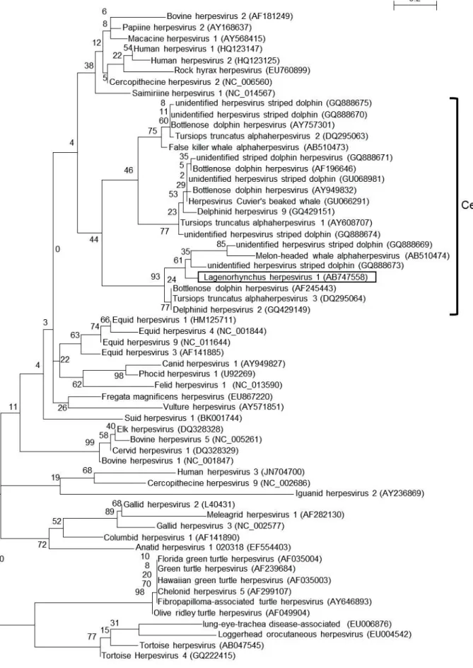
2.4.3. Phylogenetic analysis using a partial DNA polymerase gene
The ML tree using the amino acid sequences of the DNA polymerase proteins of alphaherpesviruses indicated that this Lagenorhynchus herpesvirus 1 belonged to the subfamily Alphaherpesvirinae. In addition, Lagenorhynchus herpesvirus 1 and the other cetacean alphaherpesviruses clustered together and were further divided into three groups. The Lagenorhynchus herpesvirus 1 grouped with herpesviruses detected from striped dolphins in Spain (Bellière et al., 2007), a melon-headed whale in Japan (Miyoshi et al., 2011), and bottlenose dolphins in the USA (Maness et al., 2011;
Blanchard et al., 2001) and Germany (Smolarek et al., 2006) (Fig.2-1).
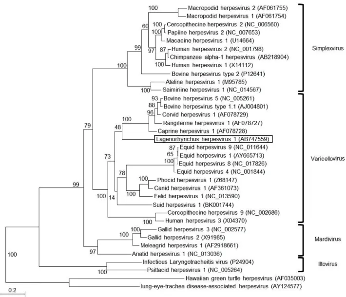
2.4.4. The sequence of the complete envelope glycoprotein B gene
To clarify further the phylogenetic relationship with other herpesviruses, a complete gene encoding the envelope glycoprotein B was recovered and analyzed. The nucleotide sequence determined by sequencing analysis was deposited in the DDBJ as accession number AB747559.
The sequence of the complete envelope glycoprotein B-encoding gene (2,757 bp) of the Lagenorhynchus herpesvirus 1 was compared to that of other herepesviruses, revealing that the predicted gB protein (918 amino acids) was most closely related (61.9% identity at the protein level) to that of Canid herpesvirus 1. Phylogenetic analysis using the amino acids sequence of envelope glycoprotein B gene indicated that the Lagenorhynchus herpesvirus 1 belonged to the genus Varicellovirus (Fig. 2-2).
2.5. DISCUSSION
To our knowledge, this is the first report of the isolation of an alphaherpesvirus from a cetacean. As a result of phylogenetic analysis of alphaherpesviral DNA polymerase proteins, this virus clustered with herpesvirus of cetaceans in the previous study (Miyoshi et al., 2011). Furthermore, phylogenetic analysis using amino acid sequences of envelope glycoprotein B indicated that the Lagenorhynchus herpesvirus 1 belonged to the genus Varicellovirus, suggesting that cetacean alphaherpesviruses should be classified into the genus Varicellovirus.
In summary, the Lagenorhynchus herpesvirus 1 described here was similar to herpesviruses detected in striped dolphins in Spain (Bellière et al., 2007), a melon-headed whale in Japan (Miyoshi et al., 2011), and bottlenose dolphins in the United States (Maness et al., 2011; Blanchard et al., 2001) and Germany (Smolarek et al., 2006) (Fig. 2-1). One of them, a herpesvirus detected from a bottlenose dolphin in USA, was associated with systemic infectious disease, as demonstrated by gross, histologic, ultrastructural, and molecular genetic analyses (Blanchard et al., 2001). Although the pathogenesis of the Lagenorhynchus herpesvirus 1 is still unknown, it is possible that this novel virus causes disease in these cetaceans.
Until then, since only partial sequences of DNA polymerase-encoding gene were available for cetacean herpesvirus, phylogenetic analysis of cetacean herpesviruses was limited to the partial DNA polymerase protein. Subsequently we succeeded in isolating the virus using the primary dolphin kidney cell culture, providing DNA in amounts sufficient to permit cloning and sequencing of the complete gB-encoding gene from the new virus. This novel isolate is expected to be useful for further genetic analysis of cetacean herpesviruses. However, further study will be required to identify cell lines capable of supporting the growth of this virus. In addition, sero-epidemiology
of cetacean herpesvirus remains poorly characterized. The predicted envelope glycoprotein B, which represents the major antigen protein of herpesvirus, will be useful for diagnosis of herpesvirus infection in white-sided dolphins and other cetaceans.
2.6. LEGENDS FOR FIGURES
Figure 2-1. Maximum likelihood (ML) tree of alphaherpesviruses using amino acid
sequences of a portion of the DNA polymerase protein. The Lagenorhynchus herpesvirus 1 is boxed. Analysis was performed by the ML method in phyML 3.0. The number along each branch indicates the bootstrap value. Designations in parentheses are GenBank accession numbers.
Figure 2-2. Maximum likelihood (ML) tree of alphaherpesviruses using amino acid
sequences of glycoprotein B. The Lagenorhynchus herpesvirus 1 is boxed. Analysis was performed by the ML method in phyML 3.0. The number along each branch indicates the bootstrap value. Designations in parentheses are GenBank accession numbers.
Figure 2-1. Maximum likelihood (ML) tree of alphaherpesviruses using amino acid sequences
Figure 2-2. Maximum likelihood (ML) tree of alphaherpesviruses using amino acid sequences
3. CHAPTER 2
The complete genomic sequence of Rhinolophus gammaherpesvirus 1 isolated from a greater horseshoe bat
3.1. ABSTRACT
In a comprehensive research project on bat viruses, we successfully isolated a novel herpesvirus from the spleen of a greater horseshoe bat (Rhinolophus ferrumequinum) in Japan using the cell line established from the kidney of the same bat. This herpesvirus was a novel gammaherpesvirus (Rhinolophus gammaherpesvirus 1; RGHV-1), which belonged to the genus Percavirus. Measurement of the growth of RGHV-1 showed that Vero and CRFK cells were more sensitive to RGHV-1 than other cell lines. Using next-generation sequencing and direct sequencing, the whole RGHV-1 genome was determined (147,790 bp). Twelve of the 84 genes predicted to contain open reading frames did not show any homology with those of other herpesviruses. One of the genes was similar to human Bcl-xl. The accumulation of information on bat viruses will be useful for understanding the emergence of bat-originated zoonotic viruses.
3.2. INTRODUCTION
Bats are the natural hosts of many viruses that cause serious diseases, such as Ebola virus, Marburg virus, Hendra virus, Nipah virus, rabies virus, and rabies-associated lyssaviruses (Calisher et al., 2006). Bats often become reservoirs of these zoonosis and emerging infectious diseases because there are many species of bats (over 1,000 species), they have a relatively long life span for their body size, they undergo hibernation and torpor, during which both virus replication and immune function are reduced, they exhibit a wide range of movements when they fly, and they live in densely populated conditions, especially cave bats (Luis et al., 2013).
Greater horseshoe bats (Rhinolophus ferrumequinum), which inhabit Palaearctic areas, including Japan (Piraccini, 2016), usually rest in caves, but sometimes they also rest in houses in Japan (Abe et al., 2008). Since severe acute respiratory syndrome (SARS)-like coronaviruses have been detected and isolated from this bat species in China, greater horseshoe bats are considered to be a natural host of SARS coronavirus (Lau et al., 2005; Ge et al., 2013). It is important to accumulate information on these viruses that infect greater horseshoe bats to better understand the emergence of bat-originated zoonotic viruses.
Herpesviruses are enveloped and double-stranded DNA viruses. As All herpesviruses derived from mammalians are classified into the family Herpesviridae, which comprises three subfamilies: Alpha-, Beta-, and Gammaherpesvirinae. The Gammaherpesvirinae subfamily is divided into four genera: Lymphocryptovirus, Macavirus, Percavirus, and Rhadinovirus (Davison et al., 2009). Although gammaherpesviruses have been isolated from and detected in many mammalian species, their pathogenicity remains unknown, with the exception of several kinds of viruses isolated from human, mouse, and sheep (Ackermann, 2006).
In our previous studies, we established several bat-derived cell lines for a comprehensive analysis of bat virus infections (Maeda et al., 2008; Maruyama et al., 2014; Maruyama et al., 2016). In addition, we succeeded in isolating a novel adenovirus derived from bats using our established bat kidney T1 (BKT1) cell line, which was derived from the kidney of a horseshoe bat (R. ferrumequinum) (Maeda et al., 2008). In the current study, a novel gammaherpesvirus was isolated from the spleen of a greater horseshoe bat using BKT1 cells, and the complete genome sequence of the virus was determined.
3.3. MATERIALS AND METHODS 3.3.1. Cell line
In this study, our established five cell lines derived from bats, BKT1, FBKT1, YubFKT1, YubFKT2 and DemKT1, and five cultured cell lines, Vero, CRFK, fcwf-4, MDCK and BHK, derived from the other mammals were used. BKT1 were established as described in 3.3.2. The other bat cell lines, FBKT1, YubFKT1, YubFKT2 and DemKT1 were derived from the kidney of a Pteropus dasymallus yayeyamae, the kidney of a Miniopterus fuliginosus, the kidney of a Miniopterus fuliginosus and the kidney of Rousettus leschenaultia, respectively. All of them were transformed with the expression plasmid DNA encoding the large T antigen of replication origin-defective simian virus 40.
All cell lines were maintained with Dulbecco's Modified Eagle's Medium (DMEM, Thermo Fisher Scientific, Waltham, Massachusetts, USA) supplemented with 10 % fetal calf serum and incubated at 37 °C in 5% CO2 under humid condition.
3.3.2. Primary cell culture and virus cultivation
One horseshoe bat, Rhinolophus ferrumquinum, was caught in a cave in Yamaguchi Prefecture in 2006 under the permission of the governor. The primary cell culture was established from the kidney and spleen. During cultivation, CPE) were appeared in the primary spleen cell culture. On the other hand, the primary cells from the kidney were established and transfected with the expression plasmid DNA encoding the large T antigen of replication origin-defective simian virus 40 (pLNCLT), kindly provided by Dr. Yasumoto. Finally, we established BKT1 cell line over 100 passages. The supernatant from the primary spleen cell culture were inoculated on BKT1 cells, the cells were passaged a few times until CPE was observed and the supernatant was
collected and used for further experiments.
3.3.3. Identification of the putative virus
DNA was extracted from the BKT1 infected with the virus using a DNeasy Bloo
as a template in the following PCR. PCR with the herpesvirus consensus primers (VanDevanter et al., 1996) and sequencing analysis was carried out as described in 2.3.2.
3.3.4. Titration
The titer of the virus suspensions of RGHV-1 was determined by the 50% tissue culture infectious dose (TCID50) assay using 96-wells flat bottom microplate. 100 µl of serial 10-fold dilution of virus suspension was applied to each well. Then, 100 µl of Vero cells (5 x 10⁴/ml) were applied to each well and incubated at 37 °C with 5 % CO2 for 8 days. Each well was observed for checking CPE, and the titer of RGHV-1 was calculated using Reed-Muench calculation (Reed et al., 1938).
3.3.5. Growth curve
Five mammalian cell lines, Vero, BHK, CRFK, fcwf-4, and MDCK, and five bat cell lines, BKT1, FBKT1, YubFKT1, YubFKT2 and DemKT1, were passaged to 6 well plates. After cells were 80% confluent, cells were inoculated with RGHV-1 at a multiplicity of infection (MOI) of 0.001. After 7 days post-infection, supernatants of cells were harvested. After centrifugation at 3,500 rpm for 5 minutes at 4 °C, the supernatants were collected and stored at -80 °C. The titer of the virus suspensions was calculated by the 50% tissue culture infectious dose (TCID50) assay using Vero cells.
For growth curve, Vero and CRFK cells were inoculated with RGHV-1 at MOI of 0.001 and the supernatant was collected every 24 hours. The viral titer was determined by TCID50 using Vero cells.
3.3.6. Extraction of genomic DNA
RGHV-1 was inoculated into monolayer of Vero and the supernatant was collected after CPE spread. After centrifugation at 3,000 rpm for 10 minutes at 4 °C, the supernatant was collected. The same operation was carried out at 8,000 rpm for 30 minutes at 4 °C, and the supernatant was recovered. For precipitating of viruses, polyethylene glycol 6,000 (PEG) was added to the virus suspension and mixed by at 4 °C overnight. After centrifugation at 8,000 rpm for 30 minutes at 4 °C, the supernatant was discarded and the pellet was suspended in PBS. This suspension was passed through a layer of 20% sucrose by ultracentrifugation at 25,000 x g for 2 hours and the pellet was suspended in PBS. DNA was extracted from the suspension using DNeasy
3.3.7. Next generation sequencing
For Preparation of fragment libraries from genomic DNA and PCR products, Fragment Library Preparation was used according to the
System according to the manufacturer's protocol. Next Generation Sequencing analysis
the next generation sequence was assembled with SPAdes (Bankevich et al., 2012). Sequences of contigs were confirmed using BWA (Li et al., 2009) and CLC workbench (QIAGEN). The positions of the contigs on the genome were predicted by referring to
the BLASTn (Altschul et al., 1990) results and the genome sequences of gammaherpesvirus.
3.3.8. PCR and sequence analysis
15 pairs of primer sets for PCR were designed based on sequences determined in the next generation sequence analysis. PCR was performed using TaKaRa LA kit (Takara Bio Inc.) and TaKaRa Gflex kit (Takara Bio Inc.). PCR products were electrophoretically analyzed and purified using QIAEXII kit. Nucleotide sequences of the fragments were determined using Sanger sequence, same as described in 2.3.2., or next generation sequence as described in 3.3.7.
3.3.9. Analysis and annotation
To identify repeat regions and inverted repeat regions, we analyzed the RGHV-1 sequence using Tandem Repeats Finder Version 4.07b (Benson et al., 1999) and Inverted Repeats Finder Version 3.07 (Warburton et al., 2004) .
The open reading frames (ORFs) and predicted proteins were analyzed using Genetyx software (version 10; Genetyx, Tokyo, Japan) and the BLASTp program (Gish et al., 1993), respectively.
We searched domains by using the Conserved Domain Database (Marchler-Bauer et al., 2017), and predicted the transmembrane domain by using the SOSUI engine (Hirokawa et al., 1998).
3.3.10. Phylogenetic analysis
All sequences of herpesviruses were collected from DDBJ/GenBank/EMBL database. Amino acid sequences were compared with the previously reported sequences
using Clustal W in MEGA 7 (Kumar et al., 2016), and the phylogenetic trees were constructed using the maximum likelihood (ML) method using MEGA 7 (Kumar et al., 2016).
3.4. RESULTS 3.4.1. Virus isolation
We attempted to establish cell lines from the spleen and kidney of a greater horseshoe bat (R. ferrumequinum). From the kidney, we succeeded in establishing cell lines of BKT1 (Maeda et al., 2008). However, CPE appeared in the primary spleen cell culture. To propagate the virus, we inoculated the supernatant to BKT1 cells. After several passages, CPE were observed in the BKT1 cells.
3.4.2. Identification of novel herpesvirus
To identify the isolate, RNA was extracted from the virus-infected cells and determination of viral RNA sequence (RDV) method was used to determine the sequences (Mizutani et al., 2007). The results indicated that one fragment possessed 51.4% homology with EHV-2 tegument protein at the amino acid sequence level. For the detection of herpesvirus genes, polymerase chain reaction (PCR) was performed using herpesvirus consensus primers (VanDevanter et al., 1996), and the nucleotide sequence was determined. The results showed that 682 bp of the amplified gene was homologous to those of gammaherpesviruses. The nucleotide sequence was deposited into the DNA Data Bank of Japan (DDBJ) under accession number AB298558. The isolated virus was tentatively designated as Rhinolophus gammaherpesvirus 1 (RGHV-1).
3.4.3. Propagation
RGHV-1 was propagated in BKT1 cells, but it was difficult to measure the virus titers using BKT1 cells, because CPE appeared in the BKT1 cells after a few passages following inoculation with RGHV-1. For the propagation of RGHV-1, we
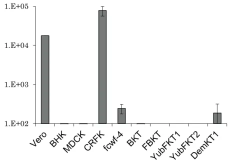
inoculated RGHV-1 to Vero cells and CPE were observed 7 days after infection without passaging of the cells. In further experiments, we used Vero cells for titrating RGHV-1. To compare the sensitivity of the cell lines, 10 mammalian cell lines, including Vero (monkey), BHK (hamster), CRFK (cat), fcwf-4 (cat), MDCK (dog), and five of our established bat cell lines, BKT1, FBKT1 (Ryukyu flying fox), YubFKT1, YubFKT2 (Eastern bent-winged bat), and DemKT1 (Leschenault's rousette), were inoculated with RGHV-1 at a multiplicity of infection of 0.001. During 7 days of observation, CPE were observed only in the Vero and CRFK cells, with Vero cells showing more CPE than the CRFK cells. Seven days after infection, supernatants were collected and the viral titers were determined using the 50% tissue culture infectious dose (TCID50) method in Vero cells. The virus titers in Vero, CRFK, fcwf-4, and DemKT1 cells were all over 100 TCID50/ml. The levels were particularly high in Vero and CRFK cells, with virus titers exceeding 1,000 TCID50/ml. In contrast, the virus titers in the other six cell lines were less than 100 TCID50/ml (Fig. 3-1).
Next, virus growth was sequentially compared between the Vero and CRFK cells. Results showed that RGHV-1 grew better in CRFK cells than in Vero cells, and the virus kept growing for 9 days (Fig. 3-2).
3.4.4. Determination of the complete genome
To determine the complete genome of RGHV-1, we extracted DNA from the viral supernatant of Vero cells infected with RGHV-1. Next-generation sequencing was performed, and 314,997 reads were obtained. Next, we performed PCR for the sequences between the contigs, and the PCR products were analyzed by direct sequencing and
confirmed by mapping.
To predict the termini of the RGHV-1 genome, we performed a search for the pac-1 signal sequence (Deiss et al., 1986). It is known that the pac-1 site is present in the vicinity of the 5'-end in equine herpesviruses 2 and 5 (Agius et al., 1992). In the
RGHV-1 genome, a pac-1-
-GGGGGGTCAACTAAGGGGGGGG-was found. Next, concatemeric DNA -GGGGGGTCAACTAAGGGGGGGG-was confirmed by PCR using primers flanking the pac-1 site and nucleotide sequence analysis. The complete genome of RGHV-1 was deposited into the DDBJ under accession number LC333428.
The complete genome of RGHV-1 was 147,790 bp, and it had a G+C content of 44.7%. The genome structure included a unique region flanked by tandem terminal repeats (4,721 bp) of identical sequences at each end (Fig. 3-3). The terminal repeats had a G+C content of 55.3%. We found several repeat regions, but did not find any of the telomeric repeats that were observed in several herpesviruses (Kishi et al., 1998).
3.4.5. ORFs in the genome
Eighty-four ORFs were identified (Fig. 3-3 and Table 3-1), and a homology search indicated that 44 ORFs were herpesvirus core genes that have been described in previous studies (Arvin et al., 2002). Of the other 40 ORFs, 28 ORFs encoded proteins that were homologues of other herpesvirus proteins, while the remaining 12 ORFs did not.
Among the 12 ORFs that did not show homology with proteins in other herpesviruses, the functions of two ORFs were predicted. The first was a protein encoded by ORF 4 that had 26% homology with the RING finger membrane protein of Cordyceps brongniartii by BLASTp analysis. The results of domain search showed that the RING-variant domain was located in the first third of the protein, and two
transmembrane helices were also in the middle of the protein. This structure was similar to that of a modulator of immune recognition (MIR) in herpesviruses (Lehner et al., 2005) despite the lack of homology, so it was speculated that this protein might function as a MIR. In addition, RGHV-1 possessed another ORF 3 encoding MIR.
The other predicted protein was encoded by ORF 13, which had 30.3% homology with human Bcl-xl, but no homology with viral Bcl-xl, and the BH-1, -2, and -4 domains were conserved.
3.4.6. The results of phylogenetic analysis
Phylogenic analysis of glycoprotein B revealed that RGHV-1 belongs to genus Percavirus (Fig. 3-5), and phylogenic analysis of the other core gene also suggested a similar classification.
3.5. DISCUSSION
To our knowledge, this is the first report of the complete sequence of a virus of the genus Percavirus derived from bats. Recently, the nucleotide sequence of Myotis herpesvirus 8 (MyHV-8) was reported using next-generation sequencing, but the terminal region could not be determined (Host et al., 2016). Within the genus Percavirus, the complete genomes of EHV-2, -5, and Felis catus gammaherpesvirus 1 have been reported, but the sizes of the terminal repeat regions and the inverted repeat sequence regions differed from those of RGHV-1 (Fig. 3-4). Thus, this RGHV-1 might have unique characteristics. A repeat region similar to the telomere-like repeats of EHV-2 was found in the terminal repeats, but telomeric sequences were not included. In this unique region, a pair of inverted repeat sequences was found, but the position appeared to differ from those of EHV-2 and -5.
Since RGHV-1 possesses several proteins related to immune evasion, it was hypothesized that these viral proteins might efficiently function in bat cell lines and persistent infection might have been established. Host et al. also reported that Myotis gammaherpesvirus 8 (MyHV-8) isolated from tumor-derived bat cultured cells did not cause any observable CPE on bat cell lines during infection (Host et al., 2016).
Susceptibility of cell lines indicated that cats might be susceptible to RGHV-1. Results showed that RGHV-1 grew better in CRFK cells than in Vero cells (Fig. 3-2). In addition, the other cat cell line, fcwf-4 cells, also supported the growth of RGHV-1 (Fig. 3-1). Further studies will be required to determine the pathogenicity of RGHV-1 in mammals, including bats and cats.
Among the herpesviruses, only human herpesvirus 8 (Lehner et al., 2005) and bovine herpesvirus 4 (Goto et al., 2003) have multiple types of MIR. These MIR homologues might suppress major histocompatibility complex expression and
contribute to the immune evasion of RGHV-1. The ORF 13 protein might have a function similar to that of Bcl-xl as an apoptosis inhibitor. In addition, RGHV-1 possessed another Bcl-2 homolog, which was encoded by ORF 8 and was similar to the BALF1 of Epstein-Barr virus that protects cells from apoptosis (Marshall et al., 1999). These two Bcl-2 homologues might have anti-apoptotic activity during RGHV-1 infection.
RGHV-1 was very close to Myotis ricketti herpesvirus 2, with 97.6% homology in the amino acid sequences, indicating that these two viruses must be the same species. The natural host of RGHV-1 needs to be clarified in further analyses.
In conclusion, we isolated a novel gammmaherpesvirus, RGHV-1, and determined its complete genome. The accumulation of information on bat viruses will be useful for understanding the emergence of bat-originated zoonotic viruses.
3.6. LEGENDS FOR FIGURES
Figure 3-1. Comparison of the sensitivity to RGHV-1 among 10 mammalian cell lines.
Vero (monkey), BHK (hamster), CRFK (cat), fcwf-4 (cat), MDCK (dog), BKT1, FBKT1 (Ryukyu flying fox), YubFKT1, YubFKT2 (Eastern Bent-winged Bat), and DemKT1 (Leschenault's rousette) cells were infected with RGHV-1 at a multiplicity of infection of 0.001. The supernatants were collected 7 days after infection, and the titers were measured by the TCID50 method.
Figure 3-2. Growth kinetics of RGHV-1 in Vero and CRFK cells. Cells were infected
with RGHV-1 at a multiplicity of infection of 0.001, and the supernatants were collected every day. The mean values from two independent experiments are shown. Filled boxes and open circles indicate the virus titers in CRFK and Vero cells, respectively.
Figure 3-3. Genome structure of RGHV-1. Arrows with diagonal stripes indicate ORFs
encoding homologues of herpesvirus proteins. Arrows with black dots on a white background indicate ORFs encoding proteins homologous to proteins other than herpesvirus proteins. White arrows indicate ORFs encoding unknown proteins.
Figure 3-4. Comparison of the gammaherpesvirus genome structures. Genome
structures of Equine herpesvirus 2 (EHV-2), Equine herpesvirus 5 (EHV-5), Human herpesvirus 4 (HHV-4), and Human herpesvirus 8 (HHV-8) were obtained from accession numbers indicated.
Figure 3-5. Maximum-likelihood (ML) tree of gammaherpesviruses constructed using
1.E+02 1.E+03 1.E+04 1.E+05
2.0 3.0 4.0 5.0 6.0 1 2 3 4 5 6 7 8 9 L o g10 ( T C ID50 /m l)
Days after infection CRFK
Vero
Table 3-1. Features of predicted protein-coding regions in the RGHV-1 genome.
Location Name of gene Strand Size of protein Note
5420-5734 ORF1 + 104 Hypothetical protein
8356-9228 ORF2 + 290 Hypothetical protein, homolog of equid gammaherpesvirus 2 E2 9508-10062 ORF3 - 184 MIR-like membrane protein
9969-10505 ORF4 - 178 MIR-like membrane protein
11155-12354 ORF5 + 399 Hypothetical protein, homolog of equid gammaherpesvirus 2 E3
12537-13016 ORF6 - 159 Hypothetical protein
13703-17575 ORF7 + 1290 Hypothetical gammaherpesvirus 2 ORF3 protein, homolog of saimiriine
17889-18440 ORF8 + 183 Bcl-2
18667-22065 ORF9 + 1132 Single-stranded DNA-binding protein 22094-24223 ORF10 + 709 DNA packaging terminase subunit 2 24214-26820 ORF11 + 868 Glycoprotein B
27170-30196 ORF12 + 1008 DNA polymerase catalytic subunit
31447-31947 ORF13 - 166 Bcl-2
32275-33285 ORF14 + 336 Hypothetical protein, homolog of equid gammaherpesvirus 2 E6
33553-34935 ORF15 + 460 Hypothetical protein, homolog of human herpesvirus 8 ORF10 35004-36320 ORF16 + 438 Hypothetical protein, homolog of human herpesvirus 8
ORF11
36390-36860 ORF17 - 156 Hypothetical protein 40400-42274 ORF18 - 624 Glycoprotein
42428-44182 ORF19 - 584 Capsid maturation protease 9508-10062 ORF20 - 184 Capsid scaffold protein
44181-44993 ORF21 + 270 Hypothetical protein, homolog of human herpesvirus 8 ORF18 44990-46645 ORF22 - 551 Hypothetical protein, homolog of human herpesvirus 8 ORF19 46494-47219 ORF23 - 241 Hypothetical protein, homolog of human herpesvirus 8 ORF20 47218-49071 ORF24 + 617 Thymidine kinase
49071-51419 ORF25 + 782 Glycoprotein H
51416-52618 ORF26 - 400 Hypothetical protein, homolog of human herpesvirus 8 ORF23 52676-54898 ORF27 - 740 Hypothetical protein, homolog of human herpesvirus 8
ORF24
54900-59036 ORF28 + 1378 Major capsid protein 59093-59995 ORF29 + 300 Capsid triplex subunit 2
60022-60516 ORF30 + 164 Hypothetical protein, homolog of human herpesvirus 8 ORF27 60652-60993 ORF31 + 113 Hypothetical protein, homolog of human herpesvirus 8 ORF28 61091-62233,
65497-66411 ORF32 - 685 DNA packaging terminase subunit 1
62460-63173 ORF34 + 237 Hypothetical protein, homolog of human herpesvirus 8 ORF31
63119-64501 ORF35 + 460 Hypothetical protein, homolog of human herpesvirus 8 ORF32 64494-65507 ORF36 + 337 Hypothetical protein, homolog of human herpesvirus 8
ORF33
66428-67396 ORF37 + 322 Hypothetical protein, homolog of human herpesvirus 8 ORF34 67383-67856 ORF38 + 157 Hypothetical protein, homolog of human herpesvirus 8
ORF35
67741-69075 ORF39 + 444 Tegument serine/threonine protein kinase 69102-70544 ORF40 + 480 Deoxyribonuclease
70499-70723 ORF41 + 74 Myristylated tegument protein 70884-72023 ORF42 - 379 Glycoprotein M
72282-73707,
73799-74361 ORF43 + 662 Helicase-primase subunit
74356-75180 ORF44 - 274 Hypothetical protein, homolog of human herpesvirus 8 ORF42 75217-76923 ORF45 - 568 Capsid portal protein
76898-79267 ORF46 + 789 Helicase-primase helicase subunit 79999-80787 ORF47 - 262 Hypothetical protein
80797-81552 ORF48 - 251 Uracil-DNA glycosylase 81509-81967 ORF49 - 152 Glycoprotein L
82060-83592 ORF50 - 510 Hypothetical protein, homolog of human herpesvirus 8 ORF48 83928-83945,
85019-86923 ORF51 + 640 Protein Rta
84089-84991 ORF52 - 300 Hypothetical protein, homolog of human herpesvirus 8 ORF49 87054-87758 ORF53 + 234 Hypothetical protein
87942-88427 ORF54 + 161 Hypothetical protein 88481-89056 ORF55 + 191 Dihydrofolate reductase
89275-90000 ORF56 + 241 Hypothetical protein, homolog of equid gammaherpesvirus 2 E7A 89997-90995 ORF57 + 332 Hypothetical protein
91035-91463 ORF58 - 142 Hypothetical protein 91523-91858 ORF59 - 111 Glycoprotein N
91972-92844 ORF60 + 290 Deoxyuridine triphosphatase
93042-93707 ORF61 - 221 Hypothetical protein, homolog of human herpesvirus 8 ORF55 93680-96292 ORF62 + 870 Helicase-primase primase subunit
96415-96466,
96986-98361 ORF63 + 475 Multifunctional expression regulator 98551-99039 ORF64 + 162 Hypothetical protein
99043-100107 ORF65 - 354 Hypothetical protein, homolog of human herpesvirus 8 ORF58 100118-101416ORF66 - 432 DNA polymerase processivity subunit
101688-102605ORF67 - 305 Ribonucleotide reductase subunit 2 102654-105047ORF68 - 797 Ribonucleotide reductase subunit 1 105106-106122ORF69 - 338 Capsid triplex subunit 1
ORF63
109128-117659ORF71 + 2843 Large tegument protein 117696-118154ORF72 - 152 Small capsid protein
118181-119413ORF73 - 410 Hypothetical protein, homolog of human herpesvirus 8 ORF66 119413-120261ORF74 - 282 Nuclear egress membrane protein
120358-120633ORF75 - 91 Hypothetical protein, homolog of human herpesvirus 8 ORF67A 120923-122374ORF76 + 483 Hypothetical protein, homolog of human herpesvirus 8 ORF68 122395-123315ORF77 + 306 Nuclear egress lamina protein
123525-124394ORF78 - 289 Thymidylate synthase 125197-125700ORF79 + 167 Hypothetical protein
127721-129001ORF80 - 426 LANA-1
129385-130365ORF81 + 326 Hypothetical protein, homolog of human herpesvirus 8 ORF74 131132-135121ORF82 - 1329 Hypothetical protein, homolog of human herpesvirus 8
ORF75
135246-135659ORF83 + 137 Hypothetical protein 139879-140217ORF84 + 112 Hypothetical protein
Table 3-2. Features of repeat regions in the RGHV-1 genome.
Location Name of repeat Feature of repeat
1-4721 Terminal repeat End of RGHV-1 genome
38259-38637 DRL Direct repeat of nt 126086 to 126455
39890-40372 IRL Inverted repeat of nt 79289 to 79762
79289-79762 IRR Inverted repeat of nt 39890 to 40372
126086-126455 DRR Direct repeat of nt 38259 to 38637
4. GENERAL CONCLUSION
Herpesviruses infect from mammals to molluscs. The site of latent infection, the pathogenicity and the proliferative property are different among subfamilies. Alphaherpesviruses, which cause various diseases in humans, companion animals, and livestock animals, have been well studied. On the other hand, betaherpesviruses and gammaherpesviruses in animals have not been unclear.
In this study, I isolated two novel herpesviruses from wild animals and analyzed.
In CHAPTER 1, I analyzed the herpesvirus isolated from the kidneys of dead domestic dolphins.
In primary cell culture of the kidney of the dolphin died in the aquarium in Japan, cytopathic effect (CPE) mainly composed of syncytia was observed. As the result of PCR using herpesvirus consensus primers and sequencing analysis, the virus was a novel herpesvirus belonging to the subfamily Alphaherpesvirinae (Lagenorhynchus herpesvirus 1). Phylogenetic analysis of the amino acid sequences of DNA polymerase region showed that the novel herpesvirus belongs to the same clade with the other alphaherpesviruses derived from cetaceans.
In order to clarify the phylogenetic relationship with the other alphaherpesviruses, the complete nucleotide sequence (2,757 bp) of the envelope glycoprotein gB gene was determined by PCR using newly designed primers and inverse PCR, suggesting that this herpesvirus belongs to the genus Varicellovirus and is closely related to herpesvirus derived from ruminants. This is the first report on the isolation of alphaherpesvirus derived from cetaceans. In addition, our results suggested
that herpesviruses evolved with their host.
In CHAPTER 2, the novel herpesvirus, RGHV-1 was isolated from the spleen of a great horseshoe bat. RGHV-1 was characterized and the complete nucleotide sequence of RGHV-1 was determined.
I successfully isolated a novel herpesvirus from the spleen of a greater horseshoe bat (Rhinolophus ferrumequinum) in Japan using the cell line established from the kidney of the same bat. The result of RDV methods indicated that this virus is the novel gammaherpesvirus (Rhinolophus gammaherpesvirus 1; RGHV-1). Vero cells and CRFK cells supported viral growth more than the other cell lines.
Using next-generation sequencing and direct sequencing, the whole RGHV-1 genome was determined (147,790 bp). The unique region of RGHV-1 was flanked by terminal direct sequences (4,721bp). In the genome, there are 84 predicted ORFs. 12 ORFs did not show any homology with those of the other herpesviruses. One of the 12 genes was similar to human Bcl-xl. The structure of the other predicted protein was similar to the structure of MIR of HHV-8. Phylogenetic analysis showed that RGHV-1 belongs to the genus Percavirus in the subfamily Gammaherpesvirinae. RGHV-1 was very close to Myotis ricketti herpesvirus 2, with 97.6% homology in the amino acid sequences, indicating that these two viruses must be the same species. The natural host of RGHV-1 needs to be clarified in further analyses.
In this study, I succeeded in isolation of one alphaherpesvirus from a cetacean and one gammaherpesvirus from a bat. Both herpesviruses are novel herpesviruses. Usage of cell cultures and cell lines derived from natural host of virus was effective for
isolation of viruses from animals. Isolation of these viruses made it possible to determine the complete nucleotide sequence of glycoprotein gB of the dolphin herpesvirus and the whole nucleotide sequence of RGHV-1.
Herpesviruses derived from cetaceans are thought to be associated with skin lesions, genital legions, and systemic symptoms. For the virus isolated in CHAPTER 1, further study including more effective viral growth is thought to lead to the diagnosis, prevention and treatment of cetaceans in the zoo.
Some proteins involved in immune evasion were identified in the genome of the RGHV-1 isolated in CHAPTER 2. Analysis of these proteins will be useful for understand of the immune system of the bat, known as the host of multiple zoonotic infections.