Effect of active application of universal adhesives with different etching modes on dentin bond durability after thermal cycling
Nobuyuki Moritake
Nihon University Graduate School of Dentistry, Major in Operative Dentistry
(Directors: Prof. Masashi Miyazaki and Assoc. Prof. Toshiki Takamizawa)
Contents
Summary P. 1 - 4
Introduction P. 5 - 7
Materials and methods P. 7-10
Results P. 11 - 14
Discussion P. 14 - 18
Conclusions P. 18
References P. 19 - 23
Tables P. 24-26
Figures P.27-29
This thesis was structured on the basis of published articles listed below with the additional data.
Moritake N, Takamizawa T, Ishii R, Tsujimoto A, Barkmeier WW, Latta MA, Miyazaki M
(2019) Effect of active application on bond durability of universal adhesives. Oper Dent 44,
188-199.
1 Summary
Universal adhesives have distinctive characteristics compared to the previous generations of adhesive systems. One of the important characteristics of universal adhesives is that they can be used with either etch-and-rinse or self-etch approaches. Universal adhesives enable the practitioner to select the optimal etching approach based on cavity conditions such as depth, size, location and the proportion of enamel and dentin. Previous studies showed that although the enamel and dentin bond strengths of universal adhesives were not higher and were often lower than those of two-step self-etch adhesives, they were equal to or greater than those of single-step self-etch adhesives. However, there is concern that the technology of universal adhesives may not offer a genuine advantage when compared to previous generations of single-step self-etch adhesives.
To enhance the bonding effectiveness of universal adhesives, active adhesive application technique is recommended in clinical situations. Active application of adhesives may lead to micromechanical interactions with the underlying mineralized tissue and penetration into the tooth substrate. Stirring the adhesives may accelerate chemical reactions between hydroxyapatite and functional monomers. However, little information is available on the effect of active application on the long term dentin bond performance of universal adhesives. The purpose of the present study was to determine the effect of active application on dentin bond durability when using different etching modes with universal adhesives, by measuring the bond strength after thermal cycling, coupled with morphological observations of treated surfaces and the resin/dentin interfaces.
Three universal adhesives, All Bond Universal (AB, Bisco), Adhese Universal (AU,
Ivoclar Vivadent), and Scotchbond Universal (SU, 3M Oral Care), were used. All these
universal adhesives are recommended for use with active application. Bovine incisors were
used as substitute for human teeth. The dentin surfaces were wet ground using a sequence of
2
silicone carbide (SiC) papers ending with #320-grit. The prepared specimens were subjected to the following surface treatments (ten specimens per each group): 1) etch-and-rinse mode with active application, 2) etch-and-rinse mode with inactive application, 3) self-etch mode with active application, and 4) self-etch mode with inactive application. The adhesive agents were applied using a micro-brush either with (active) or without (inactive) rubbing motion.
An Ultradent bonding assembly was used in this study. Following adhesive application to the dentin adherent surface, the resin composite was condensed into the mold, and light irradiated for 30 s. The samples were stored in distilled water at 37°C for 24 h and then treated with 5,000, 10,000, 30,000, or 50,000 thermal cycles (TC) between 5 and 55°C with a dwell time of 30 s. Baseline specimens were stored in distilled water at 37°C for 24 h before the shear bond strength (SBS) tests. The SBS tests were conducted using a universal testing machine.
After testing, the bonding sites on the tooth surfaces and the resin composite cylinders were observed to determine the failure mode.
Representative treated dentin surfaces, resin-dentin interfaces and de-bonded fracture
sites were observed using scanning electron microscopy (SEM). Dentin surfaces were treated
in accordance with the experimental protocol for bonding procedures, then rinsed with
acetone and water. For ultrastructural morphological observations of the resin/dentin
interfaces, bonded specimens stored in 37ºC distilled water for 24 h were embedded in epoxy
resin, and then longitudinally sectioned. The sectioned surfaces were polished to a high gloss
with SiC papers followed by diamond pastes down to a particle size of 0.25 μm. Half of the
polished specimens were etched with HCl solution and deproteinized by immersion in NaOCl
solution to visualize the internalized resin tags. All SEM specimens were dehydrated in
ascending grades of tert-butyl alcohol and then transferred to the chamber of a freeze-drying
system for 30 min. The resin/dentin interface specimens were then subjected to argon-ion
beam etching for 20 s with the ion beam directed perpendicularly to the polished surfaces.
3
The other sectioned resin-dentin interfaces were subjected to argon-ion beam etching for 40 s directed perpendicular to the polished surfaces. The de-bonded specimens from each storage condition were prepared directly for SEM. Finally, all SEM specimens were coated with a thin film of gold in a vacuum evaporator, and observations were performed under SEM at an operating voltage of 10 kV.
Regarding the etch-and-rinse mode, no significant differences were observed in the SBS values between the active and inactive application groups for any of adhesives in the baseline groups. For the TC groups, all the adhesives in the inactive application group showed lower SBS values as the number of TC increased, and significant differences were found between the active and inactive application groups at 50,000 TC.
Regarding the self-etch mode, all the adhesives in the baseline groups showed higher SBS values with the active application method, and the difference was significant for AU.
For the TC groups, all the adhesives in the active application group showed higher SBS values than with inactive application at all TC groups. However neither application method showed significant differences in SBS between the 50,000 TC and the baseline groups.
In representative SEM images of the treated dentin surfaces in etch-and-rinse mode, complete removal of the smear layer, smear plugs and open dentinal tubules were clearly observed for all the adhesives tested regardless of the application method. On the other hand, for the specimens in self-etch mode, the smear layer and the scratch marks caused by the SiC paper were clearly visible regardless of the adhesive and application method.
In representative SEM images of resin/dentin interfaces, all the adhesives showed dense resin tags longer than 50 μm in the etch-and-rinse mode with active application group.
Although resin tags with a similar length were observed in both active and inactive
application groups for AB, the resin tags for SU and AU in the inactive application group
were obviously shorter than those in the active application group. A hybrid layer of
4
approximately 1 - 2 μm was observed in etch-and-rinse mode, regardless of the adhesive or application methods. On the other hand, the resin tags in self-etch mode were sparse, thin and much shorter than those in etch-and-rinse mode and a hybrid layer was not observed, regardless of the adhesive or application method. In representative SEM images of resin/dentin interfaces after argon-ion etching, although the adhesive layers of AU and SU were approximately 10 μm, the adhesive layers of AB were 3 - 5 μm, regardless of the etching mode or application method. All the adhesives in etch-and-rinse mode showed a thin, high-density layer, a reaction layer, below the HL. In representative SEM images of the failure sites after the bond strength test, the appearance of the failure patterns was dependent on the etching mode, application method, and storage condition.
This laboratory study clearly indicated that factors of application method, type of adhesive, and TC significantly influenced the SBS values regardless of the etching mode.
Although the application method did not strongly influence the dentin bond strength in the
immediate bond strength tests, regardless of the etching mode, for dentin bond durability
under thermal cycling stress, application method had a significant influence on dentin bond
strengths in both etch-and-rinse and self-etch modes. From the results of this in vitro study, it
was concluded that the active application might be effective in enhancing the dentin bond
durability of universal adhesives.
5
Introduction
A prepared tooth consists of complex structures with more than one component and unique morphologies. It is essential for restorations to bond effectively to the external and internal walls of a prepared tooth despite the cavity complexity. One of the most significant problems with restorations is that enamel and dentin respond differently to various treatments.
Universal adhesives are versatile and capable of sensitive adaptation to a variety of cavity conditions (1-4). However, work on the best techniques to use with universal adhesive is still at an early stage.
Phosphoric acid etching of dentin before self-etch adhesive application is believed to have a negative impact on bonding (5-7). The procedure results in an incompletely hybridised region in the vicinity of the resin-dentin interface, which may be a critical factor in the bond degradation of self-etch adhesives (5,8,9). Phosphoric acid dissolves the hydroxyapatite (HAp) surrounding the collagen fibrils, causing the chemical bonds between HAp and functional monomers to decline both in quality and quantity (7,8). Therefore, when using self-etch adhesives, avoiding phosphoric acid pre-etching or selective etching only the enamel region are recommended (10,11). In the particular case of enamel bonding when using self-etch adhesives, selective etching of enamel is effective because it creates stronger micromechanical interlocking and increases surface wettability (12,13). However, it is difficult to precisely etch only the enamel region, particularly with small tooth preparations, complex configurations, or proximal surface area. Hence, concerns remain that the phosphoric acid agent may attack the dentin region of the cavity.
Universal adhesives have distinctive characteristics compared to the previous
generations of adhesive systems. These adhesives can be used with different types of
substrates, such as enamel, dentin, silica-based glass ceramics, zirconia ceramics and metal
alloys (14-17). Therefore, universal adhesives can be used not only for direct resin composite
6
restorations but also for indirect restorations. In addition, universal adhesives are expected to help simplify the bonding procedure for patch restorations used to repair aged restorations with flaws on the surface. Another important characteristic of universal adhesives is that they can be used with either etch-and-rinse or self-etch approaches (2,3). Universal adhesives enable the practitioner to select the optimal etching approach based on the cavity conditions such as depth, size, location and the proportion of enamel and dentin. In general, universal adhesives may be classified as single-step self-etch adhesives as a result of their usage and broadly similar composition to single-step self-etch adhesives.
Previous studies (2,3,18,19) showed that although the enamel and dentin bond strengths of universal adhesives were not higher and indeed were often lower than those of two-step self-etch adhesives, they were equal to or greater than those of single-step self-etch adhesives. However, there is concern that the technology of universal adhesives may not offer a genuine advantage when compared to previous generations of single-step self-etch adhesives (14,20). For example, when considering the dentin bonding of universal adhesives in etch-and-rinse mode, concerns are raised that the chemical bonds between HAp and functional monomers may be reduced as a result of lack of HAp, as in case with single-step self-etch adhesives.
To enhance the bonding effectiveness of universal adhesives, an active adhesive
application technique is recommended (1,4). Active application of adhesives may lead to
micromechanical interactions with the underlying mineralised tissue and penetration into the
tooth substrate (21). Stirring the adhesives can also accelerate chemical reactions between
HAp and functional monomers (22). That is, active application may mobilise a greater
number of H
+ions to react with mineral components (4). Imai and others (4) evaluated the
influence of adhesive application methods on enamel bond strength and surface free energy
using four commercially available universal adhesives under different etching modes and
7
concluded that although active application was effective in self-etch mode, it had a negative impact on enamel bond strength in etch-and-rinse mode. Although active application may enhance enamel bonding performance with the self-etch approach, little information is available on the effect of active application on the long term dentin bond performance of universal adhesives in different conditions.
The purpose of the present study was to determine the effect of active application on dentin bond durability when using different etching modes with universal adhesives by measuring the bond strength after thermal cycling, coupled with morphological observations of treated surfaces and the resin/dentin interfaces. The null hypothesis was that active application does not affect dentin bond durability, regardless of the etching mode used.
Materials and methods Study materials
The materials used in this study are shown in Table 1. Three universal adhesives used were: 1) All Bond Universal (AB; Bisco, Schaumburg, IL, USA), 2) Adhese Universal (AU;
Ivoclar Vivadent, Schaan, Liechtenstein) and 3) Scotchbond Universal (SU; 3M Oral Care, St.
Paul, MN, USA). These products are recommended for use with active application.
Phosphoric acid pre-etching agent used was Ultra-Etch (Ultradent Products, South Jordan, UT, USA). Clearfil AP-X (Kuraray Noritake Dental, Tokyo, Japan) was used as a restorative material for bonding to dentin. A visible-light curing unit (Optilux 501; sds Kerr, Danbury, CT, USA) was used, and the light irradiance (average 600 mW/cm
2) of the curing unit was checked using a dental radiometer (Model 100; Demetoron, Lincoln, NE, USA).
Specimen preparation
Extracted mandibular bovine incisors stored frozen for up to two weeks were used.
Approximately two-thirds of the apical root structure of each tooth was removed using a
8
diamond impregnated disc in a low-speed saw (IsoMet 1000; Precision Sectioning Saw, Buehler, Lake Bluff, IL, USA). The labial surface of each tooth was ground with wet #240- grit silicon carbide (SiC) paper (Fuji Star Type DDC; Sankyo Rikagaku, Saitama, Japan) to create a flat dentin surface. Each tooth was mounted in self-curing acrylic resin (Tray Resin II; Shofu, Kyoto, Japan) to expose the flattened area. Dentin adherent surfaces were polished using a water coolant and a series of SiC polishing papers, ending with 320-grit SiC paper (Fuji Star Type DDC). This grit was chosen for consistency with the ISO 29022 (23) Standard for the shear bond strength (SBS) tests, and with clinical conditions.
Thermal cycling (TC) and SBS tests
The prepared dentin adherent surfaces were treated in accordance with the experimental protocol for the bonding procedures (Table 2). Six hundred specimens in total were divided into 12 groups according to the type of adhesive, and subjected to the following surface treatments (10 specimens for each group): 1) etch-and-rinse mode (phosphoric acid applied for 15 s prior to application of the adhesives) with active application, 2) etch-and- rinse mode with inactive application, 3) self-etch mode (without phosphoric acid etching) with active application and 4) self-etch mode with inactive application. The adhesive agents were applied using a micro-brush either with (active) or without (inactive) a rubbing motion.
In the active application group, the adhesive was rubbed for the duration indicated by the manufacturer, whereas in the inactive application group, the adhesive was allowed to stand for the same period of time. An Ultradent bonding assembly (Ultradent Products) was used in this study. Following adhesive application to the dentin adherent surface, bonded resin composite cylinders were built on dentin surfaces with plastic moulds (Bonding Mold Insert;
2.4 mm in internal diameter, approximately 2.5 mm in height, Ultradent Products) in the
fixture (Bonding Clamp; Ultradent Products) against the dentin surfaces. The resin composite
was condensed into the mould and light irradiation was applied for 30 s with a curing unit.
9
The bonded specimens were stored in distilled water at 37°C for 24 h and then treated with 5,000, 10,000, 30,000, or 50,000 TC between 5 and 55°C with a dwell time of 30 s, creating a division into a total of 5 different storage conditions. Baseline specimens were stored in distilled water at 37°C for 24 h before the SBS tests (baseline group). The SBS of the three universal adhesives to dentin was measured using the notched-edge SBS test, as described in ISO 29022 (24). The bonded specimens were loaded to failure at 1.0 mm/min with a shearing fixture (Test Base Clamp; Ultradent Products) using a universal testing machine (Type 5500R; Instron, Canton, MA, USA). SBS values (MPa) were calculated by dividing the peak load at failure by the bonded surface area. After testing, the bonding sites on the tooth surfaces and the resin composite cylinders were observed under an optical microscope (SZH- 131; Olympus, Tokyo, Japan) at a magnification of 10× to determine the bond failure mode.
Based on the percentage of substrate area (adhesive - resin composite - dentin) observed in the de-bonded resin composites and tooth bonding sites, bond failure was classified into 1) adhesive failure, 2) cohesive failure in the composite, 3) cohesive failure in the dentin, or 4) mixed failure, defined as partially adhesive and partially cohesive.
Scanning electron microscopy (SEM) observation
Representative treated dentin surfaces, resin-dentin interfaces and debonded fracture sites were observed using SEM (ERA-8800FE; Elionix, Tokyo, Japan). Dentin surfaces were first treated in accordance with the experimental protocol for bonding procedures, then rinsed with acetone and water. For ultrastructural morphological observations of the resin-dentin interfaces to determine the penetration of the adhesives, the bonded specimens stored in 37ºC distilled water for 24 h were embedded in epoxy resin, and longitudinally sectioned using the low-speed saw. The sectioned surfaces were polished to a high gloss with SiC papers (Fuji Star Type DDC) followed by diamond pastes down to a particle size of 0.25 μm (DP-Paste;
Struers, Ballerup, Denmark). After ultrasonic cleaning for three min, the polished surface was
10
etched with HCl solution (6 mol/L) for 25 s and deproteinised by immersion in 6% NaOCl solution for three min. The de-bonded specimens from each storage condition were prepared directly for SEM. All SEM specimens were dehydrated in ascending grades of tert-butyl alcohol (50% for 20 min, 75% for 20 min, 95% for 20 min, and 100% for two h), and then transferred to a freeze-drying system (Model ID-3; Elionix.) for 30 min. Then the resin- dentin interfaces were subjected to argon-ion beam etching for 20 s.
The other sectioned resin-dentin interfaces were subjected to argon-ion beam etching for 40 s directed perpendicular to the polished surfaces. Finally, all SEM specimens were coated with a thin film of gold in a vacuum evaporator (Quick Coater Type SC-701; Sanyu Denshi, Tokyo, Japan). Observations were performed under SEM at an operating voltage of 10 kV.
Statistical analysis
A statistical power analysis indicated that at least nine samples were necessary for
effective measurement of bond strength. Therefore, this experiment was initially performed
with sample sizes of 10. After gathering the data, post hoc power tests were performed, and
these tests indicated that the sample size was adequate. Because of their homogeneity of
variance (Bartlett test) and normal distribution (Kolmogorov-Smirnov test), the data obtained
from each adhesive were subjected to analysis of variance (ANOVA) followed by Tukey
honestly significant difference test at a significance level of 0.05. Three-way ANOVA was
used for statistical analysis of the etch-and-rinse and self-etch mode SBS data separately. All
statistical analyses were performed using the Sigma Plot software (ver. 11.0; SPSS, Chicago,
IL, USA).
11 Results SBS measurements
Three-way ANOVAs for SBS in etch-and-rinse mode revealed that all the factors evaluated significantly influenced the SBS values (p < 0.001), and the three-way interactions between the evaluated factors and all the interactions were significant (p < 0.05). Three-way ANOVA for the SBS values in self-etch mode revealed that all the factors significantly influenced the SBS (p < 0.001), similar to the results for the etch-and-rinse mode. Although the three-way interaction between the factors and the interactions between the adhesives and TC significantly influenced the SBS values (p < 0.05), other pairwise interactions were not significant (application method vs adhesive: p = 0.302, and application method vs thermal cycle: p = 0.851).
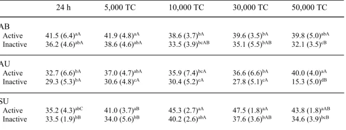
The SBS values in etch-and-rinse mode under different TC are shown in Table 3. For all the adhesives in the baseline groups (24-h water storage), no significant differences were observed in the SBS values between the active and inactive application groups. For the TC groups, all the adhesives in the inactive application group showed lower SBS values as the number of TC increased, and significant differences were found between the active and inactive application groups at 50,000 TC cycles. On the other hand, all of the universal adhesives in the active application group were observed to have reliable SBS values, in that there were no significant SBS reductions in the 50,000 TC groups when compared to those of the baseline groups.
The SBS results in self-etch mode under different numbers of TC are shown in Table
4. For the baseline groups, similar to the etch-and-rinse mode, all the adhesives showed
higher SBS values with the active application method, and the difference was significant for
AU. For the TC groups, all the adhesives in the active application group showed higher SBS
values than with inactive application at all TC conditions. However, neither application
12
method showed significant differences in SBS between the 50,000 TC and the baseline groups. When observing the SBS values under different TC conditions, all the adhesives showed similar, but not identical, patterns of change. The SBS value for AU in etch-and-rinse mode at 50,000 TC was markedly lower than that for the other adhesives.
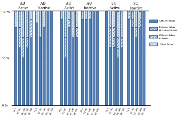
Failure mode analysis
The frequencies of different failure modes are shown in Figs. 1, 2. Adhesive failure was most commonly observed in all de-bonded baseline specimens, regardless of the etching mode, type of adhesive, or application method. When TC was applied, although mixed failure and cohesive failure in dentin were observed frequently for all the adhesives at 5,000 TC, the adhesive failure increased with higher number of TC. In particular, this trend was obvious in self-etch mode with active application regardless of the type of adhesive. When comparing the failure pattern between active and inactive application, adhesive failure was more common in the inactive application group, regardless of the TC condition.
SEM observations
Representative SEM images of the treated dentin surfaces are shown in Figs. 3, 4. In etch-and-rinse mode, complete removal of the smear layer, smear plugs and open dentinal tubules was clearly observed for all the adhesives tested regardless of the application method.
For AU, greater amounts of deposit on the etched surface were observed in the active application group than in the inactive group. On the other hand, for the specimens in self-etch mode, the smear layer and the scratch marks caused by the SiC paper were clearly visible regardless of the adhesive and application method used. However, the smear layer and smear plugs were dissolved in some locations with the active application method, in contrast to the inactive method.
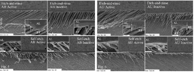
Representative SEM images of demineralised and deproteinised resin/dentin
interfaces are shown in Figs. 5, 6. For all the adhesives, dense resin tags longer than 50 μm
13
were observed in the etch-and-rinse with active application group. Although resin tags with a similar length were observed in both the active and inactive application groups for AB, the resin tags for SU and AU in the inactive application group were obviously shorter than those in the active application group. A hybrid layer of approximately 1 - 2 μm was observed in etch-and-rinse mode, regardless of the adhesive or application methods. On the other hand, the resin tags in self-etch mode were sparse, thin and much shorter than those in etch-and- rinse mode and a hybrid layer was not observed, regardless of the adhesive or application method used. Comparing the penetration status of the resin tags with active and inactive application in self-etch mode, the resin tag penetration with inactive application was much lower than that with active application.
In the SEM images of resin/dentin interfaces after argon-ion beam etching, although the adhesive layer of AU and SU were approximately 10 μm, the adhesive layer of AB was 3
- 5 μm, regardless of the etching mode or application mode. AB showed a homogeneous
adhesive layer due to the absence of inorganic fillers, while the other adhesives showed a heterogeneous adhesive layer. All the adhesives in etch-and-rinse mode showed a thin, high- density layer, as a reaction layer (RL) below the HL (Figs. 7, 8). On the other hand, all the adhesives in self-etch mode revealed rather thick, high-density layer between the adhesive layer and intact dentin. These different layers appeared to be clearer with active application than with inactive application.
Representative SEM images of the failure sites after the bond strength test are shown
in Figs. 9, 10. The appearance of the failure patterns was dependent on the etching mode,
application method, and storage condition. For the baseline groups in etch-and-rinse mode,
the failure sites primarily showed detachment at the adhesive-dentin interface, and evidence
of resin tags was clearly visible, regardless of the application method. For the 50,000 TC
specimens in etch-and-rinse mode, although evidence of resin tags was observed with both
14
application methods, they were less clear than in the baseline specimens. In particular, a flat and smooth failure site was observed with inactive application. For debonded specimens in self-etch mode, the failure site included detachment at both the adhesive-dentin and adhesive- resin composite interfaces at lower magnification, regardless of the storage condition. At higher magnification, although evidence of resin tags was barely observable with both application methods in the baseline groups, it was difficult to observe any evidence of resin tags after TC.
Discussion
In this study, no significant differences in dentin SBS were found between active and inactive application in the baseline groups, with the exception of AB in self-etch mode.
However, all the adhesives showed significantly higher dentin SBS values in the active application groups than in the inactive application groups at 50,000 TC regardless of the etching mode used. Therefore, the null hypothesis that active application would not affect the dentin bond durability, regardless of etching mode, was rejected. Active application of universal adhesives may enhance dentin bond durability as a result of the potential of tooth demineralisation, penetration, and chemical bonding with the dentin substrate.
In self-etch mode, the gel-like collagen in the dentin smear on the sound tissue can
interfere with the penetration of resin monomers contained in adhesives (24,25). SEM
observations of treated-dentin surfaces in self-etch mode showed that actively applied
adhesives can dissolve a certain amount of the smear layer compared to those applied
inactively (Figs. 3, 4). It can be speculated that unreacted H
+ions were supplied from
functional monomers in the adhesive, resulting in the progression of the demineralisation
process (4,21). Previous studies (26-28) have reported the benefits of active application for
optimal dentin bond performance and durability with self-etch adhesives. Increased dentin
15
bond strength with active application has been suggested to be due to the stirring of adhesive- inducing solvent evaporation, resulting in a higher rate of resin monomer incorporation inside the smear layer (27,28). Furthermore, the nanolayering of calcium-salt formed from HAp and the functional monomer is significantly greater with active application than with inactive application (22). This appears to be supported by the SEM observation of the resin/dentin interface in this study, where the high-density layer between the adhesive layer and intact dentin appeared to be thicker with active application than with inactive application (Figs. 7, 8). The induction of a chemical reaction between functional monomers and HAp by active application may also reduce the levels of the acidic monomer, which would contribute to amine co-initiators and enhance the photo-polymerisation of adhesives containing acidic functional monomers (29,30). Therefore, active application in self-etch mode may enhance the dentin bond durability of universal adhesives as a result of the same mechanisms seen with conventional self-etch adhesives.
On the other hand, in the etch-and-rinse mode, phosphoric acid pre-etching solubilises
not only the surface debris but also the subsurface of the dentin substrate (31). A previous
investigation of dentin bond durability after four-year water storage showed that a three-step
etch-and-rinse system resulted in higher microtensile bond strength than a two-step etch-and-
rinse system, and this tendency did not change over four years (32). The primary bonding
mechanism underlying three-step etch-and-rinse systems is thought to be micromechanical
interlocking between demineralised exposed collagen fibrils and resin monomers, leading to
hybrid layer and resin tag formation (31). With phosphoric acid pre-etching performed prior
to the application of a self-etch adhesive, concerns remains that demineralised dentin without
resin impregnation may persist at the bottom of the hybrid layer, which will act as a weaker
region in the vicinity of the resin/dentin interface (5-7). This unstable region increases the
16
risk of biodegradation and biomechanical influences for not only self-etch adhesives but also for universal adhesives (33,34).
For all the adhesives subjected to different numbers of TC with active application, SBS values in etch-and-rinse mode were equal to or greater than those in self-etch mode, with the exception of AU in early cycles. This result was consistent with those of previous studies (2,35) investigating the effects of fatigue stress on the dentin bond durability of universal adhesives in different etching modes. However, SBS values in etch-and-rinse mode with inactive application were lower than those in self-etch mode with active application regardless of the type of adhesive or cycles of TC. Hence, active application is thought to be helpful for the dentin bond durability of universal adhesives in etch-and-rinse mode, similar to the self-etch mode.
The role of resin tag formation in enhancing dentin bond performance is still controversial. SEM images of resin/dentin interfaces in etch-and-rinse mode revealed higher penetration for adhesives with active application (Fig. 6). In addition, the penetration of adhesives into the branches of dentinal tubules was observed in the active application group, in contrast to behaviour in the inactive application group. From the results of the present study, long, thick, and dense resin tags with active application contribute to the higher bond strength due to mechanical retention. Some universal adhesives were designed to have lower hydrophilicity than conventional single-step self-etch adhesives, which may explain the greater resistance of resin tags to hydrolytic degradation. In addition, deep penetration into dentinal tubules and creating branches might enhance chemical bonding between functional monomers and the HAp of internal dentinal tubules.
The penetration capability of adhesives may be closely dependent on their
composition, such as the amount of water, type of solvent, presence or absence of inorganic
fillers, and hydrophilic or hydrophobic resin monomers (36). Although the purity and quantity
17
of each ingredient in the adhesives vary, all the tested adhesives in this study contained the same functional monomer, 10-methacryloyloxydecyl dihydrogen phosphate (MDP), the hydrophilic resin monomer 2-hydroxyethyl methacrylate (HEMA), water and ethanol as a solvent. However, SEM observations of treated dentin surfaces and resin/dentin interfaces were adhesive dependent. In contrast to AB and SU, greater amounts of deposit were observed on the etched surface in AU (Fig. 4).
With regard to resin tag formation, AB showed noticeably longer resin tags than the other adhesives. This is probably due to its application method, in which the adhesive is applied twice without irradiation between the applications. Thus, the second application may push resin deeper into the substrate. For all the adhesives, no significant SBS reduction was found compared to the baseline until 30,000 TC, regardless of the etching mode or application method used. However, the SBS value for AU in etch-and-rinse mode with inactive application after 50,000 TC was markedly lower than that for all the other conditions. The long tags may explain AB’s durability, and SU contains Vitrebond copolymers that utilize
glass ionomer technology that may be biocompatible with exposed collagen fibrils (37). SEM observations revealed that resin monomer penetration in AU was lower than in the other adhesives. This observation is attributed to the presence of aggregated inorganic fillers in AU (Fig. 6). Although the incorporation of inorganic fillers enhances the mechanical properties of the adhesive layer, the ability of the adhesive to flow into tubules might decrease.
The SEM images of resin/dentin interfaces after argon-ion beam etching exhibited a
thin high-density layer below the HL. This thicker, high-density layer has been proposed as
the RL (38), and it might be an evidence of chemical interaction between functional
monomers of universal adhesive and intact dentin below the HL. In addition, this reaction
layer appears to be clearer with active application than with inactive application. It can be
inferred that active application may induce chemical interaction even when using universal
18
adhesives in etch-and-rinse mode. Further research is required to determine the role of RL in the bonding mechanism of universal adhesives in etch-and-rinse mode and their effect on dentin bond durability.
Conclusions
Within the limitations of this laboratory study, all the factors evaluated, namely application method, adhesive and thermal cycle period, significantly influenced the SBS values regardless of the etching mode used. For immediate dentin bond results, application method did not strongly influence the dentin bond strengths, regardless of the etching mode.
However, for dentin bond durability under thermal cycling stress, application method was a significant influence on dentin bond strengths in both etch-and-rinse and self-etch modes.
From the results of this in vitro study, it was concluded that the active application is effective
in enhancing the dentin bond durability of universal adhesives.
19
References
1. Loguercio AD, Muñoz MA, Luque-Martinez I, Hass V, Reis A, Perdigão J (2015) Does active application of universal adhesives to enamel in self-etch mode improve their performance?. J Dent 43, 1060-1070.
2. Takamizawa T, Barkmeier WW, Tsujimoto A, Berry TP, Watanabe H, Erickson RL, Latta MA, Miyazaki M (2016) Influence of different etching modes on bond strength and fatigue strength to dentin using universal adhesive systems. Dent Mater 32, e9-e21.
3. Suzuki T, Takamizawa T, Barkmeier WW, Tsujimoto A, Endo H, Erickson RL, Latta MA, Miyazaki M (2016) Influence of etching mode on enamel bond durability of universal adhesive systems. Oper Dent 41, 520-530.
4. Imai A, Takamizawa T, Sai K, Tsujimoto A, Endo H, Barkmeier WW, Latta MA, Miyazaki M (2017) Influence of application method on surface free-energy and bond strength of universal adhesive systems to enamel. Eur J Oral Sci 125, 385-395.
5. Van Landuyt KL, Kanumilli P, De Munck J, Peumans M, Lambrechts P, Van Meerbeek B (2006) Bond strength of a mild self-etch adhesive with and without prior acid-etching. J Dent 34, 77-85.
6. Van Landuyt KL, Peumans M, De Munck J, Lambrechts P, Van Meerbeek B (2006) Extension of a one-step self-etch adhesive into a multi-step adhesive. Dent Mater 22, 533- 544.
7. Ikeda M, Tsubota K, Takamizawa T, Yoshida T, Miyazaki M, Platt JA (2008) Bonding durability of single-step adhesives to previously acid-etched dentin. Oper Dent 33, 702- 709.
8. Sato M, Miyazaki M (2005) Comparison of depth of dentin etching and resin infiltration
with single-step adhesive systems. J Dent 33, 475-484.
20
9. Soares CJ, Castro CG, Santos Filho PC, da Mota AS (2007) Effect of previous treatments on bond strength of two self-etching adhesive systems to dental substrate. J Adhes Dent 9, 291-296.
10. Frankenberger R, Lohbauer U, Roggendorf MJ, Naumann M, Taschner M (2008) Selective enamel etching reconsidered: Better than etch-and-rinse and self-etch?. J Adhes Dent 10, 339-344.
11. Peumans M, De Munck J, Van Landuyt KL, Poitevin A, Lambrechts P, Van Meerbeek B (2010) Eight-year clinical evaluation of a 2-step self-etch adhesive with and without selective etching. Dent Mater 26, 1176-1184.
12. Erickson RL, Barkmeier WW, Kimmes NS (2009) Bond strength of self-etch adhesives to pre-etched enamel. Dent Mater 25, 1187-1194.
13. Erickson RL, Barkmeier WW, Latta MA (2009) The role of etching in bonding to enamel: A comparison of self-etching and etch-and-rinse adhesive systems. Dent Mater 25, 1459-1467.
14. da Rosa WL, Piva E, da Silva AF (2015) Bond strength of universal adhesives: A systematic review and meta-analysis. J Dent 43, 765-776.
15. Xie H, Li Q, Zhang F, Lu Y, Tay FR, Qian M, Chen C (2016) Comparison of resin bonding improvements to zirconia between one-bottle universal adhesives and tribochemical silica coating, which is better?. Dent Mater 32, 403-411.
16. Siqueria F, Cardenas AM, Gutierrez MF, Malaquias P, Hass V, Reis A, Loguercio AD, Perdigão J (2016) Laboratory performance of universal adhesive systems for luting CAD/CAM restorative materials. J Adhes Dent 18, 331-340.
17. Tsujimoto A, Barkmeier WW, Takamizawa T, Wilwerding TM, Latta MA, Miyazaki M
(2017) Interfacial characteristics and bond durability of universal adhesives to various
substrates. Oper Dent 42, e59-e70.
21
18. Tsujimoto A, Barkmeier WW, Takamizawa T, Watanabe H, Johnson WW, Latta MA, Miyazaki M (2017) Comparison between universal adhesives and two-step self-etch adhesives in terms of dentin bond fatigue durability in self-etch mode. Eur J Oral Sci 125, 215-222.
19. Fujiwara S, Takamizawa T, Barkmeier WW, Tsujimoto A, Imai A, Watanabe H, Erickson RL, Latta MA, Nakatuka T, Miyazaki M (2018) Effect of double-layer application on bond quality of adhesive systems. J Mech Behav Biomed Mater 77, 501- 509.
20. Chen C, Niu LN, Xie H, Zhang ZY, Zhou LQ, Jiao K, Chen JH, Pashley DH, Tay FR (2015) Bonding of universal adhesives to dentin - Old wine in new bottles?. J Dent 43, 525-536.
21. Loguercio AD, Stanislawczuk R, Mena-Serrano A, Reis A (2011) Effect of 3-year water storage on the performance of one-step self-etch adhesives applied actively on dentine. J Dent 39, 578-587.
22. Yoshihara K, Yoshida Y, Hayakawa S, Nagaoka N, Irie M, Ogawa T, Van Landuyt KL, Osaka A, Suzuki K, Minagi S, Van Meerbeek B (2011) Nanolayering of phosphoric acid ester monomer on enamel and dentin. Acta Biomater 7, 3187-3195.
23. International Organization for Standardization (2013) Dentistry-Adhesion-notched-edge shear bond strength test. ISO 29022; 2013, Geneva.
24. Thanatvarakorn O, Nakajima M, Prasansuttiporn T, Ichinose S, Foxton RM, Tagami J (2014) Effect of smear layer deproteinizing on resin-dentine interface with self-etch adhesive. J Dent 42, 298-304.
25. Takamizawa T, Barkmeier WW, Sai K, Tsujimoto A, Imai A, Erickson RL, Latta MA,
Miyazaki M (2018) Influence of different smear layers on bond durability of self-etch
adhesives. Dent Mater 34, 246-259.
22
26. Amaral RC, Stanislawczuk R, Zander-Grande C, Michel MD, Reis A, Loguercio AD (2009) Active application improves the bonding performance of self-etch adhesives to dentin. J Dent 37, 82-90.
27. Amaral RC, Stanislawczuk R, Zander-Grande C, Gagler D, Reis A, Loguercio AD (2010) Bond strength and quality of the hybrid layer of one-step self-etch adhesives applied with agitation on dentin. Oper Dent 35, 211-219.
28. Zhang Y, Wang Y (2013) Effect of application mode on interfacial morphology and chemistry between dentine and self-etch adhesives. J Dent 41, 231-240.
29. Tay FR, Pashely DH, Yiu CKY, Sanares AME, Wei SHY (2003) Factors contributing to the incompability between simplified-step adhesives and chemically-cured or dual-cured composites. Part I. Single-step self-etching adhesive. J Adhes Dent 5, 27-40.
30. Moszner N, Salz U, Zimmermann J (2005) Chemical aspects of self-etching enamel- dentin adhesives: a systematic review. Dent Mater 21, 895-910.
31. Pashely DH, Tay FR, Breschi L, Tjӓderhane L, Carvalho RM, Carrilho M, Tezvergil- Mutluay A (2011) State of the art etch-and-rinse adhesives. Dent Mater 27, 1-16.
32. De Munck J, Van Meerbeek B, Yoshida Y, Inoue S, Vargas M, Suzuki K (2003) Four- year water degradation of total-etch adhesives bonded to dentin. J Dent Res 82, 136-140.
33. Mazzoni A, Scaffa P, Carrilho M, Tjӓderhane L, Di Lenarda R, Polimeni A, Tezvergil- Mutlay A, Tay FR, Pashley DH, Breschi L (2013) Effects of etch-and rinse and self-etch adhesives on dentin MMP-2 and MMP-9. J Dent Res 92, 82-86.
34. Takamizawa T, Barkmeier WW, Tsujimoto A, Scheidel DD, Watanabe H, Erickson RL,
Latta MA, Miyazaki M (2015) Influence of water storage on fatigue strength of self-etch
adhesives. J Dent 43, 1416-1427.
23
35. Takamizawa T, Barkmeier WW, Tsujimoto A, Suzuki T, Scheidel DD, Erickson RL, Latta MA, Miyazaki M (2016) Influence of different pre-etching times on fatigue strength of self-etch adhesives to dentin. Eur J Oral Sci 124, 210-218.
36. Van Meerbeek B, Yoshihara K, Yoshida Y, Mine A, De Munck J (2011) State of the art of self-etch adhesives. Dent Mater 27, 17-28.
37. Nezu T, Winnik FM (2000) Interaction of water-soluble collagen with poly (acrylic acid).
Biomater 21, 415-419.
38. Takamizawa T, Imai A, Hirokane E, Tsujimoto A, Barkmeier WW, Erikson RL, Latta
MA, Miyazaki M (2019) SEM observation of novel characteristic of the dentin bond
interfaces of universal adhesives. Dent Mater 35, 1791-1804.
24 Table 1: Materials used in this study
Code Adhesive Main components Classification Manufacturer
(Lot No.) according to pH
AB All-Bond Universal MDP phosphate monomer, Bis-GMA, Ultra-mild Bisco, Schaumburg, (1300008503) HEMA, ethanol, water, initiators (pH = 3.2)* IL, USA AU Adhese Universal MDP, Bis-GMA, HEMA, MCAP, Ultra-mild Ivoclar Vivadent,
(U49302) D3MA, ethanol, water, initiator, (pH = 2.5-3.0)* Schaan, Lichtenstein silicon dioxide, stabilizers
SU Scotchbond Universal MDP phosphate monomer, Ultra-mild 3M Oral Care, (41256) HEMA, dimethacrylate resins, (pH = 2.7)* St. Paul, MN, USA
Vitrebond copolymer, filler, ethanol, water, initiators, silane
Etching agent Ultradent Products,
Ultra-Etch 35% phosphoric acid South Jordan, UT, USA
(G017)
Resin composite
Clearfil AP-X Bis-GMA, TEGDMA, silane barium glass filler, Kuraray Noritake Dental, (N416713) silane silica filler, silanated colloidal silica, Tokyo, Japan
catalysts, accelerators, CQ, pigments, others
Abbreviations: MDP, 10-methacryloyloxydecyl dihydrogen phosphate; Bis-GMA, 2,2-bis [4-(2- hydroxy-3-methacryloyloxypropoxy) phenyl] propane; HEMA, 2-hydroxyethyl methacrylate; MCAP, methacrylated carboxylic acid polymer; D3MA, decandiol dimethacrylate;
TEGDMA, triethyleneglycol dimethacrylate; CQ, dl-camphorquinone;
* References 1, 18.
25
Table 2: Application protocol for pre-etching and self-etching adhesives
Method Pre-etching protocol
Etch-and-rinse Dentin surface was conditioned with phosphoric acid for 15 s.
Conditioned surface was rinsed with water for 15 s and air-dried.
Self-etch Phosphoric acid pre-etching was not performed.
Code Application methods Adhesive application protocol
AB Active application Adhesive was applied to dentin surface (not desiccated) with rubbing action for 10-15 s per coat. No light cure between coats. Gentle stream of air applied over the liquid for at least 10 s. Light irradiation performed for 10 s.
Inactive application Adhesive was applied to dentin surface (not desiccated) without rubbing action for 10-15 s per coat. No light cure between coats. Gentle stream of air applied over the liquid for at least 10 s. Light irradiation performed for 10 s.
AU Active application Adhesive was applied to the dentin surface with rubbing action for 20 s, followed by application of medium air pressure for 5 s. Light irradiation performed for 10 s.
Inactive application Adhesive was applied to the dentin surface without rubbing action for 20 s, followed by application of medium air pressure for 5 s. Light irradiation performed for 10 s
SU Active application Adhesive was applied to the dentin surface with rubbing action for 20 s, followed by application of medium air pressure for 5 s. Adhesive light cure for 10 s.
Inactive application Adhesive was applied to the dentin surface without rubbing action for 20 s, followed by application of medium air pressure for 5 s. Adhesive light cure for 10 s.
26
Table 3: Influence of thermal cycling (TC) on shear bond strength (SBS)-Etch-and-rinse mode
24 h 5,000 TC 10,000 TC 30,000 TC 50,000 TC
AB
Active 41.5 (6.4)aA 41.9 (4.8)aA 38.6 (3.7)bA 39.6 (3.5)bA 39.8 (5.0)abA Inactive 36.2 (4.6)abA 38.6 (4.6)abA 33.5 (3.9)bcAB 35.1 (5.5)bAB 32.1 (3.5)cB
AU
Active 32.7 (6.6)bA 37.0 (4.7)abA 35.9 (7.4)bcA 36.6 (6.6)bA 40.0 (4.0)aA Inactive 29.3 (5.3)bA 30.6 (4.8)cA 30.4 (5.2)cA 27.8 (5.1)cA 15.3 (5.0)dB
SU
Active 35.2 (4.3)abC 41.0 (3.7)aB 45.3 (2.7)aA 47.5 (1.8)aA 43.8 (1.8)aAB Inactive 33.5 (1.9)bB 34.0 (5.6)bB 40.2 (2.6)abA 37.6 (3.6)bAB 34.6 (3.9)bcB
Same lowercase letters in vertical columns indicate no difference ; p > 0.05.
Same capital letters in horizontal rows indicate no difference; p > 0.05.
Table 4: Influence of thermal cycling (TC) on shear bond strength (SBS)-Self-etch mode
24 h 5,000 TC 10,000 TC 30,000 TC 50,000 TC
AB
Active 40.2 (2.0)aA 37.8 (3.0)abA 39.9 (3.9)abA 41.0 (2.7)aA 41.7 (2.2)aA Inactive 37.9 (2.0)abA 32.9 (3.2)cB 33.3 (2.9)cB 35.7 (3.8)bcAB 35.5 (4.3)bcAB
AU
Active 34.1 (4.4)bC 41.8 (5.1)aAB 43.2 (3.3)aA 37.0 (2.8)bcBC 31.3 (3.3)cdC Inactive 28.2 (4.1)cB 38.0 (2.6)abA 37.2 (2.1)bcA 34.5 (3.4)cdA 29.9 (2.6)dB
SU
Active 37.7 (5.7)abA 38.2 (3.5)abA 40.2 (2.6)abA 38.8 (2.5)abA 39.2 (2.2)abA Inactive 34.6 (3.7)bAB 34.9 (4.8)bcAB 38.1 (2.8)bA 32.8 (1.5)dB 33.4 (4.6)cdB
Same lowercase letters in vertical columns indicate no difference; p > 0.05.
Same capital letters in horizontal rows indicate no difference; p > 0.05.
27
Fig. 1: The frequencies of different failure modes in etch-and-rinse mode.
Fig. 2: The frequencies of different failure modes in self-etch mode.
28
Fig. 3: Representative SEM images of treated dentin surfaces (AB).
Upper left image: AB in etch-and-rinse mode with active application method (5,000×).
Upper right image: AB in etch-and-rinse mode with inactive application method (5,000×).
Lower left image: AB in self-etch mode with active application method (5,000×).
Lower right image: AB in self-etch mode with inactive application method (5,000×).
Fig. 4: Representative SEM images of treated dentin surfaces (AU).
Upper left image: AU in etch-and-rinse mode with active application method (5,000×).
Upper right image: AU in etch-and-rinse mode with inactive application method (5,000×).
Lower left image: AU in self-etch mode with active application method (5,000×).
Lower right image: AU in self-etch mode with inactive application method (5,000×).
Fig. 5: Representative SEM images of the resin-dentin interfaces. The visible material is indicated by abbreviations: AL, adhesive layer; HL, hybrid layer; RT, resin tag.
Upper left image: AB in etch-and-rinse mode with active application method (5000× and 20,000×).
Upper right image: AB in etch-and-rinse mode with inactive application method (5000× and 20,000×).
Lower left image: AB in self-etch mode with active application method (5000× and 20,000×).
Lower right image: AB in self-etch mode with inactive application method (5000× and 20,000×)
Fig. 6: Representative SEM images of the resin-dentin interfaces. The visible material is indicated by abbreviations: AL, adhesive layer; HL, hybrid layer; RT, resin tag.
Upper left image: AU in etch-and-rinse mode with active application method (5,000× and 20,000×).
Upper right image: AU in etch-and-rinse mode with inactive application method (5,000× and 20,000×).
Lower left image: AU in self-etch mode with active application method (5,000× and 20,000×).
Lower right image: AU in self-etch mode with inactive application method (5,000× and 20,000×).
29
Fig. 7: Representative SEM images of the resin-dentin interfaces after argon-ion etching. The visible material is indicated by abbreviations:
AL, adhesive layer; HL, hybrid layer; RL, reaction layer.
Upper left image: AB in etch-and-rinse mode with active application method (2,500× and 10,000×).
Upper right image: AB in etch-and-rinse mode with inactive application method (2,500× and 10,000×).
Lower left image: AB in self-etch mode with active application method (2,500× and 20,000×).
Lower right image: AB in self-etch mode with inactive application method (2,500× and 20,000×).
Fig. 8: Representative SEM images of the resin-dentin interfaces after argon-ion etching. The visible material is indicated by abbreviations:
AL, adhesive layer; HL, hybrid layer; RL, reaction layer.
Upper left image: SU in etch-and-rinse mode with active application method (2,500× and 10,000×).
Upper right image: SU in etch-and-rinse mode with inactive application method (2,500× and 10,000×).
Lower left image: SU in self-etch mode with active application method (2,500× and 20,000×).
Lower right image: SU in self-etch mode with inactive application method (2,500× and 20,000×).
Fig. 9: Representative SEM images of the fractured resin surface in etch-and-rinse mode under different storage conditions. The visible material is indicated by abbreviations: Ad, adhesive; De, dentin; RT, resin tag.
Upper left image: AU in etch-and-rinse mode with active application method at baseline (40× and 2,500×).
Upper right image: AU in etch-and-rinse mode with active application method at 50,000 TC (40× and 2,500×).
Lower left image: AU in etch-and-rinse mode with inactive application method at baseline (40× and 2,500×).
Lower right image: AU in etch-and-rinse mode with inactive application method at 50,000 TC (40× and 2,500×).
Fig. 10: Representative SEM images of the fractured resin surface in self-etch mode under different storage conditions. The visible material is indicated by abbreviations: Ad, adhesive; De, dentin; Rc; resin composite; RT, resin tag.
Upper left image: AU in self-etch mode with active application method at baseline (40× and 2,500×).
Upper right image: AU in self-etch mode with active application method at 50,000 TC (40× and 2,500×).
Lower left image: AU in self-etch mode with inactive application method at baseline (40× and 2,500×).
Lower right image: AU in self-etch mode with inactive application method at 50,000 TC (40× and 2,500×).