Protective effect of 3-hydroxybutyrate against endoplasmic reticulum stress-associated
vascular endothelial cell damage induced by low glucose exposure
Eri Soejima, Tsuyoshi Ohki, Yayoi Kurita, Xiaohong Yuan, Kayo Tanaka, Satomi Kakino, Kento Hara, Hitomi Nakayama, Yuji Tajiri, Kentaro Yamada*
Division of Endocrinology and Metabolism, Department of Medicine, Kurume University School of Medicine, Kurume, Fukuoka, Japan
Abstract
Aims/Hypothesis
The aim of this study was to elucidate the mechanism by which severe hypoglycemia accel- erates vascular complications. Furthermore, we assessed the possible protective effect of ketone bodies against the endothelial cell damage caused by glucose deficiency.
Methods
Human umbilical vein endothelial cells (HUVECs) were cultured at a glucose level of either 0.56 or 5.6 mmol/L with or without 3-hydroxybutyrate (3-HB) supplementation. Cell viability was assessed with a CCK-8 assay and a lactate dehydrogenase (LDH) release assay. The activity of caspases was measured using fluorogenic substrates. The expression of genes associated with endothelial cell function and endoplasmic reticulum (ER) stress was evalu- ated by real-time quantitative PCR. Protein levels of ER stress-related molecules were assessed by Western blotting.
Results
Culture of HUVECs in low-glucose medium for 24 or 48 h resulted in reduction of cell viability accompanied by activation of caspase-3/7 and caspase-8. The addition of a pan caspase inhibitor attenuated the cell death. After incubation in the low-glucose medium, we found reduced mRNA and protein levels of endothelial nitric oxide synthase. ER stress responses mediated by phosphorylation of protein kinase RNA-like ER kinase (PERK) and cleavage of activating transcription factor 6 (ATF6) were augmented, but X-box binding protein 1 (Xbp1) splicing was reduced. Most of these responses to glucose deficiency were significantly attenuated by supplementation with 3-HB.
Conclusions/Interpretation
These observations showed that exposure to low glucose induces ER stress, caspase acti- vation, endothelial cell dysfunction and cell death. The beneficial effects of 3-HB shown in a1111111111
a1111111111 a1111111111 a1111111111 a1111111111
OPEN ACCESS
Citation: Soejima E, Ohki T, Kurita Y, Yuan X, Tanaka K, Kakino S, et al. (2018) Protective effect of 3-hydroxybutyrate against endoplasmic reticulum stress-associated vascular endothelial cell damage induced by low glucose exposure.
PLoS ONE 13(3): e0191147.https://doi.org/
10.1371/journal.pone.0191147
Editor: Salvatore V Pizzo, Duke University School of Medicine, UNITED STATES
Received: December 13, 2016 Accepted: January 1, 2018 Published: March 19, 2018
Copyright:©2018 Soejima et al. This is an open access article distributed under the terms of the Creative Commons Attribution License, which permits unrestricted use, distribution, and reproduction in any medium, provided the original author and source are credited.
Data Availability Statement: All relevant data are within the paper.
Funding: The authors received no specific funding for this work.
Competing interests: The authors have declared that no competing interests exist.
this study suggest that hypoketonemic severe hypoglycemia induced by insulin injections or insulin secretagogue administration may be more harmful than hyperketonemic severe hypoglycemia.
Introduction
Severe hypoglycemia has been identified as a strong predictor of cardiovascular events in patients with type 2 diabetes. A meta-analysis of six eligible studies with 903,510 participants showed a relative risk of 2.05 for cardiovascular disease associated with severe hypoglycemia [1]. Severe hypoglycemia stimulates sympathetic neural activity, accompanied by a surge in plasma catecholamines, leading to vasoconstriction and platelet aggregation [2,3]. However, recurrent hypoglycemia attenuates sympathoadrenal responses to hypoglycemia by a phenom- enon called hypoglycemia-associated autonomic failure [4]. Thus, hypoglycemic attacks likely accelerate the development of vascular complications without the catecholamine surge in patients with diabetes.
It has been established that endothelial dysfunction characterized by the impairment of endothelium-dependent vasodilation is an early step in the development of atherosclerosis [5,6]. The impairment is attributable to the reduction of nitric oxide (NO) bioavailability resulting from augmented oxidative stress. Furthermore, endothelial cell injury and apoptosis facilitate local lipid deposition and cause plaque instability [7]. Thus hypoglycemia-induced vascular damage could be associated with the impairment of NO production.
Glucose deficiency has been shown to increase endoplasmic reticulum (ER) stress in neu- rons and various cancer cells, resulting in the augmentation of autophagy and apoptosis [8,9].
For vascular endothelial cells, a number of studies have shown that oxygen-glucose depriva- tion, as a model of ischemic stroke, induced autophagy and apoptosis through oxidative stress [10–12]. However, the effects of glucose deficiency alone on endothelial cells have not been well described, and the role of ER stress in low glucose-induced endothelial cell damage remains to be determined.
When glucose is deficient, fatty acids and ketone bodies are used as energy sources. Unlike fatty acids, ketone bodies cross the blood-brain barrier and therefore can provide an alternate fuel source for the brain during starvation [13]. The utilization of ketone bodies requiresβ- ketoacyl-CoA transferase, which converts acetoacetate to acetoacetyl-CoA. This enzyme is present in all tissues except the liver, in which ketone bodies are generated. Therefore, ketone bodies likely also protect non-neural tissues against glucose deprivation. For example, ketone bodies are efficiently used in cardiac tissue as a fuel source when glucose metabolism is dis- turbed [14]. In fact, increased ketogenesis has been implicated in the favourable effects of a sodium glucose cotransporter 2 (SGLT2) inhibitor shown in the EMPA-REG OUTCOME study [15]. In the present study, we assessed the effect of ketone body supplementation on endothelial cell injury induced by glucose deficiency.
Materials and methods
Culture of human umbilical vein endothelial cells
Human umbilical vein endothelial cells (HUVECs) were purchased from Lonza (Walkersville, MD, USA) and maintained at 37˚C under a humidified atmosphere of 95% air and 5% CO2in endothelial growth medium (Lonza). For all experiments, HUVECs were used within passage
seven. The cells were seeded at a density of 5×10⁴cells/well in 96-well plates or 10×104cells/
well in 24-well plates. Then, the cells were incubated in Medium199 (United States Biological, Salem, MA, USA) supplemented with 0.2% fetal bovine serum (Lonza) and 0.1% gentamicin sulfate/amphotericin-B (GA-1000, Lonza) at a glucose level of either 5.6 or 0.56 mmol/L with or without D-3-hydroxybutyrate (3-HB) (Sigma-Aldrich, St. Louis, MO, USA).
Cell viability measurement
Cell viability was measured with a CCK-8 assay (Dojindo, Kumamoto, Japan) according to the manufacturer’s protocol. Briefly, 10μl of CCK-8 solution was added to each well of a 96-well plate and incubated at 37˚C for 4 h. The optical density was measured at an absorption wave- length of 450 nm. Results were normalized to control levels. Cell death was also quantified by a lactic acid dehydrogenase (LDH) release assay. LDH activity was assessed by determining the amount of NADH generated in a reaction between NAD(+) and lactate (L-Type LD. J, Wako, Osaka, Japan).
Caspase-8 activity assay
The activity of caspase-8 was measured using the fluorogenic substrate Ac-LETD-AFC (Enzo Life Sciences, Farmingdale, NY, USA). Briefly, cells were lysed in lysis buffer containing 20 mmol/L Tris at pH 7.5, 150 mmol/L NaCl, 1 mmol/L EDTA, and 1% Triton X-100. Cell lysates were incubated with the fluorogenic substrate at a final concentration of 100μmol/L in the dark at 37˚C for 2 h. Caspase-8 activity was assessed by fluorescence emission at 517 nm with excitation at 400 nm using a microplate reader (EZS-FL, Asahi Techno Glass, Tokyo, Japan).
The protein concentration in cell lysates was determined using a BCA Protein Assay Kit (Thermo Fisher Scientific, Waltham, MA, USA). Bovine serum albumin was used as a standard.
Caspase-3/7 activity assay and assessment of the effect of a caspase inhibitor
Detection of caspase-3/7 was performed using a fluorogenic, no-wash indicator of activated caspase-3/7 (CellEvent Caspase-3/7 Green Readyprobes Reagent, Thermo Fisher). HUVECs were seeded on a 35 mm glass bottom dish. Then, the cells were incubated at a glucose level of either 5.6 or 0.56 mmol/L with or without 3-HB. After 24 h, cells were loaded with the reagent.
Nuclei were visualized by Hoechst 33342 (NucBlue Live Cell Stain, Thermo Fisher, Waltham, MA, USA). Positive cells were obtained with a fluorescence microscope (Eclipse Ti, Nikon, Tokyo, Japan) and analysed with CellInsight CX5 (Thermo Fisher).
A pan caspase inhibitor zVAD-fmk (Selleck, Osaka, Japan) was added at the concentration of 10μmol/L at the start of the low-glucose culture. After 24 h, cell viability was assessed with the CCK-8 assay and the LDH release assay.
Real-time quantitative RT-PCR
The gene expression of endothelial nitric oxide synthase (eNOS), protein kinase RNA-like ER kinase (PERK), activating transcription factor 6 (ATF6), inositol-requiring enzyme 1 (IRE1), CCAAT-enhancer-binding protein homologous protein (CHOP), 78 kDa glucose-regulated protein (GRP-78), protein phosphatase 1 regulatory subunit 15A (PPP1R15A), and the unspliced and spliced forms of X-box binding protein 1 (XBP1) mRNA were measured by real-time quantitative PCR. Total RNA was extracted from HUVECs using a kit (RNAeasy micro, QIAGEN, Tokyo, Japan). cDNA was synthesized from 500 ng of total RNA with a
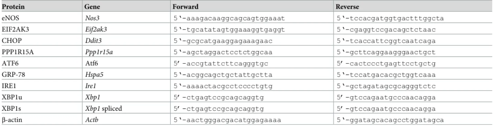
PrimeScript RT reagent kit and gDNA Eraser (Takara, Tokyo, Japan). Gene expression was determined by quantitative RT-PCR using the SYBR green-based fluorescence method (SYBR Premix Ex Taq, Takara). Amplification was performed using StepOnePlus (Applied Biosys- tems, Foster City, CA, USA). The PCR cycling conditions were 30 sec at 95˚C followed by 40 cycles of 5 sec at 95˚C and 30 sec at 58˚C/60˚C. The results were calculated as the expression of the target gene relative to the expression of theβ-actin gene. The sequences of primers used in this study are shown inTable 1.
Western blot analysis
HUVECs were lysed in ice-cold lysis buffer containing 1 mmol/l dithiothreitol, 0.0025% NP40 and a cocktail of proteinase inhibitors. The total protein concentration of the lysate was mea- sured using a kit (BCA Protein Assay, Thermo Fisher). After being heated at 100˚C for 5 min, 14μg of total protein was loaded into each well, separated by 15% SDS-PAGE (Wako), and transferred to nitrocellulose membranes. The membranes were incubated with a mouse mono- clonal antibodies against CHOP and ATF6 (Abcam, Cambridge, UK), rabbit polyclonal anti- bodies against binding immunoglobulin protein (BiP)/GRP78, PERK (Abcam) and phospho- PERK (Thr980) (Bioss, Boston, MA, USA), or rabbit monoclonal antibodies against eNOS and β-Actin (Cell Signaling, Danvers, MA, USA) at 4˚C overnight. After being washed, the mem- branes were incubated with peroxidase-conjugated goat anti-rabbit IgG (Wako) or horseradish peroxidase-linked sheep anti-mouse IgG (GE Healthcare, Buckinghamshire, UK), and then visualized using an ECL system (GE Healthcare).
Statistical analysis
Data are expressed as the means and SD. Statistical analysis was performed using SAS v.9.3 (SAS Institute, Cary, NC, USA). ANOVA and Student’st-test were used to compare the differ- ences between groups. The results with aP<0.05 were considered statistically significant.
Results
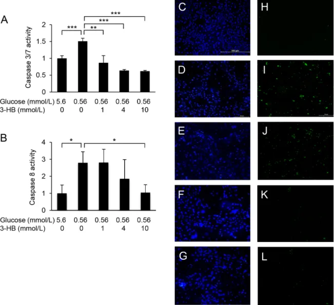
Culture of HUVECs in a low-glucose medium (0.56 mmol/L) for 24 or 48 h resulted in a reduction of cell viability (Fig 1A, 1B and 1E). The low-glucose-induced damage was attenu- ated by supplementation with 3-HB in a dose-dependent manner (Fig 1A, 1B and 1F). The protective effect of 3-HB was confirmed by the LDH release assay (Fig 1C). Furthermore, 48-h exposure of HUVECs to the low-glucose medium caused the activation of caspase 3/7 and cas- pase-8, which was suppressed by the addition of 3-HB (Fig 2). Caspase 3/7-positive cells were
Table 1. Primer sequences.
Protein Gene Forward Reverse
eNOS Nos3 5‘-aaagacaaggcagcagtggaaat 5‘-tccacgatggtgactttggcta
EIF2AK3 Eif2ak3 5‘-tgcatatagtggaaaggtgaggt 5‘-cgaggtccgacagctctaac
CHOP Ddit3 5‘-gcgcatgaaggagaaagaac 5‘-tcaccattcggtcaatcaga
PPP1R15A Ppp1r15a 5‘-agctaggactcctctggcaa 5‘-gcttcaggaagggaactgct
ATF6 Atf6 5’-accgtattcttcagggtgc 5’-cactccctgagttcctgctg
GRP-78 Hspa5 5‘-acggcagctgctattgctta 5‘-tccatgacacgctggtcaaa
IRE1 Ire1 5‘-aaaactacgcctcccctgtg 5‘-gctagatagcgcagggtctc
XBP1u Xbp1 5’-ctgagtccgcagcaggtg 5’-gtccagaatgcccaacagga
XBP1s Xbp1spliced 5’-ctgagtccgcagcaggtg 5’-gtccagaatgcccaacagga
β-actin Actb 5‘-aactgggacgacatggagaaaa 5‘-ggatagcacagcctggatagca
https://doi.org/10.1371/journal.pone.0191147.t001
increased in number after the low-glucose culture, and the increase was inhibited by the sup- plementation of 3-HB (Fig 2C–2L). The addition of a caspase inhibitor zVAD-fmk to the medium successfully protected HUVECs against the cytotoxic effect of glucose deprivation (Fig 3).
To assess the effects of low-glucose exposure on vascular endothelial cell function, we mea- sured the gene expression of eNOS (Nos3) and eNOS protein levels in HUVECs. After a 24-h incubation in the low-glucose medium, we found reductions inNos3expression and eNOS protein levels, both of which were alleviated by the addition of 3-HB (Fig 4).
Next, we analysed the gene expression of ER stress-associated genes in low-glucose-treated cells. The expression ofEif2ak3encoding PERK, an ER stress sensor, was up-regulated in HUVECs when cultured in the low-glucose medium for 6 h (Fig 5). The expression of two downstream molecules of PERK, i.e., CHOP (Ddit4) and GADD34/Ppp1r15a (Ppp1r15a), was also increased. The increase in the ER stress markers was dose-dependently suppressed by the addition of 3-HB. The expression ofAtf6was not significantly changed by the low-glucose cul- ture. However, the expression of the BiP/GRP78 gene (Hspa5) was increased by culture in low- glucose medium, and the increase was suppressed by the addition of 3-HB. In contrast, low- glucose exposure did not affect the expression ofIrel,andthe splicing ofXbp1mRNA was rather reduced by low-glucose culture.
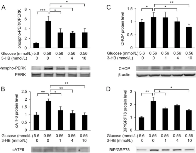
Western blot analysis showed that low-glucose exposure increased the amount of phospho- PERK and cleaved ATF6 (cATF6) (Fig 6A and 6B). Protein levels of CHOP and BiP/GRP78 were also elevated in HUVECs exposed to low glucose (Fig 6C and 6D). These low-glucose- induced elevations were attenuated by the supplementation of 3-HB (Fig 6).
Fig 1. Effects of low-glucose exposure on cell viability and protective effects of 3-hydroxybutyrate (3-HB). Human umbilical vein endothelial cells (HUVECs) were treated with 5.6 or 0.56 mmol/L glucose in the presence of various concentrations of 3-HB (0, 1, 4, 10 mmol/L) for 24 h (A) or 48 h (B). Cell viability was assessed by a CCK-8 assay (A) (B) and an LDH (C) assay. Microscopic images of HUVECs treated for 24 h (D-F). Means and SD,P<0.05,
P<0.01,P<0.001 (n = 4).
https://doi.org/10.1371/journal.pone.0191147.g001
Discussion
It has been well described that hyperglycemia-induced endothelial damage is involved in the early stage of atherosclerosis [16,17]. However, recent studies [1,18] have shown that hypogly- cemia may also play a role in the development of cardiovascular disease, although the mecha- nism remains to be elucidated. Here, we showed that culture of HUVECs in low-glucose medium resulted in a reduction of viability and impairment of endothelial cell function.
Vascular tone and platelet aggregation are regulated by NO that is generated from L-argi- nine by endothelial NO synthase (eNOS). Although multiple risk factors are involved in the development and progression of atherosclerosis, a decrease in the amount of bioavailable NO is a common feature of endothelial dysfunction [19]. Thus, the reduction of the eNOS gene (NOS3) expression and eNOS protein induced by low-glucose exposure may facilitate the development of atherosclerosis.
Fig 2. Caspase activation by low glucose and suppressive effect of 3-hydroxybutyrate (3-HB). Caspase-3/7 (A) and caspase-8 (B) activities were measured in HUVECs cultured in low-glucose medium with or without supplementation of 3-HB for 48 h. Results are exppressed as fold increase from control. Means and SD,P<0.05,P<0.01,P<0.001 (n = 4). Cell nuclei were visualized by Hoechst 33342 staining (blue) (C-G), and caspase-3/7-positive cells were stained green with a fluorogenic substrate (H-L). Bar, 100μm.
https://doi.org/10.1371/journal.pone.0191147.g002
It has been suggested that glucose deprivation induces a reduction of protein glycosylation and thereby the accumulation of unfolded proteins [20]. In this study we showed that the low- glucose-induced endothelial cell dysfunction was accompanied by the up-regulation of ER stress marker genes. ER stress activates a signalling network referred to as the unfolded protein response (UPR) to prevent cellular damage. UPR signalling is initiated by three ER-transmem- brane proteins that detect ER stress, i.e., PERK, ATF6, and IRE1 [21].
PERK is activated by oligomerization and autophosphorylation when ER stress is increased [22]. In this study PERK gene expression and phosphorylation of PERK protein were up-regu- lated in HUVECs exposed to low glucose. The expression of downstream factors of the PERK pathway, i.e., CHOP (Ddit3/Gadd153) and GADD34 (Ppp1r15a), was also increased. CHOP is a pro-apoptotic transcriptional factor which mediates ER stress-induced apoptosis [23].
GADD34 exerts feedback inhibition which may be required to enable the cells to produce pro- teins essential to survive [24].
ATF6 is activated by a sequential action of two proteases S1P and S2P upon ER stress [25].
Although no significant change was observed in the mRNA level ofAtf6, the amount of cATF6
Fig 3. Protective effect of a pan caspase inhibitor on HUVECs exposed to low glucose. HUVECs were treated with 5.6 or 0.56 mmol/L glucose with or without 10μmol/L zVAD-fmk for 24 h. Cell viability was assessed by the CCK-8 assay (A) and the LDH release assay (B). Means and SD,P<0.05,P<0.01,P<0.001 (n = 4).
https://doi.org/10.1371/journal.pone.0191147.g003
was increased in HUVECs cultured in low glucose medium. In accordance with this observa- tion, the expression ofHspa5which encodes BiP/GRP78, one of the best-characterized ER chaperones induced by ATF6, was significantly increased by the low-glucose culture. A large volume of studies has established that induction ofHspp5is a useful marker for ER stress [26].
The third response to increased ER stress is ER-associated degradation (ERAD). This signalling pathway is mediated by IRE1, which is activated by oligomerization and autophosphorylation,
Fig 4. Effect of glucose deprivation on endothelial nitric oxide synthase (eNOS). HUVECs were treated with 5.6 or 0.56 mmol/L glucose in the presence of various concentrations of 3-hydroxybutyrate (3-HB) for 24 h. mRNA (A) and protein levels (B) of eNOS were assessed by real-time PCR and Western blotting, respectively. A representative image of Western blotting is shown (C). Means and SD,P<0.05,P<0.01,P<0.001.
https://doi.org/10.1371/journal.pone.0191147.g004
Fig 5. Effects of 3-hydroxybutyrate (3-HB) on the endoplasmic reticulum (ER) stress markers. Expression of ER stress marker genes, Eif2ak3/Perk(A),Ddit3(B),Ppp1r15a(C),Atf6(D),Hsppa5(E),Ire1(F),XBP1u(G) andXBP1s(H) were assessed in HUVECs treated with 5.6 or 0.56 mmol/L glucose in the presence of various concentrations of 3-HB for 6 h. Means and SD,P<0.05,P<0.01,P<0.001 (n = 4).
https://doi.org/10.1371/journal.pone.0191147.g005
similar to PERK. Active IRE1 catalyses the splicing ofXbp1mRNA, and spliced XBP1 protein enhances the transcription of ERAD-associated genes [27,28]. In this study, however, the expres- sion ofIre1was not altered, and unexpectedly,Xbp1splicing was decreased by glucose deprivation.
Thus IRE1 activity seems to be reduced in HUVECs exposed to low glucose. The inadequate capacity of ERAD may be associated with the vulnerability of HUVECs to glucose deprivation.
Moreover, we found activation of caspase-8 and caspase-3/7 by glucose deprivation. Cas- pase-8 is a key initiator of the caspase cascade leading to apoptosis [29]. Apoptosis mediated by caspase-8 was reported in cancer cells treated with glucose deprivation [30]. On the other hand, caspase-3 is a key effector caspase in the apoptotic signalling cascade [31]. Caspase-7, which shares common substrate specificity with caspase-3, may be also involved in ER stress response induced by glucose deprivation [32]. Furthermore, zVAD-fmk, a cell-permeant pan caspase inhibitor, protected HUVECs against low-glucose-induced cell damage. According to these observations, the reduction in viability of HUVECs exposed to low-glucose medium was likely attributable to ER stress-induced caspase-mediated apoptosis. The observations in this study on low glucose-treated HUVECs are essentially in accordance with previous studies using neurons or cancer cells.
Fig 6. Western blot analysis of ER stress markers. Protein levels of PERK (A), cleaved ATF6 (cATF6) (B), CHOP (C) and Bip/GRP78 (D) were assessed in HUVECs treated with 5.6 or 0.56 mmol/L glucose in the presence of various concentrations of 3-hydroxybutyrate (3-HB) for 24 h.β-actin was used as a loading control. Representative images of Western blotting are shown. Means and SD,P<0.05,P<0.01,
P<0.001 (n = 3).
https://doi.org/10.1371/journal.pone.0191147.g006
In this study we further assessed the effects of 3-HB supplementation on HUVECs and found that 3-HB dose-dependently attenuated the induction of apoptosis, the reduction of NOS3, and the increase of ER stress induced by the low glucose exposure. Ketone bodies can serve as an energy source when glucose availability is limited [13]. 3-HB is readily picked up by the extra-hepatic tissues, independent of insulin actions, and converted to acetyl-CoA, which enters the mitochondria and is oxidized in the Krebs cycle to generate ATP. Thus, the induction of ER stress by glucose deprivation may be attributable not only to the insufficient glycosylation but to the energy deficiency. The increase in energy supply by 3-HB may be asso- ciated with the attenuation of low-glucose-induced ER stress, although the precise mechanism is still unknown.
Ketone bodies are important sources of energy for the brain, which is incapable of using FFA as fuel [33]. A high fat, low carbohydrate ketogenic diet is an established nonpharmacolo- gical treatment for epilepsy in childhood [34,35]. Several studies have shown the beneficial effects of a ketogenic diet or ketone bodies on ischemic stroke and Alzheimer’s disease [36–
38]. In rats, systemic administration of 3-HB was shown to prevent hypoglycemia-induced neuronal death [39]. Although the precise mechanism is not known, 3-HB may attenuate low- glucose-induced ER stress through the increase in energy supply.
The possible protective effects of ketone bodies on cardiovascular disease presently attract a great deal of interest because the EMPA-REG OUTCOME study recently demonstrated a remarkable effect of the SGLT2 inhibitor empagliflozin on cardiovascular and renal outcomes [13,40]. SGLT2 inhibitors increase urine glucose excretion and thereby result in the improve- ment of plasma glucose levels and body weight accompanied by an increase in plasma ketone bodies [41]. Although excessive production of ketone bodies may lead to dangerous ketoacido- sis, mild to moderate elevation of ketone bodies in plasma could result in an improvement in energy metabolism in diabetic patients with insulin resistance. The exact mechanism of the beneficial effect of empagliflozin is still unclear and may be multifaceted. However, the eleva- tion of plasma ketone bodies has been implicated as a mechanism by which empagliflozin pre- vented cardiorenal events [41], although the concentration of ketone bodies in treated patients was much lower than the doses used in this study.
In conclusion, culture of HUVECs in low-glucose medium resulted in an increase in ER stress leading to the induction of endothelial dysfunction and caspase-mediated apoptosis.
These cellular responses caused by glucose deficiency were efficiently suppressed by supple- mentation with 3-HB, a major component of plasma ketone bodies. Thus, hypoketonemic hypoglycemia induced by insulin injection or insulin secretagogue administration may be associated with a higher risk of cardiovascular events when compared with hyperketonemic hypoglycemia induced by starvation or SGLT2 inhibitor therapy. This may also be the case for other organs vulnerable to hypoglycemia, such as the brain.
Acknowledgments
The authors thank Tomoka Egashira for her excellent technical support.
Author Contributions
Conceptualization: Kentaro Yamada.Funding acquisition: Kentaro Yamada.
Investigation: Eri Soejima, Tsuyoshi Ohki, Yayoi Kurita, Kayo Tanaka, Satomi Kakino, Kento Hara.
Methodology: Xiaohong Yuan.
Supervision: Hitomi Nakayama, Yuji Tajiri, Kentaro Yamada.
Writing – original draft: Eri Soejima.
Writing – review & editing: Kentaro Yamada.
References
1. Goto A, Arah OA, Goto M, Terauchi Y, Noda M. Severe hypoglycaemia and ardiovascular disease: sys- tematic review and meta-analysis with bias analysis. BMJ. 2013; 347: f4533.https://doi.org/10.1136/
bmj.f4533PMID:23900314
2. Hutton RA, Mikhailidis D, Dormandy KM, Ginsburg J. Platelet aggregation studies during transient hypoglycaemia. J Clin Pathol. 1979; 32: 434–8. PMID:469000
3. Wright RJ, Frier BM. Vascular disease and diabetes: is hypoglycaemia an aggravating factor? Diabetes Metab Res Rev. 2008; 24: 353–63.https://doi.org/10.1002/dmrr.865PMID:18461635
4. Cryer PE. The barrier of hypoglycemia in diabetes. Diabetes 2008; 57:3169–76.https://doi.org/10.
2337/db08-1084PMID:19033403
5. Davignon J, Ganz P. Role of endothelial dysfunction in atherosclerosis. Circulation. 2004; 109: III27–
32.https://doi.org/10.1161/01.CIR.0000131515.03336.f8PMID:15198963
6. Versari D, Daghini E, Virdis A, Ghiadoni L, Taddei S. Endothelial dysfunction as a target for prevention of cardiovascular disease. Diabetes Care. 2009; 32: S314–21.https://doi.org/10.2337/dc09-S330 PMID:19875572
7. Foteinos G, Afzal AR, Mandal K, Jahangiri M, Xu Q. Anti-heat shock protein 60 autoantibodies induce atherosclerosis in apolipoprotein E-deficient mice via endothelial damage. Circulation. 2005; 112:
1206–13.https://doi.org/10.1161/CIRCULATIONAHA.105.547414PMID:16116071
8. Maier PJ, Zemoura K, Acuña MA, Ye´venes GE, Zeilhofer HU, Benke D. Ischemia-like oxygen and glu- cose deprivation mediates down-regulation of cell surfaceγ-aminobutyric acidB receptors via the endo- plasmic reticulum (ER) stress-induced transcription factor CCAAT/enhancer-binding protein (C/EBP)- homologous protein (CHOP). J Biol Chem. 2014; 289: 12896–907.https://doi.org/10.1074/jbc.M114.
550517PMID:24668805
9. Mahadevan NR, Rodvold J, Almanza G, Pe´rez AF, Wheeler MC, Zanetti M. ER stress drives Lipocalin 2 upregulation in prostate cancer cells in an NF-κB-dependent manner. BMC Cancer. 2011; 11: 229.
https://doi.org/10.1186/1471-2407-11-229PMID:21649922
10. Wang J, Alexanian A, Ying R, Kizhakekuttu TJ, Dharmashankar K, Vasquez-Vivar J, et al. Acute expo- sure to low glucose rapidly induces endothelial dysfunction and mitochondrial oxidative stress: role for AMP kinase. Arterioscler Thromb Vasc Biol. 2012; 32: 712–20.https://doi.org/10.1161/ATVBAHA.111.
227389PMID:22207730
11. Rizzo MT, Leaver HA. Brain endothelial cell death: modes, signaling pathways, and relevance to neural development, homeostasis, and disease. Mol Neurobiol. 2010; 42: 52–63.https://doi.org/10.1007/
s12035-010-8132-6PMID:20407845
12. Alluri H, Anasooya Shaji C, Davis ML, Tharakan B. Oxygen-glucose deprivation and reoxygenation as an in vitro ischemia-reperfusion injury model for studying blood-brain barrier dysfunction. J Vis Exp.
2015; 7: e52699.https://doi.org/10.3791/52699PMID:25992584
13. Cahill GF Jr. Fuel metabolism in starvation. Annu Rev Nutr. 2006; 26: 1–22.https://doi.org/10.1146/
annurev.nutr.26.061505.111258PMID:16848698
14. Aubert G, Martin OJ, Horton JL, Lai L, Vega RB, Leone TC, et al. The failing heart relies on ketone bod- ies as a fuel. Circulation. 2016; 133: 698–705.https://doi.org/10.1161/CIRCULATIONAHA.115.017355 PMID:26819376
15. Zinman B, Wanner C, Lachin JM, Fitchett D, Bluhmki E, Hantel S, et al. Empagliflozin, cardiovascular outcomes, and mortality in type 2 diabetes. N Engl J Med. 2015; 373: 2117–28.https://doi.org/10.1056/
NEJMoa1504720PMID:26378978
16. Henry RM, Ferreira I, Kostense PJ, Dekker JM, Nijpels G, Heine RJ, et al. Type 2 diabetes is associated with impaired endothelium-dependent, flow-mediated dilation, but impaired glucose metabolism is not;
The Hoorn Study. Atherosclerosis. 2004; 174: 49–56.https://doi.org/10.1016/j.atherosclerosis.2004.
01.002PMID:15135250
17. Ravikumar R, Deepa R, Shanthirani C, Mohan V. Comparison of carotid intima-media thickness, arterial stiffness, and brachial artery flow mediated dilatation in diabetic and nondiabetic subjects (The Chennai Urban Population Study [CUPS-9]). Am J Cardiol. 2002; 90: 702–7. PMID:12356381
18. Hanefeld M, Frier BM, Pistrosch F. Hypoglycemia and cardiovascular risk: Is there a major link? Diabe- tes Care. 2016; 39: S205–9.https://doi.org/10.2337/dcS15-3014PMID:27440834
19. Gimbrone MA Jr, Garcı´a-Cardeña G. Endothelial cell dysfunction and the pathobiology of atherosclero- sis. Circ Res. 2016; 118: 620–36.https://doi.org/10.1161/CIRCRESAHA.115.306301PMID:26892962 20. Benavides A, Pastor D, Santos P, Tranque P, Calvo S. CHOP plays a pivotal role in the astrocyte death induced by oxygen and glucose deprivation. Glia. 2005; 52: 261–75.https://doi.org/10.1002/glia.20242 PMID:16001425
21. Xu C, Bailly-Maitre B, Reed JC. Endoplasmic reticulum stress: cell life and death decisions. J Clin Invest. 2005; 115: 2656–64.https://doi.org/10.1172/JCI26373PMID:16200199
22. Cui W, Li J, Ron D, Sha B. The structure of the PERK kinase domain suggests the mechanism for its activation. Acta Crystallogr D Biol Crystallogr. 2011; 67: 423–8.https://doi.org/10.1107/
S0907444911006445PMID:21543844
23. Sano R, Reed JC. ER stress-induced cell death mechanisms. Biochim Biophys Acta. 2013; 1833:
3460–70.https://doi.org/10.1016/j.bbamcr.2013.06.028PMID:23850759
24. Novoa I, Zeng H, Harding HP, Ron D. Feedback inhibition of the unfolded protein response by GADD34-mediated dephosphorylation of eIF2alpha. J Cell Biol. 2001; 153: 1011–22. PMID:11381086 25. Ye J, Rawson RB, Komuro R, Chen X, Dave´ UP, Prywes R, et al. ER stress induces cleavage of mem- brane-bound ATF6 by the same proteases that process SREBPs. Mol Cell. 2000; 6: 1355–64. PMID:
11163209
26. Lee AS. The ER chaperone and signaling regulator GRP78/BiP as a monitor of endoplasmic reticulum stress. Methods. 2005; 35: 373–81.https://doi.org/10.1016/j.ymeth.2004.10.010PMID:15804610 27. Badiola N, Penas C, Miñano-Molina A, Barneda-Zahonero B, Fado´ R, Sa´nchez-Opazo G, et al. Induc-
tion of ER stress in response to oxygen-glucose deprivation of cortical cultures involves the activation of the PERK and IRE-1 pathways and of caspase-12. Cell Death Dis. 2011; 2: e149.https://doi.org/10.
1038/cddis.2011.31PMID:21525936
28. Tardif KD, Mori K, Kaufman RJ, Siddiqui A. Hepatitis C virus suppresses the IRE1-XBP1 pathway of the unfolded protein response. J Biol Chem. 2004; 279: 17158–64.https://doi.org/10.1074/jbc.
M312144200PMID:14960590
29. Cullen SP, Martin SJ. Caspase activation pathways: some recent progress. Cell Death Differ. 2009; 16:
935–8.https://doi.org/10.1038/cdd.2009.59PMID:19528949
30. Caro-Maldonado A, Tait SW, Ramı´rez-Peinado S, Ricci JE, Fabregat I, Green DR, et al. Glucose depri- vation induces an atypical form of apoptosis mediated by caspase-8 in Bax-, Bak-deficient cells. Cell Death Differ. 2010; 17: 1335–44.https://doi.org/10.1038/cdd.2010.21PMID:20203689
31. Woo M, Hakem R, Soengas MS, Duncan GS, Shahinian A, Ka¨gi D, et al. Essential contribution of cas- pase 3/CPP32 to apoptosis and its associated nuclear changes. Genes Dev. 1998; 12: 806–19. PMID:
9512515
32. de la Cadena SG, Herna´ndez-Fonseca K, Camacho-Arroyo I, Massieu L. Glucose deprivation induces reticulum stress by the PERK pathway and caspase-7- and calpain-mediated caspase-12 activation.
Apoptosis. 2014; 19: 414–27.https://doi.org/10.1007/s10495-013-0930-7PMID:24185830 33. Tildon JT, Rodeder LM. Transport of 3-hydroxy [3-14C] butyrate by dissociated cells rat brain. Am J
Physiol. 1988; 255: C133–9.https://doi.org/10.1152/ajpcell.1988.255.2.C133PMID:3407758 34. Shinha SR, Kossoff EH. The ketogenic diet. Neurologist 2005; 11: 161–70.https://doi.org/10.1097/01.
nrl.0000160818.58821.d2PMID:15860138
35. Cervenka MC, Henry B, Nathan J, Wood S, Volek JS. Worldwide dietary therapies for adults with epi- lepsy and other disorders. J Child Neurol 2013; 28: 1034–40.https://doi.org/10.1177/
0883073813488671PMID:23670244
36. Puchowicz MA, Zechel JL, Valerio J, Emancipator DS, Xu K, Pundik S, et al. Neuroprotection in diet- induced ketotic rat brain after focal ischemia. J Cereb Blood Flow Metab. 2008; 28: 1907–16.https://
doi.org/10.1038/jcbfm.2008.79PMID:18648382
37. Krikorian R, Shidler MD, Dangelo K, Couch SC, Benoit SC, Clegg DJ. Dietary ketosis enhances mem- ory in mild cognitive impairment. Neurobiol Aging 2010; 33:425.e19-27.https://doi.org/10.1016/j.
neurobiolaging.2010.10.006PMID:21130529
38. Van der Auwera I, Wera S, Van Leuven F, Henderson ST. A ketogenic diet reduces amyloid beta 40 and 42 in a mouse model of Alzheimer’s disease. Nutr Metab. 2005; 2: 28.https://doi.org/10.1186/
1743-7075-2-28PMID:16229744
39. Julio-Amilpas A, Montiel T, Soto-Tinoco E, Gero´nimo-Olvera C, Massieu L. Protection of hypoglycemia- induced neuronal death byβ-hydroxybutyrate involves the preservation of energy levels and decreased production of reactive oxygen species. J Cereb Blood Flow Metab. 2015; 35: 851–60.https://doi.org/10.
1038/jcbfm.2015.1PMID:25649993
40. Mudaliar S, Alloju S, Henry RR. Can a shift in fuel energetics explain the beneficial cardiorenal out- comes in the EMPA-REG OUTCOME study? a unifying hypothesis. Diabetes Care. 2016; 39: 1115–22.
https://doi.org/10.2337/dc16-0542PMID:27289124
41. Ferrannini E, Baldi S, Frascerra S, Astiarraga B, Heise T, Bizzotto R, et al. Shift to fatty substrate utiliza- tion in response to sodium-glucose cotransporter 2 inhibition in subjects without diabetes and patients with type 2 diabetes. Diabetes. 2016; 65: 1190–5.https://doi.org/10.2337/db15-1356PMID:26861783