Acta Med. Nagasaki 39: 131-136
Iron absorption in Crohn's Disease
Masaya NAKATA, Kazuya MAKIYAMA
The Second Department of Internal Medicine, Nagasaki University of Medicine
Anemia is observed in a considerable number of patients with Chohn's disease (CD). Iron absorption was investi- gated, using radioactive iron (59FeCl3), in 12 normal subjects (controls), seven patients with iron deficiency anemia (ID), and 23 patients with CD. After oral administration of 1 µ Ci of 59Fe with 4 mg of FeSO4 as a carrier, serum 59Fe levels were determined for approximately three hours with a liquid scintillation counter. The proportion of iron remaining in the body two weeks after administration was also determined as the absorption ratio by whole-body counting. In investigat- ing the factors affecting the absorption ratio, erythrocyte count, hemoglobin, total protein, albumin, serum iron (Fe), serum transferrin (TS), and serum ferritin were used as dependent variables, and were subjected to a multiple regres- sion analysis for the prediction of the absorption ratio. The analysis revealed that hemoglobin and ferritin levels corre- lated significantly with the absorption ratio. The absorption ratio was not useful in differentiating between ID and CD patient, while both TS and ferritin were useful in differentiat- ing between CD, ID and normal subjects. The time-course change in serum 59Fe after administration of 59Fe showed slightly higher levels in ID and CD than in controls.
Key words : Crohn's disease, iron absorption, ferritin, transferrin, anemia
Introduction
Crohn's disease (CD) is increasing in Japan. Several clinical features of CD have been previously investigated, including iron absorption. In the present study, radioactive iron (59FeC13) was administered orally, and the serial changes in serum 59Fe were determined. Iron absorption was also investigated by measuring the proportion of iron remaining in the whole body two weeks after oral admini- stration (residual whole-body ratio). This level was termed as the absorption ratio.
Subjects and methods 1. Subjects
A total of 23 patients (13 males and 10 females) were admitted to the Second Department of Internal Medicine,
Nagasaki University School of Medicine for the treatment of CD between January 1990 and November 1993. Thier ages at the time of examination ranged from 15 to 54 years (mean, 23.7 years). Six of the patients were positive for fecal occult blood. The diagnosis of CD was made based on radiological, endoscopic and histopathological findings.
Seven patients with iron deficiency anemia (ID ; mean age, 32 years ; range, 25-47 years) were also included in the study. Twelve normal adults (7 males and 5 females ; mean age, 30.1 years ; range, 22-39) served as coutrols. All normal subjects were healthy except for one who had a clinically confirmed duodenal ulcer.
2. Methods
Each subject was instructed to fast from after the evening meal on the days before the examination until the end of the test. On the examination day, erythrocyte count, hemoglobin, and serum protein, albumin, Fe, unsaturated iron binding capacity, transferrin (TS), ferritin, vitamin B12, folic acid, reticulocyte count, and C-reactive protein (CRP) were determined. FeSO4 (4 mg) was used as a carrier, and 1 ,u Ci of 59Fe was administered orally with 15 ml of deionized water. An equal amount of deinoized water was given twice'). Blood samples were collected 30, 60, 90, 120, 150 and 180 minutes after admini- stration of "Fe, and changes in serum 59Fe levels were
determined serially with a liquid scintillation counter (Aloka ; Automatic well-type scintillation counter, ARC 500). The residual whole-body ratio was determined by whole-body counting 60 minutes and about two weeks after administration of 59Fe as the absorption ratio. The count- ing system used NaI (Harshaw Chem.) as the detector. To
balance sensitivity, reciprocating scanning and correction of scanning speed were performed 2). The background 59Fe count and previous count were determined in the subjects, and physical corrections were also performed. In the statistical analysis the Kruskal-Wallis test was used for overall comparisons and the Mann-Whitney test for inter-group analysis in multiple groups. Multiple regres- sion analysis was used to identify factors contributing to the absorption ratio. A p value of <0.05 reflected the presence of a statistically significant difference between tested parameters.
Results
1. Patient population
Table 1 shows age, sex, hemoglobin, and serum albumin, Fe, TS, ferritin and absorption ratios of patients with CD.
Anemia was determined based on the level of hemoglobin.
Male subjects with Hb levels below 12 g/dl and female subjects with levels below 11 g/dl were considered anemic.
The level of hemoglobin in CD patients varied from 7.8 g/
dl to 14.1 g/dl, and 19 of the patients (82.6 %) were anemic. Eight patients had CD lesions affecting the colon, seven patients with lesions in the small intestine, and eight patients with ileocolic involvement. The normal serum Fe level in our hospital is 80 ,u g/dl in male subjects and 60,u g/dl in females. Seventeen CD patients (74 %) had low levels of serum Fe.
2. Test results according to disease
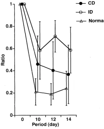
As shown in Fig. 1, there was a significant difference in the absorption ratio between ID patients and control subject (p < 0.05). The absorption ratio in CD patients was not significantly different from that of ID patients or controls. There was no significant difference in erythro-
Fig. 1 Comparison of iron absorption between patients with Crohn's disease (CD), iron deficiency anemia (ID) and normal subjects (NP).
Table 1. Clinical data in patients with Crohn's disease
Case No. age Sex RBC Hb TP Alb Fe Transferrin Ferritin Absorption Site of*
ratio Lesion*
1 19 F 369 9.8 5.9 4.1 67 354 10 0.67 c
2 27 M 371 11.4 4.5 2.3 37 40 41 0.41 i
3 26 F 379 13.1 6.7 4.4 61 234 256 0.3 1
4 54 F 415 10.6 5.6 3.1 26 184 11 0.74 i
5 16 F 396 9.8 5.7 3.4 23 234 8 0.33 i
6 29 M 447 11.1 6.7 4.7 25 208 24 0.39 is
7 16 M 390 10.7 7.3 4.4 27 390 21 0.83 c
8 15 M 417 11.7 5.9 3.3 32 199 60 0.03 c
9 18 F 424 10 6.2 3.5 35 277 13 0.57 c
10 36 M 240 9 5.7 3.7 121 193 188 0.1 c
11 34 F 427 9.4 6.6 3.8 43 375 22 0.47 i
12 16 F 400 11.1 5.9 3.5 41 268 103 0.29 is
13 18 M 451 12 6.5 3.9 32 303 9 0.4 c
14 40 M 379 11.9 7.1 3.6 17 237 253 0.02 i
15 19 F 423 7.8 5.4 3.1 15 263 9 0.67 is
16 29 M 466 9.8 6.3 4 8 259 5 0.83 i
17 16 F 452 9.6 6.2 3.5 10 298 14 0.63 i
18 20 M 433 14.1 6.6 4.2 111 271 34 0.12 is
19 22 M 440 12.5 6.3 4.4 70 208 75 0.09 c
20 24 M 496 11.5 5.6 3.5 8 295 10 0.15 is
21 18 M 422 11.2 7.1 3.8 55 191 425 0.13 is
22 18 M 457 13.1 6.3 4 103 389 235 0.06 is
23 16 F 428 11.5 7.6 4.3 77 354 67 0.31 c
mean±SD 23.7±9.7 M:F=13:10 414±49.8 11±1.5 6.2±0.7 3.8±0.5 45.4±32.8 261.9±80.8 82.3±111.7 0.4±0.3
*i = small intestinal type
, is = ileocolic type, c = colonic type
Fig. 2 Iron absorption ratio
Fig. 3 Elimination of unabsorbed "Fe.
cyte count between the different groups. However, there were significant intergroup differences among the three groups in hemoglobin, serum protein, serum albumin, and serum Fe levels. Furthemore, there were significant differences in TS and ferritin between CD and ID and between ID patients and controls. When the data for the female subjects alone were examined, no significant differences between ID patients and controls were observed in absorption tatio, serum protein or serum albumin.
There were no significant differences between female CD and ID patients in serum TS or ferritin. The presence or absence of differences from the controls in hemoglobin levels was determined in seven CD patients and seven ID patients whose hemoglobin levels were lower than 10 g/dl.
There were significant differences in TS, total protein or albumin, but no significant differences in absorption ratio, Fe or ferritin.
3. Iron absorption ratio
Fig. 2 shows the curves for residual whole-body ratio after oral administration of 'Fe to each patients group.
There were no changes in absorption ratios 10, 12 or 14 days after administration. The absorption ratio ranged from 7 to 60 % in the controls. One patient in this group exhibited latent iron deficiency. When this patient was excluded, the ratio ranged from 7 to 43 %. Although the
Fig 4 The relationship between serum iron, transferrin and ferritin, in normal subjects, CD and ID patients.
Fig 5 Time course of 59Fe counts after oral administration
ratio in one patient with ID was 24 %, others had high absorption ratios ranging from 44 to 86 %. Nineteen (82.6 %) CD patients had also anemia, and two of these patients had absorption ratios of 2 % and 3 %, but the ratio in other patients ranged from 6 to 83 %. Eight CD patients had high (> 44 %) absorption ratios. The absorp- tion ratio in CD patients without anemia ranged from 9 to 31 %. Fig. 3 shows the mean residual whole-body ratios 10, 12 and 14 days after the administration of "Fe according to the type of disease. Since the levels on day 10 and 12 after the intake of "Fe administration were not complete, absorption ratios at 10, 12 and 14 days after administra- tion were different. A multiple regression analysis was performed to predict the absorption ratio. Erythrocyte count, hemoglobin, total protein, albumin, Fe, TS and ferritin were used as the dependent variables. Hemoglobin and ferritin were found to contribute significantly to the absorption ratio, and that other factors had a negligible effect (y = 0.974-0.047 X Hb-0.001 X ferritin, R = 0.687, n = 41). Since the effect of sex may influence the statisti- cal analysis, we selected female subjects only for analysis.
As a result, hemoglobin was found to be the only contribu- tor to the absorption ratio (y - 1.174 -0.067 X Hb, R = 0.646, n=21).
4. Assessment of three factors (ferritin, TS and Fe) Fig. 4 shows the distribution of TS, Fe and ferritin in the three groups. The correlation between ferritin and Fe indicated that the level of ferritin and Fe tended to be low in ID patients. A similar relationship was observed in CD patients. The correlation between ferritin and TS and between Fe and TS, ID patients tended to have high TS values but low ferritin levels.
5. Serial changes in serum 59Fe after the oral administra-
tion of 59Fe
As shown in Fig. 5, the level of serum 59Fe rose to approximately 10 cpm in ID and CD patients, while it increased only to an average of 5 cpm in control subjects throughout the course of the observation period. The level of serum 'Fe was slightly higher in ID than in CD patients even 60 min after administration, but the mean level was lower at 120 min after the intake of 59Fe.
Discussion
The incidence of CD is increasing in Japan. Its clinical features have been discussed in western countries for a long time'"). Several studies have also recently investi- gated the characteristics of CD in Japan's 9)
Furthermore, several reports have demonstrated the presence of anemia in CD'o>, il)Our results support these
earlier studies by demonstrating a high incidence of anemia in CD patients (83 %).
It is well established that iron metabolism depends on hematopoietic capacity") and stored iron. In general, iron absorption is not necessarily increased by the control of inflammation of intestinal mucosa, although hemat- opoiesis does increase in anemia. The results of our investi- gation regarding iron absorption in patients with CD revealed a significant difference in the absorption ratio between ID patients and controls, but no significant difference between ID and CD patients or between CD patients and controls. When seven CD patients with severe anemia (Hb < 10 g/dl) were compared with seven ID patients, significant differences were observed in total protein, albumin and TS. However, there were no signifi- cant differences between the two population samples in the absorption ratio, serum Fe or serum ferritin. These differences are most likely due to differences in nutritional status.
The multiple regression analysis used in the present study to predict the absorption ratio, using erythrocyte count, hemoglobin, total protein, albumin, Fe, TS and ferritin as dependent variables, indicated that hemoglobin and ferritin are useful in predicting the absorption ratio.
Our results supported the notion that ferritin serves as an indicator of iron absorption and stored Fe, as suggested by Bartels et al."). In general, iron deficiency occurs as a result of blood loss, but iron absorption is though to increase due to homeostasis. Our results indicated that anemic CD patients (due to blood loss through the intesti- nal tract, patients 4 and 7) had markedly higher absorp- tion ratios. The determination of serial changes in serum Fe after administration of 59Fe also revealed higher peak levels in ID and CD patients compared with controls. In other words, while the level in ID and CD patients was not significantly different from that of controls, the absorp- tion of iron appeared to have increased in ID and CD patients. The level was also higher in ID than in CD patients, even 60 min after administration of 59Fe, but started to decrease 120 min after administration in ID patients. Absorption appeared to increase in ID patients, and the ratio was even higher in these patients than in CD patients. A certain number of CD patients with anemia exhibited a mechanism similar to that in ID ; since absorbed iron is rapidly incorporated into erythroblasts and used for erythropoiesis, resulting in a fall in serum Fe.
Iron deficiency is also believed to develop as a result of chronic inflammation").") While we were able to deter- mine iron absorption after inflammation had subsided, in patients 8 and 14, CRP was 3.97 and 1.07, respectively, indicating the presence of inflammation, and the absorp- tion ratios were low, 0.03 and 0.02, respectively. Condi- tions favoring iron-deficiency may be caused by inflam- mation. For example, interleukin-1 (IL-1) can act as a causative agent of inflammation"), and its level may be
increased in CD`)-`). Although the pathophysiological mechanisms of inflammation-induced anemia are still unclear, IL-1 seems to impair the release of stored Fe in anemic CD patients.
Not only inflammation but disorders of intestinal absorption may also be present in anemic CD patients. For example, a reduction in the intracellular transfer of Fe due to edema has to be considered. With regard to the iron deficiency observed in CD, Child et al.') proposed a full evaluation of true iron deficiency including bone marrow puncture, while Thomson et al.") suggested evaluation of ferritin levels. Our study also indicated a cause-effect relationship between iron and ferritin. Measurement of ferritin is considered important in the diagnosis and treatment of latent iron deficiency. Iron is absorbed in the intestinal mucosa, processed by intramucosal transport mechanisms and is transported in the blood. Various theories have been proposed concerning the mechanism of iron absorption by the intestinal mucosa22)-26>, but certain aspects of the physiological process are still not clear.
Iron-binding protein may also be involved during the intramucosal transport of iron, and hypoproteinemia may retard iron absorption.
As described above, the anemia in CD may result from a variety of underlying conditions, such as bleeding, inflam- mation and hypoproteinemia. In the present study, it was not possible to distinguish between the anemia of CD and ID patients based on the absorption ratio, but TS and ferritin were useful in differentiating between control subjects, CD, and ID patients. The correlations between hemoglobin and other factors was investigated in the present study. Our results demonstrated that serum iron, erythrocyte count and absorption rate correlated with the severity of anemia. Furthemore, the absorption ratio correlated with the severity of anemia in CD patients. As pointed out by Makiyama et a127)., patients with high serum Fe levels have long-term remission of CD. Several studies examining the prognosis of CD have been reported")-'), and anemia may also become an important factor in the future. It should be stressed that anemia of CD may has to be treated carefully, taking into considera- tion various contributing factors. Intestinal absorption disorders should also be considered with a rapid improve- ment of inflammation and correction of hypoproteinemia.
Finally, intravenous injection of iron may occasionally be necessary.
Conclusions
1) Iron absorption was investigated using radioactive iron (59FeC13) in 12 normal subjects, seven patients with iron deficiency anemia (ID), and 23 patients with Crohn's
disease (CD).
2) Hemoglobin and ferritin were found to influence Fe
absorption ratio.
3) The absorption ratio did not help in defferentiating between ID and CD, but transferrin and ferritin appeared to be useful.
4) The severity of anemia in CD patients was dependent on the absorption ratio.
5) There were no significant differences in serial changes in serum 'Fe between ID and CD. Iron absorption in ID patients was slightly higher than that in CD patients.
6) Anemia in CD is attributed to various factors.
Treatment of anemia should also include treatment of its causes, and intestinal iron absorption disorders must be considered. Intravenous injection of iron is occasionally necessary.
Acknowledgments
We express our thanks to Professor Kohei Hara for his constant quidance, and to Professor Yutaka Okumura and Tatsuya Shimasaki for their supports.
REFERENCES
1) Saito, H., Sargent, T., Parker, H. G., and Lawrence, J. H.: Normal iron absorption in man. Proceedings of the 9 congress of the international
society of hematology 3 : 511-522, 1960.
2) Okajima, S., Norimura, T.: Studies on Whole-Body Counter. Nipppon acta radiologica 34: 169-178, 1974. (Japanese)
3) Best, W. R., Becktel, J. M., Singleton, J. W. and Kern, F.: Development
of a Crohn's disease activity index Gastroenterology 70: 439-444, 1976.
4) Farmer, R. G., Hawk, W. A. and Turnbull, R. B.: Clinical Patterns in Crohn's disease. A statistical study of 615 cases. Gastroenterology 68:
627-635, 1975.
5) Fielding, J. F.: Clinical feature of Crohn's disease in Ireland. Am. J.
G astroent. 81 : 524-528, 1986.
6) Mekhjian, H. S., Switz, D. M., Melnyk, C. S., Rankin, G. B., and Brooks R. K.: Clinical features and natural history of Crohn's disease.
Gastroenterology 77: 898-906, 1979.
7) Andersson, H., Dotevall, G., Gillberg, R., Jagenburg, R., and Kock, N.
G.: Absorption studies in patients with Crohn's disease and in patients
with ulceritive colitis. Acta med. scand. 190: 407-410, 1971.
8) Okada M., Yao T., Fuchigami T., lida M. and Date H.: Anatomical
involvement and clinical features in 91 Japanese patients with Crohn's disease. J. Clin. Gastroenterol. 9: 165-171, 1987.
9) Yao T., Okada M., Fuchigami T., lida M., takenaka K., Date K., and Fujita K.: The relationship between the radiological and clinical
features in patients with Crohn's disease. Clin. radiol. 40: 389-392, 1989.
10) The Japanese Reserch Committee for Crohn's disease: Gastroent. Jpn.
14: 366, 1979.
11) Dyer, N. H., Child, J. A., Mollin, D. L. et al.: Anemia in Crohn's disease. Q. J. Med., 164: 419, 1972.
12) Finch, C. A.: Review: Erythropoiesis, erythropoitin and iron. Blood.
60: 1241, 1982.
13) Bartels, U., Pederson, N. S. and Jarnum, S.: Iron absorption and serum ferritin in chronic inflammatory bowel disease. Scand J.
Gastroenterol., 13: 649-656, 1978.
14) Cartwright, G. E., Lauristen, M. A., Jones, P. J., Merrill, I. M. and Wintrope, M. M.: The anemia of infection. I. Hypoferremia, hyper-
cupremia, and alterations in porphyrin metabolism in patients. J. Clin.
Invest., 25: 65-80, 1946.
15) Cartwright, G. E.: The anemia of chronic disorders. Semis. Hemat., 3 : 351-375, 1968.
16) Simeon, E. G., Donald, A. C, Michael, J. and Craig, J. M.: Interleukin 1-induced depression of iron and zinc : role of granulocytes and
lactoferrin. Am. J. Physiol., 252: E27-E32, 1987.
17) Mahida, Y. R., Wu, K. and Jewl, D. P.: Enhanced production of interleukin 1- R by mononuclear cells isolated from mucosa with active
ulcerative colitis or Crohn's disease. Gut., 30: 835-838, 1989.
18) Ligumsky, M., Simon, P. L., Karmeli, F., Rachmilewitz, D.: Role of interleukin 1 in inflammatory bowel disease-enchanced production
during active disease. Gut., 31: 686-689, 1990.
20) Child, J. A., Brozoric, B., Dyer, N. H.: The Diagnosis of iron deficiency in patients with Crohn's disease. Gut., 14: 1973.
21) Thomson, A. B. R., Brust, R., Ali, M. A. M., Mant, M. J., and Valberg, L. S.: Iron deficiency in inflammatory bowel disease. Diagnostic
efficacy of serum ferritin. Dig. Dis., 23: 705-709, 1978.
22) Granick, S.: Protein apoferritin and ferritin in iron feeding and absorption. Sicence. 103: 107, 1956.
23) Conrad, M. E., and Crosby, W. H.: Intestinal mucosal mechanism controlling iron absorption. Blood, 22: 406, 1963.
24) Huebers, H. A., Huebers, E., Csiba, E., Rummel, W. and Finch, C. A.:
The significance of transeferrin for iron absorption. Blood, 61 : 283-290,
1983.
25) Conard, M. E. et al.: Small intestinal regulation of iron absorption in the rat. J. Lab. Clin. Med. 110: 48, 1987.
26) Banerjee, D., Franagan, P. R., Cluett, J. and Valberg, L. S.: Transfer- rin receptors in the human gastrointestinal tract. Relationship to body
iron stores. Gasroenterol., 91 : 861-869, 1986.
27) Makiyama, K., Itsuno, M., Murata, I. et al.: Clinical Course of 63 Patients with Crohn's Disease following Surgical or Conservative
Therapy Clinical Features of Patients in Long-term Remission-. J.
Jpn. Soc. Colo-proctol., 44 : 198-205, 1991. (Japanease)
28) Imamura, T., Okada, M., Seo, M. and Yao, T.: Quality of life 106 Japanese Patients with Crohn's Disease. J. Jpn. Soc. Colo-proctol., 46:
136-146, 1993. (Japanease)
29) Kufman, S., Chalmer, B., Heilman, R. and Beeken, W.: A Prospective Study of the Course of Crohn's Disease. Digestive Diseases and Siences,
24: 269-276, 1979.
30) Bergman, L. and Kruse, U.: Crohn's disease-A long term study of the clinical course in 186 patients. Scandinavian Journal of Gastro-
enterology, 12: 937, 1977.