Introduction
Oviraptorosaurs are generally regarded as non- avian theropod dinosaurs (Osborn, 1924; Bars- bold, 1976, 1977, 1981, 1983, 1986, 1997; Gau- thier, 1986; Smith, 1992; Sereno, 1999; Barsbold, Currie et al., 2000; Barsbold, Osmólska et al., 2000; Holtz, 2000, 2001; Norell et al., 1994;
Norell et al., 1995; Norell, Clark, and Makovicky, 2001; Xu, Norell et al., 2002). They are medium-sized theropods characterized by a short, deep skull with toothless jaws in derived forms, teeth present in primitive forms such as Incisivosaurus gauthieri(Xu, Cheng et al., 2002) and Caudipteryx zoui (Ji et al., 1998), pneu- matilized caudal vertebrae present in derived forms, anteriorly concave pubic shaft, and a pos- teriorly curved ischium present in Oviraptoridae (Barsbold and Osmólska, 1990; Barsbold, Osmólska et al., 2000; Makovicky and Sues, 1998). Oviraptorosauria is here regarded to in- clude three families: Oviraptoridae, Caenagnathi-
dae, and Caudipterygidae (Barsbold, 1976; Stern- berg, 1940; Currie, 2000; Clark et al., 2001; Ji et al., 1998; Zhou and Wang, 2000; Zhou et al., 2000).
The Oviraptorosauria are mainly distributed in Mongolia (Osborn, 1924; Barsbold, 1976, 1977, 1981, 1983, 1986, 1997; Osmólska, 1976; Norell et al., 1994, 1995; Wester, 1996; Clark et al., 1999, 2001, 2002), China (Dong and Currie, 1996; Ji et al., 1998; Lü et al., 2000; Xu, Cheng et al., 2002; Lü, 2003) and North America (Sternberg, 1940; Cracraft, 1971; Currie and Russell, 1988; Sues, 1997). Additionally, ovirap- torosaurs from the Southern Hemisphere have been reported (Frey and Martill, 1995; Currie et al., 1996; Frankfurt and Chiappe, 1999), al- though validity of the taxonomic assignment is doubtful.
In the summer of 1996, an incomplete ovirap- torid skeleton with a nearly complete skull was discovered in the Nemegt Formation of Nemegt Basin, southwestern Mongolia by the Mongolian
New Oviraptorid Dinosaur (Dinosauria: Oviraptorosauria) from the Nemegt Formation of Southwestern Mongolia
Junchang Lü1, Yukimitsu Tomida2, Yoichi Azuma3, Zhiming Dong4and Yuong-Nam Lee5
1Institute of Geology, Chinese Academy of Geological Sciences, Beijing 100037, China
2National Science Museum, 3–23–1 Hyakunincho, Shinjukuku, Tokyo 169–0073, Japan
3Fukui Prefectural Dinosaur Museum, 51–11 Terao, Muroko, Katsuyama 911–8601, Japan
4Institute of Paleontology and Paleoanthropology, Chinese Academy of Sciences, Beijing 100044, China
5Korea Institute of Geoscience and Mineral Resources, Geology & Geoinformation Division, 30 Gajeong-dong, Yuseong-gu, Daejeon 305–350, South Korea
Abstract Nemegtia barsboldigen. et sp. nov. here described is a new oviraptorid dinosaur from the Late Cretaceous (mid-Maastrichtian) Nemegt Formation of southwestern Mongolia. It differs from other oviraptorids in the skull having a well-developed crest, the anterior margin of which is nearly vertical, and the dorsal margin of the skull and the anterior margin of the crest form nearly 90°; the nasal process of the premaxilla being less exposed on the dorsal surface of the skull than those in other known oviraptorids; the length of the frontal being approximately one fourth that of the parietal along the midline of the skull. Phylogenetic analysis shows that Nemegtia barsboldiis more closely related to Citipati osmolskaethan to any other oviraptorosaurs.
Key words : Nemegt Basin, Mongolia, Nemegt Formation, Late Cretaceous, Oviraptorosauria, Nemegtia.
Highland International Dinosaur Project team. It was initially described by Lü et al.(2002) as In- geniasp. The skull morphology (vertical anterior margin of the developed crest on the skull, less exposed nasal process of the premaxilla, and a process on the quadrate bearing a convex surface projecting into the cotyla on the medial surface of the quadratojugal) indicates that it is distinct from named oviraptorosaurs. It represents a new taxon from the Late Cretaceous.
Systematic Paleontology Theropoda Marsh, 1881 Oviraptorosauria Barsbold, 1976 Family Oviraptoridae Barsbold, 1976
Nemegtiagen. nov.
Diagnosis: As for the only known species.
Etymology: Nemegtia refers to the fossil lo- cality, the Nemegt Basin of southwestern Mon- golia.
Nemegtia barsboldisp. nov.
(Figs. 1, 3, 5, 6) Ingeniasp. Lü et al., 2002
Etymology: The species is named in honor of Dr. R. Barsbold, the Mongolian vertebrate pale- ontologist, one of the leaders of the Mongolian Highland International Dinosaur Project.
Type locality and horizon: The Nemegt lo- cality of Gradzinski et al.(1968), Nemegt Basin, southern Gobi Desert, Mongolia; Nemegt Forma- tion, mid-Maastrichtian (Jerzykiewicz and Rus- sell, 1991) of the Upper Cretaceous.
Holotype: GIN100/2112 (formerly PC100/
2112, see Lü et al., 2002): an incomplete skele- ton with a nearly complete skull. Specimen stored in the Paleontological Center of the Mon- golian Academy of Sciences, Ulan Bator, Mon- golia.
Diagnosis: An oviraptorid displaying the fol- lowing derived characters. The skull with a naso- premaxillary crest, anterior margin (the nasal process of the premaxilla) nearly vertical; the
nasal process of the premaxilla, which forms part of the crest, being barely exposed in dorsal view;
the anteroposterior length of the frontal is ap- proximately one quarter of that of the parietals; a presence of prefrontal; process on the quadrate bears a convex surface projecting into the cotyla on the medial surface of the quadratojugal.
Mandibular condyles of the quadrate situated rostrally to the occipital condyle.
Description 1. Skull and mandible
The skull (Fig. 1) is deep, narrow and short, and has a naso-premaxillary vertical crest. The jaws are toothless, and the articular has a medial process. These characters indicate that Nemegtia barsboldiis a derived oviraptorosaur.
Skull: The premaxillae, vomers and maxillae form a hard palate as in other oviraptorid di- nosaurs (Fig. 1C), and the premaxillae form the main part of the crest. Both premaxillae are fused. The central palatal part of the joined pre- maxillae is strongly concave. There is a cleft on the central part of the palate. Anteriorly, small foramina distributed on the lateral sides of the suture between the premaxillae may represent nutrient openings. The nasal processes of the pre- maxillae are perpendicular to the palatal surface of the upper jaw, and extend dorsally, connecting with the nasals at the highest point of the crest (Fig. 1A), and is barely exposed in dorsal view of the skull. The crest extends posteriorly and ven- trally, and forms a round arc at its highest point.
The antorbital cavity consists of a large inter- nal antorbital fenestra posteriorly and a small maxillary fenestra (see Witmer, 1997) anteroven- trally. The internal antorbital fenestra is sub-oval, and the long axis is vertical. The external surface of the maxilla is smooth, and moderately con- cave. Ventrally, there are two longitudinal, round- ed ridges on each maxilla as in other ovirap- torids. In addition to the lateral ridge, there is a longitudinal groove. Posteriorly, there is a ven- trally directed tooth-like process on the palatal shelf of each maxilla as in other oviraptorids.
po p
p
stf
f
f l
m ec
j qj
pt ch
t v
n ec
q pt qj
sq
po
j m
I n
pm
cav antorbnar
pm mf
or o
q
lat sof eo
eo oc
oc bs
q par pa
t
v pm
C
B A
Fig. 1. The skull of Nemegtia barsboldigen. et sp. nov. (GIN100/2112). In lateral (A), dorsal (B), and ventral views (C). Scale bar5 cm. Abbreviations, as in appendix 1.
Both nasals are completely fused posteriorly with no trace of the suture. Anteriorly, the nasals are not fused, forming a “” shaped cleft, which receives the nasal processes of the premaxillae (Fig. 1B). The dorsal portion of the lacrimal is crescent shaped in lateral view. The shaft of the lacrimal is stout, and forms the posterior margin of the antorbital cavity. There is a large lacrimal foramen in the middle of the anterior edge of the orbit. The posterodorsal part of the lacrimal is wedged between the prefrontal and frontal. The lateral surface of the lacrimal is smooth. Pos- terodorsal to the antorbital cavity, there is a large recessed pneumatic pocket (6 mm high, 4 mm wide), similar to that in Citipati osmolskae (Clark et al., 2002).
Anterodorsal to the left orbit, there is a small triangular bone, which is located between the frontal and lacrimal. It should be a prefrontal, al- though this bone has not been reported in other oviraptorosaurs.
The jugal is a triradiate bone. The posterior part of its anterior branch is stout and plate-like.
Anteriorly, it gradually becomes rod-like and then ends as a thin plate (Fig. 2), where it inserts between the maxilla, lacrimal and ectopterygoid.
The postorbital process of the jugal inclines pos- terodorsally, and forms almost 2/3 of the height of the postorbital bar, similar to other ovirap- torosaurs (Barsbold et al., 1990). The process be- comes widened, and slightly twisted, and its me- dial surface is smooth, near the position where it contacts the descending process of the postor- bital. The dorsal part of the postorbital process of the jugal is tightly attached to the posteroventral surface of the postorbital. The posterior branch of the jugal contacts the anterior process of the quadratojugal. The angle of the postorbital branch and posterior branch of the jugal is 90°, which form the anterior and ventral margins of the large lateral temporal fenestra, respectively.
The width of the frontal is much greater than its length. In lateral view, the whole central part of the frontal surface is convex, thus the trans- verse cross-section of the frontal is arc-shaped.
The anteroposterior length of the frontal is ap- proximately one quarter of that of the parietals.
Anteriorly, the suture of the frontal and the nasal is relatively straight, while posteriorly, the suture with the parietal is convex anteriorly (Fig. 1B).
There is a deep groove along the suture with the nasal. When compared with other oviraptorids,
Medial view of the quadratojugal
cross-section of the jugal Posterior view of
the quadrate
Fig. 2. Skull of Nemegtia barsboldigen. et sp. nov. (GIN100/2112) showing the structure of the jugal and the relationship between the quadrate and the quadratojugal.
the frontal is very short medially and widens lat- erally towards orbital margin.
The parietals are fused. Their dorsal surface is smooth. A sagittal parietal ridge is present. This ridge extends anteriorly and disappears at the level of the anterior corner of the supratemporal fossa, not reaching the frontoparietal suture.
The right squamosal is almost completely pre- served. It curves posteriorly. There is a notch, which holds the distal lateral head of the quadrate on the middle of the lateral margin. The anterior process of the squamosal forms the pos- tolateral margin of the sub-oval supratemporal fossa. The long axis of the suboval supratemporal fossa points anterolaterally, making an angle with the long axis of the skull of nearly 30°.
The postorbital is a triradiate bone. The frontal process of the postorbital is upturned about 90 degrees. Its descending process occupies 2/3 of the height of the postorbital bar. The anterior sur- face of this process is smooth. The angle of the anteromedial process and the posterior squamos- al process is greater than 90°. The frontal process covers the orbital process of the frontal. The squamosal process extends posteriorly with a sharp dorsal margin and overlies the anterior process of the squamosal. This process does not reach the posterolateral corner of the supratem- poral fossa. The upper part of the postorbital bar forms most of the anterolateral margin of the supratemporal fossa.
A shallow groove separates the articular sur- face into two parts in the quadrate. In ventral view, the width of the mandibular articular sur- face of the quadrate is greater than its length.
The mandibular condyles are situated rostrally to the occipital condyle. The articular surface for the quadratojugal is convex, while that on the quadratojugal for the quadrate is concave. This joint forms a mobile junction (Fig. 2), which is similar to the condition in most birds and other oviraptorosaurs (Maryanska and Osmólska, 1997). The convex quadrate and concave quadra- tojugal joint is reversed from the condition in other oviraptorosaurs (Maryanska and Osmólska, 1997).
In ventral view, the vomer has a sharp process, which projects ventrally. This tooth-like process is also formed in part by processes of the maxil- lae. The vomers are completely fused anteriorly.
The posterior parts of the vomers are separated from each other by the cultriform process of the parasphenoid, which wedges rostrally between the vomers as in all oviraptorids. The middle part of the vomer is relatively long, its surface is smooth, and its dorsal part is fused with the ante- rior part of the cultriform process. The posterior part of the vomer is covered by the anterior part of the palatal ramus of the pterygoid. The fused part of the vomer projects ventrally, and the mid- dle part has a round ventral surface, while its posterior part is relatively flat.
The palatine forms the posterior and lateral margins of the elliptic choana. It connects with the vomer medially, and with the maxilla antero- laterally. The posterolateral part of the palatine contacts the anteromedial side of the ectoptery- goid, and its posteromedial part contacts the palatal ramus of the pterygoid.
Ventrally, the pterygoids have elongated palatal processes, which do not meet along the middle line until they contact the vomers. Each palatal process has a longitudinal groove, which divides the palatal branch of the pterygoid into two parts anteriorly: the lateral part connects with the ectopterygoid, while the medial one con- nects with the palatine and quadrate.
The ectopterygoid is located rostral to the pterygoid. It extends vertically, similar to other derived oviraptorids, whereas in other theropods, it extends horizontally. The posterior end of the dorsal part disappears near anterior end of the jugal. There is an opening within ectopterygoid, which is close to the maxilla. This opening may be homologous to the dorsal ectopterygoid recess of dromaeosaurids (Witmer, 1997).
The epipterygoid is present on both sides of the skull. It is a thin plate-like bone with an ir- regular triangular shape, whose free vertex angle projects dorsally. It tightly ties to the pterygoid branch of the quadrate. Its anterior margin is sharp and free.
The basioccipital condyle is ball-shaped, with a weak neck region. The articular surface of the occipital condyle faces posteroventrally. The an- terior part of the basioccipital projects ventrally and laterally, forming the posterior part of the basal tubera. The suture of the basioccipital and basisphenoid is clear. Its middle part projects posteriorly; therefore, in the ventral view, the su- ture has a weak “W” shape.
The basisphenoid forms the posteroventral part of the floor of the braincase. The sutures with the pterygoid, and the parasphenoid are not clear. In ventral view, it is narrow anteriorly and broad posteriorly. The portion comprising the basal tu- bera is strongly developed. Its middle portion is strongly depressed, and has a large opening. This opening is probably the Vidian canal for the pas- sage of the internal carotid artery and the palatal branch of nerve VII, which entered the pituitary fossa through the basisphenoid. The basiptery- goid process is absent, similar to other derived oviraptorosaurs (Clark et al., 2002).
The tall parasphenoid process is delicate. It is shallow anteriorly and deep posteriorly. Its depth gradually lessens anteriorly, as it connects with the vomer. Its posterodorsal part extends dorsally between the epipterygoid and pterygoid, contact- ing the laterosphenoid. In ventral view, it extends forward into the large space called interpterygoid vacuity. Its length is about 1/4 of that of the skull.
The laterosphenoid is a thin plate-like bone, anterior to the floor of the braincase. The or- bitosphenoid is also a thin plate like bone, anteri- or to laterosphenoid portion of the floor of the braincase.
The suture between the exoccipital and supraoccipital is not clear, but the trend of the su- ture shows that both exoccipitals connect above the oval foramen magnum. Thus, they separate the supraoccipital from the foramen magnum, which is larger than the occipital condyle.
Mandible: The lower jaw is short and deep (Fig. 3). The dorsal margin of the external mandibular fenestra is formed by the surangular and the dorsal arch of the dentary. The surangular has a middle branch, which projects into the ex-
ternal mandibular fenestra, as in other ovirap- torids. The ventral surface of the lower jaw is convex, and the mandibular symphysis is short.
In dorsal view, the jaw slightly projects laterally in the middle part, and both branches of the lower jaw are parallel posteriorly from the level of the posterior margin of the mandibular symph- ysis (Fig. 3A). The dentaries have a dorsal pro- trusion near the posterior margin of the mandibu- lar symphysis. The large external mandibular fenestra is located at the anterior part of the lower jaw. The coronoid process is posterior to the mandibular fenestra.
The mandibular symphysis is short and deep.
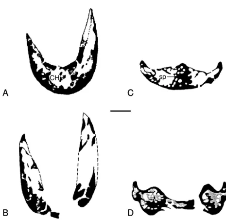
No evidence shows that a coronoid is present in the present specimen. CT scanning shows that the dorsal margin of the mandibular symphysis is thin, while its lower margin (lower part) is stout, the symphysis is highly pneumatized, and the spaces are uniform in size and distribution (Figs.
4A, B). There is no bony construction between the hollow spaces of the right and left dentaries.
The cross section near the mandibular symphysis shows that both sides are steep, displaying a “V”
shape. It is “U” shaped in Caenagnathus and there is a bar of bone between the dorsal and ven- tral surface supporting the midline ridge on the lingual surface of the symphysial shelf (Currie et al., 1993; Figs. 4C, D). Around the anterior mar- gin of the external mandibular fenestra, the den- taries are deflected inwards, forming a shallowly flattened surface, thus producing a thin and sharp anterior edge on the external mandibular fenes- tra. The ventral process of the denary is relatively weak, and it tightly extends along the ventrolater- al part of the anterior portion of the angular, and disappears at the level of the coronoid eminence.
The dorsal process of the dentary is comparative- ly massive, and dorsally, its distal end is divided into lateral and medial prongs, with the anterior branch of the articular-surangular-coronoid com- plex (ASC) wedged between them. It disappears anterior to the coronoid eminence. The ventral process of the dentary extends more posteriorly than that of the dorsal process. Only the external surfaces of both processes of the dentaries are
emf
A
B
C
d
an pre
ar asc
Fig. 3. Lower jaw of Nemegtia barsboldigen. et sp. nov. (GIN100/2112). In dorsal (A), ventral (B), and lateral (C) views. Scale bar2 cm. Abbreviations as in appendix 1.
smooth. Anterior to the external mandibular fen- estra and the lateral edges of the mandibular symphysis, irregular small holes are densely dis- tributed. A row of four small foramina is sym- metrically distributed on both sides of the serrat- ed suture of the mandibular symphysis. This row of small foramina extends posterolaterally, form- ing a nearly 30° angle with the serrated symphy- seal suture. The lateral surface of the ventral part of the mandibular symphysis is convex, and there is a process centrally.
The surangular and articular are fused in the posterior part of the lower jaw, with no clear su- ture between them. The fused part occupies the position, which corresponds to the position of the articular, surangular, and coronoid in other theropods. Currie et al. (1993) named it as the articular-surangular-coronoid complex (ASC).
There is a longitudinal rounded ridge on the ar- ticular surface. This ridge corresponds to the lon- gitudinal sulcus on the articular surface of the quadrate condyle. The ridge separates the articu- lar surface into two parts: a medial one, called the internal mandibular process by Sternberg (1940), is large and semicircular, and a lateral one, called the external mandibular process (Sternberg, 1940), is smaller and sub-oval. The surface of the lateral one slightly slants laterally.
The lengths of both processes are nearly equal.
The retroarticular process is slender, extending posteroventrally from the articular surface. Its ventral and medial surfaces are covered by the prearticular. Its lateral surface is covered by artic- ular-surangular-coronoid complex. The angular does not cover the prearticular, and it projects posteromedially. The articular-surangular-coro-
C
sp
D B
CH
A
Fig. 4. Comparison CT scans of the corresponding positions of the lower jaws of Nemegtia(GIN100/2112) and Caenagnathus. A, C: cross section through anterior part of the symphyseal region; B, D: cross section through the posterior portion of the symphyseal region. Scale bar10 mm (C and D are chosen from Currie et al., 1993, Fig. 7). Abbreviations as in appendix 1.
noid complex occupies the major part, posterior to the posterior margin of the external mandibu- lar fenestra. There is a deep, longitudinal groove on the lateral surface of the anteroventral part of the articular bone. This depression should be the insertion area for the external mandibular adduc- tors. The coronoid process projects posteromedi- ally.
The angular wedges anteriorly between the splenial and the ventral process of the dentary, and disappears anteriorly to the anterior margin of the external mandibular fenestra. Medially, the suture with the prearticular is clear, but, laterally, the suture with the surangular is not clear.
The prearticular is located at the ventromedial surface of the articular. It is a thin plate-like bone, which covers a part of the medial surface of the lower jaw.
The splenial is strap-like, wider anteriorly than posteriorly. It contacts the dentary anteriorly, but it does not take part in the formation of the mandibular symphysis. Its medial surface is
smooth. It becomes thin and sharp posteriorly, and disappears at the level of 1/3 length of the lower jaw from the retroarticular process.
2. Postcranial skeleton
A nearly complete cervical series, part of the dorsal vertebrae, a nearly complete sacrum, both ilia, the proximal ends of the pubis and ischium, the proximal end of the femur, parts of the humerus, and the complete right radius are pre- served.
Cervical series: The cervical series (Fig. 5) includes 12 of 13 vertebrae naturally articulated (including a small part of the axis). Although sometimes, it is difficult to distinguish the poste- rior cervical vertebrae from the anterior dorsal vertebrae, the presence of a weak median ventral keel and a hypapophysis on the last of the natu- rally articulated 13 vertebrae indicate that this last vertebra should be the first dorsal vertebra.
Thus, with the atlas, the number of the cervical vertebrae is 13. The measurements of the pre-
A
B
Fig. 5. The cervical vertebrae and the anterior dorsal vertebra (the last one) of Nemegtia barsboldigen. et sp.
nov. (GIN100/2112). In lateral view (A), and dorsal view (B). Scale bar5 cm. Abbreviations as in appendix 1.
served 13 vertebrae are given in Table 1.
Only the posterior part of the axis is preserved, and it is fused with the third cervical vertebra.
Two postzygapophyseal facets are preserved, and the distal end of the postzygapophysis is relative- ly sharp. There is no epipophysis on the dorsal surfaces of postzygapophyses.
The anterior end of the 3rd cervical vertebra is almost twice as wide as the posterior end. The central part of the lateral surface is strongly de- pressed with a well-developed elongated pleuro- coel. In the lateral view, the centrum is wedge- shaped. There is a weak epipophysis on the pos- terodorsal surface of the postzygapophysis. The posterior articular end of the centrum is concave.
The neural arch of the 4th cervical vertebra is obviously larger than that of the 3rd centrum, but the length of the centrum is slightly shorter than the anterior one. From the lateral view, the anteri- or articular end slants anterodorsally at 45°, cor- responding to the posterior end of the anterior connected centrum. The anterior margin of the prezygapophysis slightly outruns the anterior margin of the centrum, whereas the posterior margin of the postzygapophysis is at the level of the posterior end.
In dorsal view, the distance between the postzygapophyses of the 5th cervical vertebra is greater than that between the prezygapophyses.
The epipophysis is well developed. The anterior- ly slanted spine is much lower than the previous one. The anterior margin of the prezygapophysis extends far beyond the margin of the anterior end of the centrum. The posterior margin of the postzygapophysis is located anterodorsally to the posterior articular end of the centrum. Compared with the 4th vertebra, the relative position of the neural arch on the centrum is greatly changed,
the position of the neural arch is much more for- ward.
The distance between the prezygapophyses and that of the postzygapophyses of the 6th cer- vical vertebra is nearly equal to the length of the centra, thus forming a square outline. This is dif- ferent from that of other centrum in that the ante- rior and posterior widths are equal in dorsal view, the diapophyseal facets are larger than that of the anterior ones, but smaller than that of the posteri- or ones.
In dorsal view, the distance between the prezy- gapophyses of the 7th cervical vertebra is longer than that of the postzygapophyses. The lateral surface of the centrum is concave, with a well- developed pleurocoel on the central part. The an- terior margin of the pleurocoel is sharp, its pos- teroventral margin is not clear. The size, position and the morphology of epipophysis are similar to that of the 6th cervical vertebra.
The neural arch of the 8th cervical vertebra is obviously smaller than those of the anterior ones.
The distance between the prezygapophyses is slightly greater than that of the postzygapophy- ses. The epipophysis is relatively small and locat- ed anteriorly. The facet of the postzygapophysis faces posteroventrally.
In the 9th to 13th cervical vertebrae, the size of each vertebra is nearly equal. The position of pleurocoels on the lateral surface of the centra gradually moves posteriorly from the 9th cen- trum to the 13th centrum. It arrives at the central part of the centrum in the 12th cervical vertebra, and the pleurocoel is oval. The radius of the pos- terior margin of the pleurocoel is larger than those of the anterior ones, while the radius of the anterior margin of the pleurocoel is larger than the posterior one in the anterior vertebrae. There
Table 1. The measurements of the 12 (including part of axis but not the atlas) articulated cervical vertebrae and the first dorsal vertebra (mm).
C2 C3 C4 C5 C6 C7 C8 C9 C10 C11 C12 C13 D1
Length of the centrum ? 33 29 32 31 31 33 35 37 37 36 33 31
Width of the centrum ? 15 27 30.5 39 39 40 42* 42* 43 48 52 52*
“*” represents estimation, “?” missing.
are clear fossae posteroventral to the prezy- gapophyses of the 11th and the 12th cervical ver- tebrae. The infradiapophyseal fossa is reduced in the posterior vertebrae. There is no infraprezy- gapophyseal fossa. The distance between the prezygapophyses of the five vertebrae is equal to that of the postzygapophyses, but this distance is shorter than the length of the centrum. Therefore, their outlines are rectangle. The facet of the postzygapophysis faces postolaterally. The poste- rior part of the ventral surface of the centrum is flat and slightly expanded.
The neural spines of the cervical vertebrae are short and centered on neural arch, giving neural arches an “X” shaped appearance (Fig. 5B), as that in Caenagnathidae, Microvenator celer, and other oviraptorids (Makovicky and Sues, 1998).
The vertebrae in the middle of the cervical series are the largest (5th, 6th and 7th). The facets of the postzygapophysis from the 3rd to 8th cervical vertebrae face posteroventrally, while those from the 9th to 13th vertebrae face posterolaterally. An infradiapophyseal fossa appears in the 11th and 12th cervical vertebrae. In dorsal view, the out- line formed by the four zygapophyses in the sixth cervical is nearly square, it is rectangle (the angle formed by the right-left prezygapophysis with the centered neural arch is greater than 90°) in the 4th and 5th cervical vertebrae, but the angle is less than 90° after the seventh cervical vertebrae.
There are also variations in the pleurocoels. They are relatively small in anterior vertebrae. The an- terior margin of the oval pleurocoel is larger than that of the posterior margin from the 3rd to 9th cervical vertebrae, whereas the pleurocoel of the 10th is circular. From the 11th to the last cervical vertebrae, the posterior margin of the pleurocoel is larger than its anterior margin.
Almost all the cervical ribs are preserved. The axis rib is slender, rod-like and it is the longest one among the cervical ribs. It extends to the an- terior end of the 4th cervical rib. The 3rd cervical rib is also rod-like, longer than the length of the 3rd centrum.
The 5th and the 6th cervical ribs are very simi- lar. The rib head is fused with the parapophysis,
while the rib tubercle is not fused with the cen- trum. The rib tubercle of the 5th cervical rib is smaller than that of the 6th cervical rib.
In the 7th to 12th cervical ribs, the rib tubercle and rib head are completely fused with the di- apophyses and parapophyses of the centra. The tubercle of the 13th cervical rib is clearly not fused with parapophysis, and its morphology is similar to the anterior ones.
Dorsal vertebrae: Only the anterior one and a half dorsal vertebrae, and three posterior dorsal vertebrae and two neural arches, which articulat- ed with the sacral vertebrae, are preserved. The count of dorsal vertebrae is uncertain.
The pleurocoel in the 1st dorsal vertebra is much larger than those of cervical vertebrae. The distance between the postzygapophyses becomes shorter. There is a weak ventral keel. A clear pro- jection on the middle part of the ventral margin of the anterior end of the vertebra represents the hypapophysis. According to Sues (1997), the presence of the hypapophysis generally appears to be restricted to the first three or four dorsal vertebrae in non-avialan theropods. Compared with the cervical vertebrae, the first dorsal verte- bra is short and high, and the posterior articular end is flat. The spine is triangular in anterior and posterior views. The anterior margin of the prezygapophyseal facet approaches the middle of the anterior vertebra, unlike that of the cervical vertebrae. Only the anterior part of the 2nd dor- sal vertebra is preserved, and the anterior articu- lar surface is strongly concave.
On the five preserved posterior dorsal verte- brae, only the neural arches of the anterior two posterior dorsals are preserved. The base of the transverse process is wider than on posterior ver- tebra. The pleurocoel in the middle two is large and circular. The neural spine is wide, thin and plate-like. Its width is nearly 2/3 of the length of the vertebra.
The last dorsal vertebra is located medial to the anterior end of the ilium, and has a free rib, but its transverse process contacts the ilium. The ventral surface of the centrum is round. There is a large pleurocoel in the middle of the lateral sur-
face of the centrum. The posterior articular end is not fused to the first sacral vertebra and the ar- ticular surface is slightly concave.
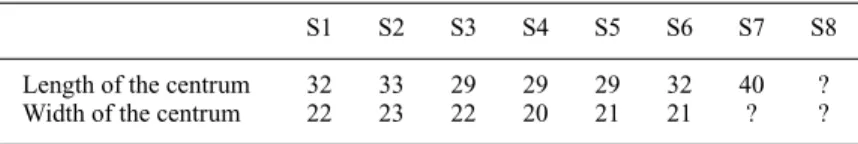
Sacral vertebrae: There are 8 sacral verte- brae (Fig. 6). The anterior six are completely pre- served, the 7th sacral vertebra is partly preserved, and the 8th has preserved only a small anterior
part and its neural arch. Whether their spines are fused together or not is uncertain, due to their being covered by the matrix, but it is clear that the distal ends of the spines do not rise above the dorsal margins of the ilia. The measurements of the sacral vertebrae are given in Table 2.
The width of the anterior end of the 1st sacral
A
B
C
Fig. 6. Sacrum and pelvic girdle of Nemegtia barsboldigen. et sp. nov. (GIN100/2112).
vertebra is greater than its posterior end. The an- terior articular end is strongly concave. The ven- tral surface of the centrum is smooth. The 2nd sacral vertebra is similar to the 1st, except that the pleurocoel is elongated and enlarged, and it occupies nearly 1/2 the length of the centrum.
The width of the anterior end of the 3rd centrum is larger than its posterior end, while in the 4th and 5th centra, the width of both ends is equal.
There is a shallow groove on the anterior portion of the ventral surface in the 4th centrum. This is clearly different from other centra. The pleuro- coel becomes smaller, and the sacral rib of the 4th vertebra is located at the same level as the is- chiadic peduncle. The anterior articular end is larger than the posterior in the 6th centrum, and the contact area of the rib with the centrum is the largest among the sacrals, occupying nearly the whole length of the centrum. Only a small part of its anterior end is fused with the posterior end of the 5th centrum. The small pleurocoel is located on the mid-posterior part of the centrum. The distal end of the 7th sacral rib is divided into two parts, which contact the ilium. Posterior to where the sacral rib is fused with the sacral centrum, there are two fenestrae, which are nearly parallel to each other, and they are much larger than the anterior pleurocoels. The right sacral rib of the 8th sacral vertebra is fused with the transverse process of the centrum and is nearly rod-like.
The middle of the rib shaft is relatively slender, but both ends are widened. Its distal end tightly contacts the internal surface of the ilium. The preserved part shows that the fusion with the connected centra is weaker than that of other cen- trum.
Caudal vertebrae: Only the neural arches of the first two caudal vertebrae are preserved. The nearly vertical facet of the prezygapophysis faces
inwards, while the postzygapophysis faces later- ally.
Shoulder girdle and the fore limb: The proxi- mal end of the left scapula is preserved. It is sim- ilar to those of other oviraptorosaurs. Distal parts of both humeri are preserved (Figs. 7A, B). The preserved portions are similar to other ovirap- torosaurs, but there is a fossa on the anterior sur- face (Fig. 7A, fmb) occupying a similar position to the fossa m. brachialis(Baumel and Witmer, 1993) in modern birds. This is not observed in other oviraptorosaurs.
The right radius is completely preserved (Figs.
7C, D). It is straight, and the cross section of the shaft is oval. Both ends are slightly expanded, but the proximal is larger.
Pelvic girdle and the hind limb(Fig. 6): The preserved length of the ilium is 280 mm. In later- al view, the dorsal margin of the ilium is straight, and the depth of the preacetabular process is the same as that of the postacetabular process. The anterodorsal part of the preacetabular process is concave, while its ventral part is convex. The pubic peduncle is slender and projects down- wards and its articular end is triangular. The is- chiadic peduncle is relatively stout with a round articular end. Dorsally, the ilia nearly meet, but are not fused. The same condition is also found in other oviraptorosaurs, such as Nomingia gobi- ensis(Barsbold, Osmólska et al., 2000).
Only the proximal part of both ischia is pre- served. The process that contacts the pubis is smaller than that which articulates with the ilium.
The proximal ends of both pubes are preserved.
Only the proximal end of the femur is pre- served. There is a clear neck between the femoral head and the shaft of the femur. The angle of the neck and the shaft is about 90°. There is a shal- low groove between the greater trochanter and
Table 2. The measurements of the sacral vertebrae (mm).
S1 S2 S3 S4 S5 S6 S7 S8
Length of the centrum 32 33 29 29 29 32 40 ?
Width of the centrum 22 23 22 20 21 21 ? ?
“?” missing.
the femoral head. The lesser trochanter is smaller and fingerlike. It connects tightly with the greater trochanter. The broken surface shows that many bony struts are distributed near the lateral wall of the shaft.
Comparison and Discussion
The high, narrow and short skull with tooth- less jaws and the medial process of the articular indicate that Nemegtia barsboldiis a derived ovi- raptorosaur (Barsbold and Osmólska, 1990;
Barsbold, 1997). Nemegtiais different from Ovi- raptorin the skull crest. The anterior margin of the skull crest is vertical and the highest point of the crest is located between the nasals and pre- maxillae in Nemegtia barsboldi. The dorsal ante- rior margin of the premaxilla projects forward and the ventral anterior margin of the crest is concave posteriorly in the specimen GIN 100/42, which was described as Oviraptor philoceratops by Barsbold et al. (1990). The highest point of
the crest is located above between the lacrimals in O. mongoliensis, although the anterior margin of the crest is nearly vertical (Barsbold and Osmólska, 1990). Dorsally, the premaxillae are barely exposed, and the anteroposterior length of the parietal is greater than the frontal in Nemeg- tia barsboldi, but the exposed nasal process of the premaxilla is much larger than the nasals, and the lengths of the parietal and the frontal are nearly equal in O. philoceratops (Barsbold and Osmólska, 1990). An additional opening appear- ing on the wall within the antorbital cavity in Ne- megtia barsboldi is smaller than that of the O.
philoceratops. The posterior margin of the quadrate condyle is distinctly posterior to the an- teroventral margin of the occipital condyle in O.
philoceratops. The shape of the ilium, and the number of the sacral vertebrae are also different between Nemegtiaand Oviraptor.
Nemegtia barsboldi has eight sacral vertebrae whereas Ingenia yanshini has six or seven. Ne- megtia barsboldi has a distinct crest in contrast fmb
A B C D
Fig. 7. The left humerus in anterior view (A) and posterior view (B) and the right radius in medial view (C) and lateral view (D) of Nemegtia barsboldigen. et sp. nov. (GIN100/2112). Scale bar4 cm. Abbreviations as in appendix 1.
to Ingenia yanshini. The ischiadic peduncle of the ilium is relatively stout, and the pubic pedun- cle is relatively weak in Nemegtia barsboldi, as opposed to the condition in Ingenia yanshini.
Nemegtia barsboldi is different from Con- choraptor gracilis in having a developed crest, while the skull has no crest in Conchoraptor gra- cilis.
Nemegtia barsboldi is different from Citipati osmolskae, in that the convex crest is higher than the dorsal surface of the skull, and the occiput and the quadrate are vertical, while the top of the skull crest is below the dorsal surface of the
skull, the occiput and the quadrate sloped an- terodorsally in Citipati osmolskae (Clark et al., 2001; 2002), the width and the length of the cer- vical vertebrae are not so different in Nemegtia barsboldi, but the length of the cervical vertebrae is about two times longer than their width in Citi- pati osmolskae. The phylogenetic analysis shows that Nemegtia barsboldiis more closely related to Citipati osmolskae than to any other ovirap- torosaurs (Fig. 8).
Nemegtia barsboldi differs from Khaan mckennai (Clark et al., 2001) in having both a distinct skull crest and a parietal crest, which are
Allosauroidea Ornithomimidae Dromaeosauridae Troodontidae
Archaeopteryx lithographica Avimimus portentosus Chirostenotes pergracilis Nomingia gobiensis
Ingenia yanshini Conchoraptor gracilis Heyuannia huangi Nemegtia barsboldi Citipati osmolskae GIN 100/42
"Oviraptor" mongoliensis
Oviraptor philoceratops Khaan mckennai Microvenator celer Caudipteryx zoui Incisivosaurus gauthieri Fig. 8. One most parsimonious tree found by PAUP.
lacking in Khaan. The dorsal process of the pre- maxilla is vertical in Nemegtia barsboldi, but it extends posterodorsally in Khaan mckennai, the jugal extends further anteriorly in Nemegtia bars- boldi, but it extends further posteriorly in Khaan mckennai.
Nemegtia barsboldiis different from Nomingia gobiensis in that having eight sacral vertebrae, compared to five in Nomingia gobiensis; the dor- sal margin of the ilium is straight in Nemegtia barsboldi, but convex in Nomingia gobiensis; the pubic peduncle is more slender and weaker in Nemegtia barsboldi, compared to Nomingia gob- iensis. Nomingiahas a pygostyle (Barsbold, Cur- rie et al., 2000).
Nemegtia barsboldidiffers Microvenator celer in having one pleurocoel on the cervicals and hy- pophysis on the ventral surface of dorsal verte- brae in Nemegtia, compared with two pleuro- coels and no hypophysis in Microvenator celer;
the number of sacral vertebrae is at least 8 in Ne- megtia barsboldi, it is less than 6 in Microvena- tor celer.
Nemegtia barsboldi differs from Caudipteryx zouiin that Nemegtia barsboldihas no premaxil- lary teeth, while Caudipteryx zoui has premaxil- lary teeth. Nemegtia has a well-developed crest in the skull, but the skull of Caudipteryx zoui lacks a crest. Additionally, the pubic peduncle is slender and weaker than ischiadic peduncle in Nemegtia barsboldi, but it is stronger in Caudipteryx zoui.
Nemegtia barsboldi differs from Heyuannia huangi(Lü, 2003) in that the articular surface for the quadratojugal is convex, while it is more groove-like in Heyuannia huangi. Nemegtia barsboldi has a developed skull crest, whereas Heyuannia huangi does not. In the lateral view, the dorsal margin of the ilium is straight through- out its length, while it is convex in Heyuannia huangi.
The relative position of the external nasal opening and the antorbital cavity varies in differ- ent oviraptorosaurs. The apparent primitive state is with the dorsal margin of the external nasal opening below or near the level of the dorsal
margin of the antorbital cavity (Fig. 9), such as in Incisivosaurus gauthieri. In Caudipteryx sp.
(IVPP V 12430, Zhou et al., 2000), although the skull is heavily crushed, it still can be inferred that the dorsal margin of the external nasal open- ing is below the dorsal margin of the antorbital cavity. Derived forms have the dorsal margin of the external nasal opening above the dorsal mar- gin of the antorbital cavity, a larger distance being more derived. This condition in Nemegtia barsboldi shows that Nemegtia barsboldi is a more derived oviraptorosaur.
The morphology of the lower jaw of Nemegtia shows that it is similar to those of derived ovirap- torosaurs, Oviraptor philoceratops and O. mon- goliensis, Ingenia yanshini (Barsbold et al., 1990), Citipati osmolskae (Clark et al., 2002), Khaan mckennai (Clark et al., 2001), and Heyuannia huangi(Lü, 2003), in which the lower jaw is short and deep, the upper margin of the ex- ternal mandibular fenestra is formed by the surangular and the dorsal branch of the dentary, the surangular has a middle branch, which pro- jects into the external mandibular fenestra. The ventral surface of the lower jaw is convex, and the mandibular symphysis is short. This is differ- ent from that of Caenagnathus collinsi (Stern- berg, 1940; Cracraft, 1971; Currie et al., 1993), where the lower jaw is slender and shallow, the mandibular symphysis is also longer, the external mandibular fenestra is elongate with a dorsal margin, which is formed by the surangular, and the surangular had no middle branch. Inci- sivosaurus gauthieri (Xu, Cheng et al., 2002) also has a longer lower jaw, similar to that of Caenagnathus collinsi, but it bears teeth. It may represent a primitive caenagnathid ovirap- torosaur.
Eight sacral vertebrae are present in Nemegtia barsboldi, similar to some derived oviraptors but more than most known non-avian theropod dinosaurs. More basal theropods, such as Compsognathus longipes (Ostrom, 1978), and Dilophosaurus wetherilli (Welles, 1984) have four sacrals, five in the relatively advanced theropods-Ceratosaurus (Gilmore, 1920), Al-
A E
B
C
D
F
G
H
0 4CM
5CM
Fig. 9. Skulls of (A) Incisivosaurus gauthieri, (B) GIN 100/2112, (C) Khaan mckennai (from Clark et al., 2001), (D) Oviraptor philoceratops(reversed, from Clark et al., 2002), (E) Conchoraptor gracilis, (F) “Ovi- raptor” mongoliensis, (G) Citipati osmolskae (from Clark et al., 2002), and (H) Oviraptor philoceratops (GIN 100/42) (from Barsbold, 1986). Scale bar5 cm in A, B, E and F, 2 cm in C and D, 4 cm in H.
losaurus fragilis(Madsen, 1976), Nomingia gob- iensis (Barsbold, Osmólska et al., 2000), six or seven in Oviraptor (Barsbold et al., 1990), and eight in Heyuannia huangi(Lü, 2003).
Nemegtia barsboldi independently acquired the avian characters such as the fused premaxil- lae, as in Confuciusornis sanctus(Chiappe et al., 1999), toothless jaws, and the presence of nutri- ent openings on the premaxilla and the maxilla as in other oviraptorids.
Sharp ventrolateral margins of the premaxillae indicate that a keratinous structure probably cov- ered the end of the rostrum, as in Erlicosaurus andrewsi (Clark et al., 1994), ornithomimids (Norell, Makovicky, and Currie, 2001; Kobayashi and Lü, 2003), and birds. The rod-like jugal is similar to that of Confuciusornis sanctus and other birds (Elzanowski, 1999), the mobile con- dition between the quadrate and the quadratoju- gal is similar to the condition of most birds, and so are the increased cervical and sacral vertebral counts.
Phylogenetic Analysis
In order to determine the phylogenetic status of Nemegtia barsboldi among oviraptorosaurs, 20 taxa and 200 characters (107 cranial and 93 postcranial characters) (Appendices 2 and 3) are used for this analysis. All characters are equally weighted and unrooted. The character/matrix are modified from Maryanska et al.(2002). Five new genera of Oviraptorosauria are added to the ma- trix; these include Khaan mckennai(Clark et al., 2001), Citipati osmolskae (Clark et al., 2001, 2002), Incisivosaurus gauthieri (Xu, Cheng et al., 2002), Nemegtia barsboldi, and Heyuannia huangi(Lü, 2003). The aim of this analysis is to determine the phylogenetic position of Nemegtia barsboldi among oviraptorosaurs, so the most primitive forms such as Herrerasaurus is- chigualastensis, Coelophysis bauri, and the spe- cialized forms such as Tyrannosauridae and Al- varezsauridae, which were used by Maryanska et al. (2002) as the outgroup in their analysis, are excluded from the present analysis. Although Mi-
crovenator celer and Troodontidae were exclud- ed in Maryanska et al.’s analysis due to the large amount of missing data they thought (Maryanska et al., 2002), taxa should not be excluded a priori from phylogenetic analysis based only on the number of preserved characters (Kearney and Clark, 2003). Therefore, in the present analysis, these taxa are still employed. Most characters are from Maryanska et al. (2002). New characters and their sources in appendix 2 are in bold.
Phylogenetic analysis was performed using MacClade 3.08 (Maddison and Maddison, 1992) and PAUP 4.0b (Swofford, 1998). Because of the large data set (20 taxa) and many missing charac- ter states, a Heuristic Search was used (Swofford and Begle, 1993), with branch-swapping options of the TBR swapping algorithm method. The analysis resulted in one most parsimonious tree (tree length481; consistency index0.5073; re- tention index0.6269) (Fig. 8). This tree shows that Oviraptorosauria forms a monophyletic group. Nineteen unambiguous synapomorphies support this clade. These characters are 1(1), 9(1), 23(1), 37(1), 40(1), 41(2), 43(1), 46(1), 47(1), 55(1), 58(1), 69(1), 71(1), 76(1), 77(1), 81(1), 84(1), 86(1) and 88(1). The primitive forms, such as Incisivosaurus gauthieri, Caudipteryx zoui, Microvenator celer, and Avim- imus portentosus form an ascending sequence from most basal to more derived in the tree.
Avimimus portentosusis a basal form of Caenag- nathoidea. Following Barsbold (1981; 1983), Maryanska et al.(2002) used GIN 100/42 (IGM 100/42) as the representative of Oviraptor philo- ceratopsin their phylogenetic analysis. The pre- sent analysis shows that GIN100/42 and Ovirap- tor philoceratops are closely related. GIN100/42 either belongs to a different species of the same genus as O. philoceratops or the same species, the latter differs from Clark et al.’s judgment (Clark et al., 2002). Nemegtia barsboldiis close- ly related to Citipati osmolskae.
Conclusion
Nemegtia barsboldiis distinguished by at least
five autapomorphies from other known ovirap- torosaurs (vertical skull crest, anteroposterior length of the frontal approximately 25% of that of the parietals in dorsal view, less exposed nasal process of the premaxilla on the dorsal surface of the skull, a process on the quadrate projecting into the cotyla of the quadatojugal, mandibular condyles of the quadrate situated rostrally to the occipital condyle). Phylogenetic analysis shows that Nemegtia barsboldi is closer to Citipati os- molskaethan to other oviraptorosaurs.
Acknowledgments
The authors would like to thank Drs. Louis L.
Jacobs, Dale A. Winkler (SMU, USA), H.
Osmólska (Poland), R. E. Molnar (USA), R.
Barsbold (Mongolia), and Y. Kobayashi (Japan) for their valuable comments on the early versions of the manuscript, and to acknowledge the mem- bers of the Mongol Highland International Di- nosaur Project. Special thanks go to Y.
Kobayashi, who found this beautiful specimen in 1996. Thanks also go to Drs. Zhou, Z. and Xu, X. (IVPP of Chinese Academy of Sciences, Bei- jing, China), for access of the specimens in their care. This project is supported by Chunichi Shin- bun Co. Ltd., Kyoto Kagaku Co. Ltd., Chukyo TV Broadcasting Co. Ltd., and Tokai Bank Ltd.;
plus Institute for the Study of Earth and Man at Southern Methodist University, the Jurassic Foundation, and the Chang Ying-Chien Science Grant for USA-China Collaborative Field Re- search to Junchang Lü.
References
Barsbold, R., 1976. [On a new Late Cretaceous family of small theropods (Oviraptoridae fam. n.) of Mongolia].
Doklady Akademia Nauk SSSR, 226(3): 685–688 (in Russian).
Barsbold, R., 1977. Kinetism and peculiarity of the jaw apparatus of oviraptors (Theropoda, Saurischia). Trans- actions, Joint Soviet-Mongolia Geological Expedition, 4: 37–47 (in Russian).
Barsbold, R., 1981. Toothless carnivorous dinosaurs of Mongolia. Trudy Sovmestnoi Sovetsko-Mongol’skoi Pa-
leontologicheskoi Ekspeditsii, 15: 28–39.
Barsbold, R., 1983. Carnivorous dinosaurs from the Cre- taceous of Mongolia. Trudy Sovmestnoi Sovetsko-Mon- gol’skoi Paleontologicheskoi Ekspeditsii, 19: 5–120.
Barsbold, R., 1986. [Raubdinosaurier Oviraptoren]. In Vorobyeva, E. I. (ed). Herpetologische Untersuchungen in der Mongolischen Volksrepublik. Pp. 210–223. Akad.
Nauk. S. S. S. R. Inst. Evolyucionnoy Morfologii i Ekologii Zhivotnykh im. A. M. Severtsova, Moskva (in Russian with German summary).
Barsbold, R., 1997. Oviraptorosauria. InCurrie, P. J. and Padian, K. (eds.). Encyclopedia of Dinosaurs. Pp.
505–509. Academic Press, San Diego.
Barsbold, R., Currie, P. J., Myhrvold, N. P., Osmólska, H., Tsogtbaatar, K. & Watabe, M., 2000. A pygostyle from a non-avian theropod. Nature, 403: 155–156.
Barsbold, R., Maryanska, T. & Osmólska, H., 1990. Ovi- raptorosauria. In Weishampel, D. B., Dodson, P. and Osmólska, H. (eds.). The Dinosauria. Pp. 249–258.
University of California Press, Berkeley.
Barsbold, R. & Osmólska, H., 1999. The skull of Veloci- raptor(Theropoda) from the Late Cretaceous of Mon- golia. Acta Palaeontologica Polonica, 44(2): 189–219.
Barsbold, R., Osmólska, H., Watabe, M., Currie, P. J. &
Tsogtbaatar, K., 2000. A new oviraptorosaur (dinosaur, theropod) from Mongolia: the first dinosaur with a py- gostyle. Acta Palaeontologica Polonica, 45(2): 97–106.
Baumel, J. J. & Witmer, M., 1993. Osteologia. InBaumel, J. J., King, A. S. et al.(eds.). Handbook of avian anato- my: nomina anatomica avium, second edition. Pp.
45–132. Cambridge, Massachusetts.
Bonaparte, J. F., Novas, F. E. & Coria, R. A., 1990.
Carnotaurus sastreiBonaparte, the horned, lightly built carnosaur from the Middle Cretaceous of Patagonia.
Contributions in Science Nat. History Museum Los An- geles County, 416: 1–42.
Chiappe, L. M., 1996. Late Cretaceous birds of southern South America: Anatomy and systematics of enantior- nithes and Patagopteryx deferrariisi. Munchner Ge- owiss. Abh.(A), 30: 203–224.
Chiappe, L. M., 2001. Phylogenetic relationships among basal birds. InGauthier, J. and Gall, L. F. (eds.). New perspectives on the origin and early evolution of birds, Proceedings of the International Symposium in Honor of John H. Ostrom. Pp. 125–139. Yale Peabody Muse- um.
Chiappe, L. M., 2002. Basal bird phylogeny: Problems and solutions. In Chiappe L. M. and Witmer L. M.
(eds.). Mesozoic birds above the heads of dinosaurs.
Pp. 448–472. University of California Press. Berkeley.
Chiappe, L. M., Ji, S-A., Ji, Q. & Norell, M. A.,1999.
Anatomy and systematics of the Confuciusornithidae (Theropoda: Aves) from the late Mesozoic of north- eastern China. Bulletin of the American Museum of
Natural History, 242: 1–89.
Chiappe, L. M., Norell, M. A. & Clark, J. M., 1996. Phy- logenetic position of Mononykus(Aves: Alvarezsauri- dae) from the Late Cretaceous of the Gobi Desert.
Memoirs of the Queensland Museum, 39(3): 557–582.
Chiappe, L. P., Norell, M. A. & Clark, J. M., 1998. The skull of a relative of the stem-group bird Mononykus.
Nature, 392: 275–278.
Clark, J. M., Altangerel, P. & Norell, M. A., 1994. The skull of Erlicosaurus andrewsi, a Late Cretaceous seg- nosaur (Theropoda: Therizinosauridea) from Mongolia.
American Museum Novitates, 3115: 1–39.
Clark, J. M., Norell, M. A. & Barsbold, R., 2001. Two new oviraptorids (Theropoda: Oviraptorosauria), Upper Cretaceous Djadokhta Formation, Ukhaa Tolgod, Mon- golia. Journal of Vertebrate Paleontology, 21(2): 209–
213.
Clark, J. M., Norell, M. A. & Chiappe, L.M., 1999. An oviraptorid skeleton from the late Cretaceous of Ukhaa Tolgod, Mongolia, preserved in an avian-like brooding position over an oviraptorid nest. American Museum Novitates, 3265: 1–36.
Clark, J. M., Norell, M. A. & Rowe, T., 2002. Cranial anatomy of Citipati osmolskae (Theropoda, Ovirap- torosauria), and a reinterpretation of the holotype of Oviraptor philoceratops. American Museum Novitates, 3364: 1–24.
Colbert, E. H. & Russell, D. A., 1969. The small Creta- ceous dinosaur Dromaeosaurus. American Museum Novitates, 2380: 1–49.
Cracraft, J., 1971. Caenagnathiformes: Cretaceous birds convergent in jaw mechanism to dicynodont reptiles.
Journal of Paleontology, 45(2): 805–809.
Cracraft, J. 1986. The origin and early diversification of birds. Paleobiology, 12: 383–399.
Currie, P. J., 1985. Cranial anatomy of Stenonychosaurus inequalis (Saurischia, Theropoda) and its bearing on the origin of birds. Canadian Journal of Earth Sciences, 22: 1643–1658.
Currie, P. J., 1995. New information on the anatomy and relationships of Dromaeosaurus albertensis (Di- nosauria: Theropoda). Journal of Vertebrate Paleontol- ogy, 15: 576–591.
Currie, P. J., 2000. Theropods from the Cretaceous of Mongolia. InBenton, M. J., Shishkin, M. A., Unwin, D.
M. and Kurochkin, E. N. (eds.). The age of dinosaurs in Russia and Mongolia. Pp. 434–455. Cambridge Uni- versity Press, Cambridge.
Currie, P. J., Godfrey, S. J. & Nessov, L., 1993. New cae- nagnathid (Dinosauria: Theropoda) specimens from the Upper Cretaceous of North America and Asia.Canadi- an Journal of Earth Sciences, 30: 2255–2272.
Currie, P. J. & Russell, D. A., 1988. Osteology and rela- tionships of Chirostenotes (Saurischia, Theropoda)
from the Judith River (Oldman) Formation of Alberta, Canada. Canadian Journal of Earth Sciences, 25: 972–
986.
Currie, P. J., Vickers-Rich, P. & Rich, T. H., 1996. Possi- ble oviraptorosaur (Theropoda, Dinosauria) specimens from the Early Cretaceous Otway Group of Dinosaur Cove, Australia. Alcheringa, 20: 73–79.
Currie, P. J. and Zhao, X.-J., 1993a: A new carnosaur (Di- nosauria: Theropoda) from the Jurassic of Xinjiang, People’s Republic of China. Canadian Journal of Earth Sciences, 30: 2037–2081.
Currie, P. J. & Zhao, X.-J., 1993b. A new troodontid (Di- nosauria, Theropoda) braincase from the Dinosaur Park Formation (Campanian) of Alberta. Canadian Journal of Earth Sciences, 30: 2231–2247.
Dong, Z.-M. & Currie, P. J., 1996. On the discovery of an oviraptorid skeleton on a nest of eggs at Byan Man- dahu, Inner Mongolia, People’s Republic of China.
Canadian Journal of Earth Sciences, 33: 631–636.
Elzanowski, A., 1999. A comparison of the jaw skeleton in theropods and birds, with a description of the palate in the Oviraptoridae. InOlson, S. L. (ed.). Avian Pale- ontology at the Close of the 20thCentury: Proceedings of the 4thInternational Meeting of the Society of Avian Paleontology and Evolution, Washington, D.C., 4–7 June 1996. Smithsonian Contributions to Paleobiology, 89: 311–323.
Frankfurt, N. G. & Chiappe, L. M., 1999. A possible ovi- raptorosaur from the Late Cretaceous of northwestern Argentina. Journal of Vertebrate Paleontology, 19(1):
101–105.
Frey, E. & Martill, D. M., 1995. A possible ovirap- torosaurid theropod from the Santana Formation (Lower Cretaceous, ?Albian) of Brazil. Neues Jahrbuch für Geologie und Paläontologie Monatshefte, 7: 397–
412.
Gauthier, J., 1986. Saurischian monophyly and the origin of birds. InPadian K. (ed.). The origin of birds and the evolution of flight. Memoirs of the California Academy of Sciences8. San Francisco, Pp. 1–55.
Gilmore, C. W., 1920. Osteology of the carnivorous Di- nosauria in the United States National Museum, with special reference to the genera Antrodemus (Al- losaurus) and Ceratosaurus. United States National Museum, Bulletin, 110: 1–154.
Gradzinski, R., Kazmierczak, J. & Lefeld, J., 1968. Geo- graphical and geological data from the Polish-Mongo- lian Palaeontological Expeditions. Palaeontologia Polonica, 19: 33–92.
Holmgren, N., 1955. Studies on the phylogeny of birds.
Acta Zoologica, 36: 243–328.
Holtz, T. R. Jr., 1994. The arctometatarsalian pes, an un- usual structure of the metatarsus of Cretaceous Theropoda (Dinosauria: Saurischia). Journal of Verte-