1
A rare case of a severe ocular complication as an initial presentation of adolescent-onset systemic lupus
erythematosus: a case report
Yuki Kotani, Kohei Miyazaki, Takuji Enya, Tomoki Miyazawa, Mitsuru Okada, and Keisuke Sugimoto
Department of Pediatrics, Kindai University Faculty of Medicine, Osaka-Sayama, Japan
Abstract Background: Systemic lupus erythematosus (SLE)
is an autoimmune disease that causes severe com- plications in multiple organs. Ocular manifesta- tions are critical in patients with SLE because of the vision-threatening risk of such complications.
However, severe retinopathy as an initial presen- tation of SLE is uncommon, even when the severity of SLE is mild, especially in children.
Case presentation: We encountered a 14-year-old female patient with rapidly progressive impair- ment of her left vision that was determined to be caused by severe retinopathy secondary to SLE.
Her other systemic symptoms, including a mild renal disorder, were not severe. The patient showed positivity for anticardiolipin antibody, although she did not have antiphospholipid anti- body syndrome. She was initially treated with
high-dose methylprednisolone, antiplatelet ther- apy, and anticoagulation therapy because of the high possibility of vision loss; this was followed by tacrolimus as maintenance therapy. Photo- coagulation therapy was also performed to pre- vent vitreous hemorrhage and retinal detach- ment. Early diagnosis of SLE based on the rapid deterioration of her vision allowed for early in- terventions and a good clinical course with recov- ery of her vision.
Conclusions: Importantly, regardless of the se- verity of the systemic symptoms in patients with SLE, ocular involvement is critical and requires aggressive treatment.
Key words:children systemic, lupus erythema- tosus, retinopathy
Introduction
Systemic lupus erythematosus (SLE) is an au- toimmune disease that may affect multiple organ systems, including the eye 1. The male:female ra- tio is 1:9; thus, SLE overwhelmingly affects women, and the age of onset is also more common in younger women aged 20 to 40 years. Childhood onset accounts for about 20% of all cases of SLE, the percentage estimated as 29% of all cases of rheumatic disease in registered children, followed by juvenile idiopathic arthritis which estimated as
51% 2, 3, 4. Among the initial symptoms, arthritis
and arthralgia occur in about 67% of patients, fol- lowed in frequency by skin lesions and fever. SLE Received May 11, 2018; Accepted September 7, 2018
is characterized by various clinical manifestations of systemic organs such as the kidneys, nerves, pleura, and pericardium 5. A meta-analysis of 16 studies was performed to investigate clinical dif- ferences between children and adults with SLE, and the authors reported that fever, lymphade- nopathy, butterfly rash, ulcers, renal impairment, seizures, hemolytic anemia, and thrombocytope- nia were more common in children 6. Children have more aggressive disease course, and one- third of pediatric patients with SLE have atypical clinical features 7. Ocular lesions are found in 30% of all patients with SLE 8, and they are more frequent particularly in cases complicated by an- tiphospholipid antibody syndrome (APS) and
central nervous system lupus 9, 10, 11. The inci- dence of retinal involvement in patients with SLE ranges from 7% to 26% 12. However, severe reti- nopathy is very rare, even when the severity of SLE is mild, especially in children. We herein de- scribe a patient whose diagnosis of SLE was trig- gered by ocular impairment.
Case report
A 14-year-old female patient presented with decreasing left vision. She was diagnosed with left retinal vasculitis with macular edema, and her left best corrected visual acuity (BCVA) had de- creased to 0.3. A sub-Tenon steroid injection and betamethasone ophthalmic solution were applied to treat the lesion. Subsequently, however, the pa- tient’s left BCVA decreased to 0.1. Furthermore, her antinuclear antibody (ANA) titer increased.
These findings led to suspicion of collagen dis- ease, and she was admitted to our institution.
No family history of autoimmune renal disease and other autoimmune diseases. Oliguria, ascites, and edema were absent. Her height was 159.7cm and her weight was 48.7 kg. Physical findings upon admission included a blood pressure of 115/75 mmHg, pulse of 86/min, and body temper- ature of 37ºC. Her consciousness was clear and her whole-body condition was good. No ulcers were found in the oral cavity. Skin examination showed depilation of the head, butterfly erythema (Figure 1a), and erythema of the finger pulp (Fig- ure 1b). Her BCVA had decreased to 0.09 in the left eye and not changed 1.5 in the right eye, in- dicating severe damage especially in the left eye.
Her bilateral intraocular pressures were normal, and corneal abnormality and iritis were absent.
Fundus examination revealed soft leukoplakia (Figure 2a), and fluorescencein angiography (FA) showed retinal arteriovenous vasculitis, strong is- chemia in the macular region, no reflux area, and leakage from the blood vessel (Figure 2b). Opti- cal coherence tomography (OCT) revealed marked macular edema (Figure 2c), and Gold- mann visual field testing indicated the scotoma in the vicinity of the center. The right fundus was normal. Her chest sounds were clear with no fric- tion rubs or murmurs. Her abdomen was soft without muscle guarding or rebound tenderness.
No coldness or ulcerative lesions were present in the peripheral limbs. Dull red spots were ob- served on the ventral side of the bilateral upper limbs.
On laboratory examination, mild to moderate proteinuria (0.5 g/day) and hematuria (1+) were noted. The pH was 6.0 in urine and urinary cylinder was (1+). Her peripheral white cell count and red cell count were normal. The platelet (23.4x10⁵/µl;
reference range, 16.7x10⁵ - 36.2x10⁵/ µl) was nor- mal. Her peripheral white cell count was normal.
Blood coagulation function test results were normal.
The results of most laboratory blood examinations, including measurements of electrolytes, serum creatinine, and blood urea nitrogen, were normal.
Total protein (8.5 g/dl; reference range, 6.5- 8.2g/dl) was mild heigh, albumin (4.6 g/dl; refer- ence range, 3.6-5.5 g/dl) was normal. The concen- trations of aspartate aminotransferase and alanine aminotransferase were normal. The lactate dehy- drogenase and creatinine kinase concentrations were also normal. The serum concentrations of fi- brinogen (267 mg/dL; reference range, 150–400 mg/dL), fibrin and fibrinogen degradation prod- ucts (2.2 μg/dL; reference range, 5.0 μg/dL), thrombin–antithrombin III complex (1.0 ng/dL;
reference range, 0.0–3.0 ng/dL), and D-dimers (0.6 μg/mL; reference range, 0.0–1.0 μg/mL) were normal. The prothrombin time and activated partial thromboplastin time were also normal, in- dicating no signs of disseminated intravascular coagulation. Serologic analysis showed an ele- vated ANA titer (x320; reference range, 0–x40).
The anti-double-strand DNA titer (12 IU/L; ref- erence range, 0–12 IU/L) was negative. The anti- single-strand DNA titer (73 IU/L; reference range, 0–25 IU/L) was elevated. Anticardiolipin anti- body was identified (42 IU/mL; reference range, 0–10 IU/mL). Anticoagulant and anticardiolipin β2 glycoprotein I antibodies (2 IU/mL; reference range, 0–10 IU/mL) were negative. Myeloperox- idase antineutrophil cytoplasmic antibodies were negative. The serum complement component C3 activity (73 mg/dL; reference range, 82–145 mg/dL) was low, while the C4 activity (13 mg/dL;
reference range, 12–33 mg/dL) and total comple- ment activity (CH50 screening test) (29.1 U/mL;
reference range, 24.2–52.8 U/mL) were normal.
Circulating immune complexes (1.5 μg/mL; ref- erence range, 0.0–3.0 µg/mL) were also normal.
The serum immunoglobulin G (IgG) concentra- tion (2009 mg/dL; reference range, 810–1780 mg/dL) was high. The IgA, IgM, and IgE concen- trations were normal. An infectious work-up came back negative for hepatitis B and C. No findings of optic neuritis were observed by short T1 inversion recovery. Ultrasonographic findings
of the renal size and shape were normal, and mass lesions were absent.
Figure 1 Skin lesions on admission. (a) Erythema of the finger pulp and (b) butterfly erythema were present.
Figure 2 Fundus photograph of the left eye at the first visit. (a) Retinal arteriovenous vasculitis, soft vitiligo, an area of nonperfusion, and macular edema were observed. (b) Fluorescein leak- age from the vessel and ischemia in the macular region were seen by fluorescein angi- ography. (c) Marked macular edema was observed by optical coherence tomography.
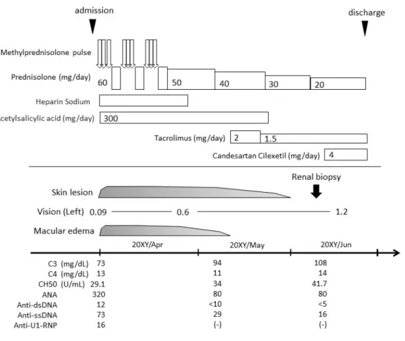
The patient met 5 of the 12 SLE diagnostic cri- teria according to Guidance to diagnosis of SLE in childhood, and the complication of SLE reti- nopathy was confirmed based on the FA. The pa- tient’s total SLE Disease Activity Index score was 16 points (8 for the vision disorder, 2 for the new skin rashes, 2 for hair loss, 2 for low-com- plement blood disease, and 2 for anti-DNA anti- body elevation). In an attempt to decrease the dis- ease activity and prevent visual loss in the left eye, three courses of high-dose methylprednisolone (1000 mg/day) were administered along with oral prednisolone (PSL) at 60 mg/day with a gradual decrease of 10 mg/day every 2 weeks. The grad- ual decrease in PSL was followed by maintenance
therapy with tacrolimus (Tac) at 2 mg/day. Alt- hough the patient was negative for lupus antico- agulant and anticardiolipin β2 glycoprotein I anti- bodies, we suspected APS based on the presence of anticardiolipin antibody and the severe find- ings of a thrombotic tendency, stenosis, and in- farction in the fundus; therefore, we administered a combination of intravenous heparin sodium so- lution as an anticoagulant and oral aspirin (300 mg/day) as antiplatelet therapy. After these treat- ments, the macular edema of the left fundus and retinal arteriovenous vasculitis improved (Figure 3), and the left BCVA recovered from 0.09 to 1.2.
The butterfly erythema and erythema of the ex- tremities also improved. The ANA titer decreased
from 320 to x80, and the anti-single-strand DNA antibody decreased from 73 to 16 IU/L. Percuta- neous renal biopsy was performed on the 50th day of disease because of the complication of lupus nephritis. Histologic examination of a renal bi- opsy specimen revealed mild to moderate prolif- eration of mesangial cells but no thickening of the glomerular capillary walls (Figure 4). Interstitial fibrosis, atrophy or other injury of the tubular cells, and necrosis were absent. Immunofluores- cence showed IgG, IgA, IgM, C3, C4, C1q, and
fibrinogen reactivity in portions of the mesan- gium and capillary walls. Electron microscopy re- vealed nodular deposits in the mesangial and ca- pillary walls. These findings indicated lupus ne- phritis (histopathologic class II according to the International Society of Nephrology/Renal Pa- thology Society). She was discharged on the 59th day of disease with PSL, Tac, and an angiotensin receptor antagonist. She continued to undergo follow-up therapy including laser treatment for the non-reflux area in the ocular fundus. The clin- ical course is summarized in Figure 5.
Figure 3 Fundus photograph of the left eye on day 32 of admission. (a) The retinal arteriovenous vasculitis had improved. The soft vitiligo, the nonperfused area, and macular edema had also improved, but were still present. (b) Fluorescein leakage from the vessel in the macular region had decreased on fluorescein angiography, and (c) no macular edema was present on optical coherence tomography.
Figure 4 Histologic findings of the renal specimen. Dif- fuse mild mesangial cell proliferation in the glomeruli and the lack of thickening of the glo- merular basement membrane indicated the pres- ence of diffuse proliferative lupus nephritis (In- ternational Society of Nephrology/Renal Pa- thology Society class II) (periodic acid-Schiff stain, ×400).
Figure 5 Clinical course.
Discussion
Ocular manifestations of SLE are variable and include abnormalities of the eyelid and ocular ad- nexa, keratoconjunctivitis sicca, retinal vasculitis, vaso-occlusive disorders, choroidopathy, scleritis and uveitis, and optic neuropathy 8, 13, 14. SLE ret- inopathy is characterized by soft vitiligo, retinal vasculitis, vitreous hemorrhage, development of avascular areas, and appearance of new blood ves- sels 15; these abnormalities are consistent with the findings in our case. The pathology of SLE reti- nopathy, as well as the onset mechanism of sys- temic vasculitis, are attributed to the impairment of the vascular endothelium and basement mem- brane caused by complement activation and auto- antibodies with deposition of antigen–antibody complexes on the blood vessel walls; this results in insufficiency of the peripheral circulation with progression to vascular occlusion. Furthermore, hypercoagulability and high blood viscosity ac- companied by the disease activity of SLE pro- motes thrombus formation and vascular occlusion.
APS is well recognized in patients with SLE 11.
The risk factors for progression of SLE retinopa- thy include complications such as APS, lupus ne- phritis, and central nervous system lupus 9, 10, 15. However, no apparent findings indicative of blood hypercoagulability or hyperviscosity were present in our case. Additionally, the patient had no clinical findings or symptoms specific to cen- tral nervous lupus, such as convulsions. One pro- spective study showed that the incidence of renal dysfunction in patients with SLE without reti- nopathy was 59.3%, but that in patients with ret- inopathy was 100.0% 15. In addition, renal in- volvement in patients with SLE will generally lead to secondary hypertension. When hyperten- sion is prolonged, it usually affects the retina and choroid by inducing retinal arterial narrowing and changes at the arteriovenous crossing. Our patient had the complication of lupus nephritis, but the histopathologic lesions were mild and the patient had no hypertension. However, the presence of hyperphospholipid antibodies indicated the ten- dency to form thrombi.
SLE retinopathy is classified into three types:
(1) focal retinal ischemia, (2) retinal vascular oc- clusive disease, and (3) the proliferative lupus type, depending on the degree of the fundus le- sion 16. In the present case, the patient exhibited the vascular occlusion type, which was mainly caused by vasculitis and retinal arteriovenous oc- clusion. This resulted in rapid and severe vision deterioration. Hydroxychloroquine is an effective medication for SLE with few side effects 17. How- ever, usage of hydroxychloroquine should be avoided in case of ocular complication because of the possibility of exacerbating side effects. Ster- oid therapy is still a key drug in Japan, although clinicians must be alert to its side effects, espe- cially in children 7. PSL is essential for suppres- sion of vasculitis and minimization of vascular occlusion. Recanalization is reportedly difficult even with steroid pulse therapy in patients with the vascular occlusion type 10, 18. After the appear- ance of ocular symptoms, our patient underwent early consultation and intervention including steroid therapy, leading to rapid reduction of the vasculitis and minimization of the obstructive le- sions. Methylprednisolone therapy was success- ful in Indian patients who exhibited a clinical course similar to that of our patient 19. In our case, photocoagulation therapy was performed early af- ter discharge because the proliferative changes and angiogenesis had resulted in the retinal lesion, causing vitreous hemorrhage in addition, espe- cially in the presence of antiphospholipid anti- bodies. However, the optimal timing of and crite- ria for photocoagulation therapy are unclear be- cause vision impairment may still progress after treatment 10, 16, 20. Especially in children, eye com- plications such as visual acuity problems deterio- rate the quality of school and daily life, leading to restrictions in the scope of the patient’s activity.
We should consider the patient’s age, disease se- verity, and similar factors in each individual case.
Ocular complications often occur after a cer- tain period of time has passed since the diagnosis of SLE, although they are reportedly correlated with the disease activity and degree of SLE 21, 22. Even patients with SLE who have no visual symp- toms or visible retinal abnormalities have demon- strated abnormal lesions of the fundus by fluores- cein angiography. To the best of our knowledge, few cases of visual impairment as the initial symptom of SLE have been reported, especially in children 19, 23. This suggests that visual impair- ment is an underlying subclinical condition 9. It seems difficult to predict the occurrence of ocular
symptoms based on a skin rash, dysfunction of other organs, and laboratory test results. However, in a study of the prevalence of clinical and labor- atory features in 10 patients with “classic” reti- nopathy, 60% of patients showed a skin rash, 40%
presented with nephritis, and 30% had alopecia and anemia 24. This emphasizes that high possi- bility of sudden deterioration of ocular complica- tions even while the SLE disease activity is stable.
Because our patient mainly had skin lesions such as butterfly erythema, hair loss, and erythema at the limb extremities with only minor disorders of other organs, the severity of the ocular complica- tions was difficult to predict. With regard to maintenance therapy, immunosuppressant ther- apy was added in the early stage to alleviate the side effects of steroids over a long period of time.
Immunosuppressive agents are selected depend- ing on the patient’s disease severity and patholog- ical condition, and cyclophosphamide is consid- ered to be effective for ocular complications 25. However, few studies have been performed to ex- amine the efficacy of immunosuppressants in de- tail. In our case, Tac was given because of the se- verity of the ocular complications and to preserve the patient’s fecundity. To the best of our knowledge, however, the efficacy of Tac for ocu- lar manifestations in patients with SLE has not been proven. The use of Tac allowed us to mini- mize the PSL dosage because of the good lesion response. The patient was followed-up as an out- patient and continued to receive laser treatment.
Conclusion
We encountered a rare case involving sudden visual deterioration, resulting in a diagnosis of SLE. Prediction of the onset of ocular complica- tions in patients with SLE is difficult; however, periodic follow-up is important because ocular complications might influence the patient’s prog- nosis of eye function regardless of the severity of systemic symptoms. Furthermore, suspicion of SLE and early medical intervention are needed when encountering a patient who exhibits rapid deterioration of visual acuity, even if his or her systemic symptoms are mild.
Declarations
Ethics approval and consent to participate Consent to publish the patient’s data, including individual details and photographs, was obtained
from the patient’s parents. Tissue staining of the renal specimens was performed with written in- formed consent from the patient’s parents.
Consent for publication
This manuscript has been seen and approved by all authors and is not under consideration for pub- lication elsewhere in a similar form, including in abstract form, in any language.
Availability of data and materials
All data generated during this case report are included in this published article.
Competing interests
The authors declare that they have no compet- ing interests.
Funding
The report was not supported by any funding.
Authors’ contributions
YK analyzed and interpreted the patient data regarding the SLE and retinopathy. KS performed the histological examination of the kidney and was a major contributor to the writing of the man- uscript. KM, TE, and TM helped to draft the man- uscript and acquire renal biopsy for histological images for illustration. MO and KS conceived the idea of the study and did the final proofing of the manuscript. All authors read and approved the fi- nal manuscript.
Acknowledgements
We thank the ophthalmologists (Dr. Aya Takahashi and Dr. Kazuki Kuniyoshi) who par- ticipated in the care of this patient and provided helpful advice. We also thank Angela Morben, DVM, ELS, from Edanz Group (www.edanzedit- ing.com/ac) for editing a draft of this manuscript.
References
1.Papagiannuli E, et al. (2016) Systemic lupus erythe- matosus: An update for ophthalmologists. Survey of ophthalmology 61: 65-82
2.Fujikawa S, Okuni M (1997) A nationwide surveil- lance study of diseases among Japanese children. Acta Paediatr Jpn 39: 242-244
3 .Klein-Gitelman M, Reiff A, Silverman ED (2002) Systemic lupus erythematosus in childhood. Rheum Dis Clin North Am 28: 561-577
4.Barsalou J1, Levy DM, Silverman ED (2013) An up- date on childhood-onset systemic lupus erythematosus.
Curr Opin Rheumatol 25: 616-622
5.Pons-Estel BA, et al. (2004) Grupo Latinoamericano de Estudio del Lupus. The GLADEL multinational Latin American prospestive inception cohort of 1214 patients with systemic lupus erythematousus: ethnic and disease heterogeneity among “Hispanics”. Medi- cine (Baltimore) 83: 1-17
6.Livingston B, Bonner A, Pope J (2011) Differences in clinical manifestations between childhood-onset lu- pus and adult-onset lupus: a meta-analysis. Lupus 20:
1345-1355
7.Brunner HI, et al. (2008) Difference in disease fea- tures between childhood-onset and adult-onset sys- temic lupus erythematosus. Arthritis Rheum 58: 556- 562
8.Palejwala NV, Walia HS, Yeh S (2012) Ocular man- ifestations of systemic lupus erythematosus: a review of the literature. Autoimmune Dis 2012: 290898 9.Au A, O’Day J (2004) Review of severe vaso-occlu-
sive retinopathy in systemic lupus erythematosus and the antiphospholipid syndrome: associations, visual outcomes, complications and treatment. Clin Exp Oph- thalmol 32: 87-100
10.Jabs DA, et al. (1986) Severe retinal vaso-occlusive disease in systemic lupus erythematous. Arch Ophthal- mol 104: 558-563
11.Petri M (2000) Epidemiology of the antiphospholipid antibody syndrome. J Autoimmu 15: 145-151
12.Read RW (2004) Clinical mini-review: systemic lu- pus erythematosus and the eye. Ocul Immunol Inflamm 12: 87-99
13.Silpa-archa S, Lee JJ, Foster CS (2016) Ocular man- ifestations in systemic lupus erythematosus. Br J Oph- thalmol 100: 135-141
14.Peponis V, et al. (2006) Ocular manifestations of sys- temic lupus erythematosus: a clinical review. Lupus 15: 3-12
15.Kharel Sitaula R, Shah DN, Singh D (2016) Role of lupus retinopathy in systemic lupus erythematosus. J Ophthalmic Inflamm Infect 6: 15
16.Vine AK, Barr CC (1984) Proliferative lupus reti- nopathy. Arch Ophthalmol 102: 852-854
17.Tang C, Godfrey T, Stawell R, Nikpour M (2012) Hydroxychloroquine in lupus: emerging evidence sup- porting multiple beneficial effects. Intern Med J 42:
968-978
18.Kayazawa F, Honda A (1981) Severe retinal vascular lesions in systemic lupus erythematosus. Ann Ophthal- mol 13: 1291-1294
19.Bandyopadhyay SK1, Moulick A, Dutta A (2006) Ret- inal vasculitis--an initial presentation of systemic lupus erythematosus. J Indian Med Assoc 104: 526-527 20.Kleiner RC, et al. (1989) Vaso-occlusive retinopathy
associated with antiphospholipid antibodies (lupus an- ticoagulant retinopathy). Ophthalmology 96: 896-904 21.Stafford-Brady FJ, et al. (1988) Lupus retinopathy.
Patterns, associations, and prognosis. Arthritis Rheum 31: 1105-1110
22.Klinkhoff AV, Beattie CW, Chalmers A (1986) Ret- inopathy in systemic lupus erythematousus: Arthritis and Rheum 29: 1152-1156
23.Wong K, Ai E, Jones JV, Young D (1981) Visual loss as the initial symptom of systemic lupus erythematosus.
Am J Ophthalmol 92: 238-244
24.Giorgi D, et al. (1999) Retinopathy in systemic lupus erythematosus: pathogenesis and approach to therapy.
Hum Immunol 60: 688-696
25.Galindo-Rodríguez G, et al. (1999) Cyclophospha- mide pulse therapy in optic neuritis due to systemic lu- pus erythematosus: an open trial. Am J Med 106: 65- 69