学位論文
学位論文
学位論文
学位論文(要約)
Applications of aminoacylation ribozymes that recognize the 3’-end of
tRNA via two consecutive base pairs
(
(
(
(tRNA の
の
の 3’末端を塩基対合によって認識する
の
末端を塩基対合によって認識する
末端を塩基対合によって認識する
末端を塩基対合によって認識する
アミノアシル化リボザイムの応用)
アミノアシル化リボザイムの応用)
アミノアシル化リボザイムの応用)
アミノアシル化リボザイムの応用)
平成
平成
平成
平成 26 年
年
年 12 月博士(理学)申請
年
月博士(理学)申請
月博士(理学)申請
月博士(理学)申請
東京大学大学院理学系研究科
東京大学大学院理学系研究科
東京大学大学院理学系研究科
東京大学大学院理学系研究科
化学専攻
化学専攻
化学専攻
化学専攻
寺坂
寺坂
寺坂
寺坂 尚紘
尚紘
尚紘
尚紘
学位論文
学位論文
学位論文
学位論文
(部分公表版)
(部分公表版)
(部分公表版)
(部分公表版)
Applications of aminoacylation ribozymes that recognize the 3’-end of
tRNA via two consecutive base pairs
(
(
(
(tRNA の
の
の 3’末端を塩基対合によって認識する
の
末端を塩基対合によって認識する
末端を塩基対合によって認識する
末端を塩基対合によって認識する
アミノアシル化リボザイムの応用)
アミノアシル化リボザイムの応用)
アミノアシル化リボザイムの応用)
アミノアシル化リボザイムの応用)
平成
平成
平成
平成 26 年
年
年 12 月博士(理学)申請
年
月博士(理学)申請
月博士(理学)申請
月博士(理学)申請
東京大学大学院理学系研究科
東京大学大学院理学系研究科
東京大学大学院理学系研究科
東京大学大学院理学系研究科
化学専攻
化学専攻
化学専攻
化学専攻
寺坂
寺坂
寺坂
寺坂 尚紘
尚紘
尚紘
尚紘
1
Abstract (abridged)
In this thesis, I applied aminoacylation ribozyme “flexizyme” to discover the novel interaction between a small non-coding RNA (ncRNA) and a small molecule, and to develop the orthogonal translation machinery consisting of the pair of ribosome and tRNAs bearing mutations at the peptidyl transferase center. Flexizymes have following unique characteristics; (i) substrate RNA is recognized by two consecutive base pairs between 3′-end of substrate RNA and 3′-end of flexizyme, (ii) these base pairs can be substituted with other base pairs and (iii) various activated amino acids can be used as substrates including both canonical and noncanonical amino acids. This flexible aminoacylation of RNAs by flexizymes were used not only as a method for labeling endogenous tRNAs but also as a tool to prepare various aminoacyl-tRNAs bearing mutations at 3′-end to engineer the translation machinery.
Chapter 1 is a general introduction of this thesis. Transfer RNA, translation and small non-coding RNA such as miRNA are firstly introduced, and the problems and questions in these research field are also described. Then aminoacylation ribozyme “flexizyme” having unique characteristics is introduced, which has a potential to overcome these problems.
In chapter 2, novel interaction between human small ncRNA and a small molecule is described.
In chapter 3, orthogonal translation machinery consisting of ribosome-tRNAs pair bearing compensatory mutations at the peptidyl transferase center is reported. The method to easily prepare various aminoacyl tRNA bearing mutations at 3′-end was developed by using compensatory mutated flexizymes. Next, translation activity of several mutant ribosome and tRNAs were investigated using FIT (Flexible In vitro Translation) system. Consequently, it was discovered that a certain mutant pair of ribosome and tRNAs had comparable translation activity and orthogonality to the wild-type pair of ribosome and tRNAs. Finally two different peptides was expressed simultaneously in one pot from a single mRNA template using the mutant and wild-type ribosome–tRNA pair.
Chapter 4 is the conclusion of the entire thesis. Achievements of the studies and new insights obtained in this thesis are included. Finally, perspective of the potential application of aminoacylation by flexizymes for other studies is discussed.
2
Chapter 2 and some parts of chapter 1 and 4 are not shown due to the reasons involving submission of the paper. All references are included in this text.
4
Table of contents
Abstract (abridged) ··· 1
Chapter 1 General introduction (abridged) ··· 7
1.1. Transfer RNA ··· 8
1.2. Other non-coding RNAs than tRNAs ··· 12
1.3. Aminoacylation ribozyme “flexizyme” ··· 13
1.4. (Removed for the reasons involving submission of the paper) ··· 18
1.5. Development of orthogonal translation machinery (a brief
introduction of chapter 3) ··· 18
Chapter 2 (Removed for the reasons involving submission
of the paper) ··· 21
2.1. Introduction ··· 22
2.2. Results and discussions ··· 26
2.3. Conclusion ··· 37
2.4. Materials and methods ··· 39
Chapter 3 Orthogonal ribosome-tRNAs pair by engineering
of peptidyl transferase center ··· 51
3.1. Introduction ··· 52
3.2. Results and discussions ··· 61
3.2.1. Aminoacylation of CCA-mutated tRNAs ··· 61
3.2.2. Preparation of tagged ribosomes ··· 67
3.2.3. Translation activity of wild-type/mutant ribosome-tRNA pairs ··· 70
3.2.4. Simultaneous expression of two different peptides from single mRNA template ··· 79
3.3. Conclusion ··· 87
5
Chapter 4 General conclusion (abriged) ···101
References ···106
7
Chapter 1
8
1.1. Transfer RNA
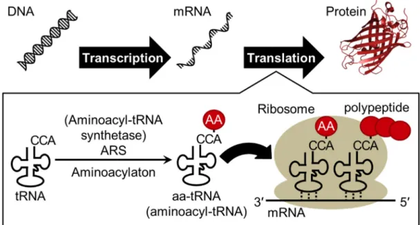
Genetic information is transcribed from DNA into mRNA (messenger RNA) and mRNA is translated into protein, and this process is called as central dogma (Figure 1). By human genome project, it was reported that only the 2% region of human genomic DNA is transcribed and translated to about 20,000-25,000 proteins1, but transcriptome study revealed that about 70% region of DNA is transcribed to RNA. In addition, widespread transcription of non-coding RNAs with important functions throughout the genome have been recently discovered2.
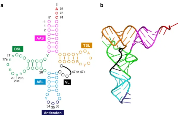
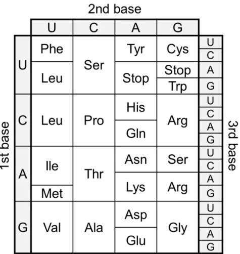
Transfer RNA (tRNA) is one of the most abundant and popular non-coding RNAs, which works as an adaptor molecule linking the language of nucleotide to the language of amino acid in the translation step. Canonical tRNA forms cloverleaf secondary structure and L-shaped tertiary structure as shown in Figure 2. Aminoacyl-tRNA sythetase (ARS) charge corresponding amino acid onto the 3′-end of specific Aminoacyl-tRNA to produce aminoacyl-tRNA (aa-tRNA) and aa-tRNA is recruited into ribosome by translation factors (Figure 1). Three consecutive nucleotides called as anticodon of tRNA forms base pairs with the sequential triplet codons of mRNA in the ribosome, and corresponding amino acid is incorporated into nascent peptide chain (Figure 1 and 2). The correspondence between codons and amino acids are defined as genetic code (Figure 3). This genetic code is well conserved in the most of all organisms (some exceptions have been found3) and accurate translation of mRNA to protein is essential for living organisms.
CCA sequence at the 3′-end are universally conserved in all three domains, prokaryote, eukaryote and archaea. During the translation reaction, CCA-3′ end interacts with ribosome and translation factors, and it is important for efficient translation. In the aminoacylation step, ARSs strictly recognize the body sequences of substrate tRNAs to charge specific amino acids onto specific tRNAs. In addition, because most ARSs also interact with the universally conserved CCA-3′ end of tRNAs4,5, it is difficult to prepare
the various aa-tRNAs bearing mutations in the CCA-3′ end by ARSs6-8. To prepare such
aa-tRNAs, so-called “chemical acylation” method where chemically synthesized aminoacyl-nucleotide was ligated to tRNA lacking the 3′-end by T4 RNA ligase9,10 may
be usable. However, in this method, it is laborious work that various aminoacyl-nuleotides are prepared by chemical synthesis. If the easy method to prepare various aa-tRNA bearing mutation in the CCA-3′ end, it is usable for analyzing the role of CCA-3′ end during the translation and engineering the translation machinery.
9
Figure 1 | Schematic illustration of central dogma. “AA” in the red circle indicate amino acid. An image of protein was prepared from crystal structure of GFP (green fluorescent protein) (PDB ID: 1GFL)11.
10
Figure 2 | Structure of tRNA. (a) Clover leaf structure of tRNA. Conserved bases are described, circles represent non-conserved bases, and numbers indicate the nucleotide position. Positions 34 to 36 (dark blue) form corresponds to the anticodon and position 74 to 76 (red) are universally conserved as CCA. The sequences composing the intron and the extra bases presenting in the variable loop (position noted 47 to 47k) are shown as black line. AAS (magenta), the amino acid-accepting stem; DSL (green), the dihydrouridine stem and loop; ASL (cyan), the anticodon stem and loop; VL (black); the variable loop, TSL (orange); the thymidine stem and loop, Y; pyrimidine, R; purine, H; not G, D; not C. The n bases at position 17, 17a, 20a and 20b are optional bases not present in all tRNAs. Position -1 bases are found in all cytoplasmic mature tRNAHis
GUG from the three biological
domains. This figure was adapted from the previous paper12. (b) L-shaped
tertiary structure of tRNA. Each color indicates the same region described in Figure 2a. This image was prepared from crystal structure of Saccharomyces
cerevisiae tRNAPhe (PDB ID: 1EHZ)13.
11
Figure 3 | Standard genetic code. All 64 codons were assigned to corresponding amino acids or termination signal (termed as “Stop” in this figure).
12
1.2. Other non-coding RNAs than tRNAs
As I mentioned above, tRNA is one of the most abundant and popular non-coding RNA (ncRNA). In addition to tRNA, ribosomal RNAs (rRNAs) are also known as popular ncRNA, which constitute a ribosome. There are three kinds of rRNAs (5S, 16S and 23S rRNAs) in prokaryote, and four kinds of rRNAs (5S, 5.8S, 18S and 28S rRNA) in eukaryote. Peptidyl transfer reaction, which makes a peptide bond, is catalyzed by 23S rRNA or 28S rRNA. These tRNA and rRNA are much abundant and account for about 95% of all RNAs in mammalian cells14.
The various other ncRNAs than tRNA and rRNA have been identified mainly in mammalian15. These ncRNAs are roughly classified into two groups; long ncRNA (longer
than 200 nt) and small ncRNA (smaller than 200 nt)16,17. Unlike small ncRNA such as
microRNA, function of long ncRNA has been less studied. However, recent studies revealed the broad functional repertoire including roles in high-order chromosomal dynamics, telomere biology and subcellular structural organization18,19.
The class of small ncRNA includes various RNAs such as snRNAs (small nuclear RNAs), snoRNAs (small nucleolar RNAs) and miRNAs (micro RNAs)20. snRNA makes complex with protein to form snRNP (ribonucleoprotein) and consists of spliceosome involving the reaction of mRNA splicing. snoRNA generally ranges from 60 to 300 nucleotides in length and guide the site-specific modification of nucleotide in target RNA21. In the past few years, about 22 nt short ncRNA (miRNA) has been widely studied. miRNA is loaded into Ago protein to form a RISC (RNA induced silencing complex)22. A RISC binds to target mRNA via base pairs between miRNA and 3′-UTR (untranslated region) of mRNA to inhibit the translation and suppress the expression of the target gene. To date more than 1,800 miRNAs in human are registered in databases (miRbase23, http://www.mirbase.org/) and are estimated to regulate the expression of more than 60% of genes to control a wide range of biological processes including proliferation, differentiation, apoptosis and development15.
13
1.3. Aminoacylation ribozyme “flexizyme”
In order to achieve these requirements described in former sections; I focused attention on the unique characteristics of flexizymes which is in vitro selected aminoacylation ribozyme24,25. First, I briefly summarize how to develop the artificial ribozyme.
A ribozyme which is an RNA having catalytic activity which was first discovered in nature in 1980s26,27. These discoveries indicated RNA can store information like DNA
and catalyze chemical reaction like proteins to strongly support the “RNA world hypothesis” that the origin of life may have relied on RNA28. However, the chemical
reactions catalyzed by natural ribozymes are limited to the cleavage or ligation of the RNA phosphodiester backbone except for peptidyl transfer by ribosome29. To expand the
variety of chemical reactions catalyzed by RNA, in vitro selection of ribozyme have been developed. SELEX (Systematic Evolution of Ligands by EXponential enrichment, or in
vitro selection) is a method based on molecular evolution engineering to identify the
active molecules having specific activities from pools of various compounds30,31. It was
first reported from two groups in 1990 and they discovered RNA aptamers binding to specific target molecules from random RNA library. In addition to discovery of aptamers, the molecules having catalytic activities such as ribozyme have also been identified32. A
general scheme of SELEX of active RNAs was shown in Figure 4. DNA library containing T7 promoter sequence, 5′-constant sequence, random sequences and 3′-constant sequence is prepared by PCR. These 3′-constant sequences are used for following reverse transcription and PCR reaction. The DNA templates are transcribed to RNAs by T7 RNA polymerase in vitro. From the RNA library, portion of RNAs are selected based on their ability to carry out a specific function (e.g. binding affinity and catalytic activity). Selected RNAs are converted to cDNAs by reverse transcription. These cDNAs are amplified by PCR and the same procedure is repeated. After the several times repeats of this process, the resulting RNA fraction will be enriched with functional molecules with the desired activity. By SELEX, many artificial ribozymes have been discovered33 (e.g.
RNA polymerization33-35, alcohol oxidation36, Diels-Alder reaction37 and
aminoacylation38-42). Among them, aminoacylation ribozymes were important because
these are candidates as key molecules which link the ancient RNA world with modern protein world.
In nature, ARS catalyze aminoacylation reaction. Aminoacylation by ARS involves two steps; (i) activation of carboxyl group amino acids by adenylation using ATP to yield aminoacyl-adenosine monophosphate (aminoacyl-AMP), and (ii) condensation
14
of aminoacyl-AMP and tRNA to yield aminoacyl-tRNA (aa-tRNA). Although no ribozyme catalyzing these both reactions have been discovered in nature, some artificial ribozymes catalyzing (i) or (ii) step were identified by in vitro selection. The KK13 ribozyme catalyze the activation of amino acid by using the 5′-triphosphate of RNA instead of ATP43. The #29 ribozyme family catalyzes cis-acyl transfer reaction using
aminoacyl-AMP to produce both aminoacyl- and peptidyl-tRNAs38,44,45, and the C3
ribozyme family consisting of only three essential nucleotides catalyze both cis- and
trans-acyl transfer reaction using aminoacyl-AMP42,46. Flexizyme is also
trans-acyl-transfer ribozyme using activated amino acids, which was developed by SELEX method and was engineered to aminoacylate various activated amino acids. There are three types of flexizymes, dFx (dinitrobenzyl flexizyme)41, eFx (enhanced flexizyme)41 and aFx
(amino flexizyme)47 (Figure 5). dFx charges wide variety of amino acid whose carboxyl
group is activated with 3,5-dinitrobenzyl ester (DBE), eFx charges aromatic amino acids activated with cyanomethyl ester (CME) or nonaromatic amino acids with 4-chlorobenzyl thioester (CBT), and aFx charges substrates activated with benzyl thioester group bearing a protonated primary amine (ABT).These flexizymes can charge wide variety of amino acids including noncanonical amino acids (ncAAs) such as N-methyl-amino acids48-50,
N-alkyl-glycines51, cyclic N-alkyl amino acids52, N-acyl-amino acids53, exotic peptides54, α-hydroxy acids49,55, and D-amino acids56 onto tRNAs.
In addition to the characteristic to charge diverse kinds of amino acids, flexizymes have the flexibility to aminoacylate the substrate tRNA bearing any body sequence because flexizymes recognize the CCA-3′ end by two consecutive base pairs with GGU-3′ of flexizymes40,57,58 (Figure 5 and 6). The crystal structure of flexizyme fused with substrate minihelix RNA docked onto the tRNAs indicated the no interaction between flexizyme and body sequence of substrate tRNA57 (Figure 6). This is because, fleixizyme can aminoacylate other RNAs different from tRNAs even if those have single strand CCA-3′ end. In addition, in the experiment using prototype of flexizymes “pre-24”, these two consecutive base pairs are important for aminoacylation and this base pairs can be substituted with certain base pairs40,58.
In summary, flexizymes have following unique characteristics; (i) substrate RNA is recognized by two consecutive base pairs between 3′-end of substrate RNA and 3′-end of flexizyme, (ii) these base pairs can be substituted with other base pairs and (iii) various activated amino acids can be used as substrates including both canonical and noncanonical amino acids. These features are different from natural protein ARSs and other aminoacylation ribozymes. From these characteristics of flexizymes, I considered
15
that flexizymes enable to achieve the requirements described early in this section.
Figure 4 | General scheme of SELEX (in vitro selection) of functional RNAs. Black sequences are constant regions including the promoter sequence for T7 RNA polymerase and the 5′/3′-constant sequences necessary for reverse transcription and PCR amplification. Colored (blue, orange and red) sequences indicate the random region. This figure is adapted from the previous review25.
16
Figure 5 | Schematic illustration of aminoacylation by three derivatives of flexizymes and acyl-donor substrates with cognate leaving group. This figure is adapted from previous review25. Lines denote the Watson-Crick base
pairs and bullets denote wobble base pairs. R represents amino acid side chains including noncanonical ones. X represent a-functional groups, such as amino, N-alkylamino, and hydroxy groups. LG; leaving group, dFx; dinitrobenzyl flexizyme, eFx; enhanced flexizyme, aFx; amino flexizyme, DBE; dinitrobenzyl ester, CME; cyanomethyl ester, CBT;
17
Figure 6 | Structures of protein and RNA aminoacyl-tRNA synthetases. (a) Crystal structure of Escherichia coli glutamiyl-tRNA synthetase (GlnRS; orange) and tRNAGln complex (PDB ID: 1GTS)59. (b) Crystal structure of
flexizyme (wheat color) docked onto E. coli tRNAGln by superimposing the
minihelix on the ASL of the tRNA (PDB ID: 3CUL)57.
18
1.4. Removed for the reasons involving submission of the paper (a brief
introduction of chapter 2)
1.5. Development of orthogonal translation machinery (a brief
introduction of chapter 3)
The CCA-3′ sequence of tRNA is conserved among all organisms and is important for aminoacylation and translation. In bacteria, the CCA-3′ end makes Watson-Crick base pairs with bases of 23S rRNA in the peptidyl transferase center (PTC) in the classical state62 and translocation63 during translation. These base pairs are important for translation activity, and the compensatory mutations in these base pairs are tolerated during peptidyl transfer reaction64 and translocation63. However, it is yet empirically unknown whether such mutations accommodates translation in its entirety.
In chapter 3, I will report development of orthogonal translation machinery65. First, I developed method to easily aminoacylate various amino acids onto tRNAs bearing mutation (or mutations) in CCA-3′ end using compensatory mutated flexizymes. Then the translation activity of the PTC-mutated ribosomes and tRNAs were measured. Consequently, I developed the mutant ribosome and tRNA pair which had comparable translation activity and orthogonality to the wild-type ribosome and tRNA pair. Finally two different peptides was expressed simultaneously in one pot from a single mRNA template using the mutant and wild-type ribosome–tRNA pair.
19
21
Chapter 2
51
Chapter 3
Orthogonal ribosome-tRNAs pair by engineering of peptidyl
transferase center
52
3.1. Introduction
Assignment of 20 canonical amino acids to codons, is achieved by specific acylation of tRNA with cognate amino acid catalyzed by aminoacyl-tRNA synthetase (ARS). Since each tRNA has an anticodon that pairs with the codon, the codons on mRNA can be decoded by the cognate aminoacyl-tRNAs (aa-tRNAs) according to the genetic code; and thus ribosome is able to catalyze the formation of peptide bond along the mRNA template, to yield a polypeptide with the encoded sequence. While 20 canonical amino acids are used in the natural translation system, it has been demonstrated that more than hundreds of different noncanonical amino acids (ncAAs) can be incorporated into nascent polypeptide chain by engineering of the genetic code113. Assignment of ncAAs in the genetic code has been achieved by two methodologies: genetic code expansion114,115 and genetic code reprogramming116. The former method generally assigns a ncAA (or multiple ncAAs) to a nonsense codon(s) such as stop codons and an artificially programmed quadruplet codon(s). Genetic code expansion has been applied for both in
vivo and in vitro expression of proteins with ncAAs.
When the genetic code expansion is performed in vivo, availability of ncAA-tRNA generally relies on an exogenously introduced ARS specifically paired with an orthogonal tRNA. The orthogonal tRNA should not be aminoacylated by endogenous ARSs. At the same time, the exogenous ARS should be engineered to specifically charge the ncAA onto the orthogonal tRNA, but not onto endogenous tRNAs. Several orthogonal ARS–tRNA pairs have been successfully developed113, and thus far demonstrated
expression of proteins containing ncAAs. However, the choices of usable ncAAs are yet limited to a few subgroups, because the majority examples of the engineered ARSs are based on Methanococcus jannaschii TyrRS115, Methanosarcina barkeri PylRS117,118, or
Methanosarcina mazei PylRS119,120; therefore, the usable ncAAs for these mutant
enzymes are limited to Phe or Lys analogs.
In addition to orthogonal ARS–tRNA pair, orthogonal ribosome–mRNA pair has been developed121. In this system engineered ribosome containing orthogonal 16S rRNA which does not recognizes endogenous mRNA but recognizes engineered mRNA bearing mutated Shine-Dalgarno (SD) sequence, was used121. Decoding center of this orthogonal 16S rRNAs have been further evolved for efficient amber codon decoding122 and quadruplet codon123. These orthogonal ribosome–mRNA pairs enabled to express specific protein containing ncAAs in vivo124. However, the numbers of usable ncAAs and codons were still limited.
53
On the other hand, in the genetic code reprograming, multiple sense codons can be simultaneously reassigned with various ncAAs. Genetic code reprogramming has been dominantly utilized in in vitro reconstituted translation system125 to produce short
polypeptides containing diverse exotic ncAAs such as D-amino acids56 and
N-methyl-amino acids48-50. Although in the genetic code reprogramming method, multiple ncAAs
can be incorporated, some canonical amino acids cannot be used simultaneously. It is because the canonical amino acids originally assigned in natural genetic code need to be removed to suppress the incorporation of canonical amino acids instead of ncAAs126. To
overcome this problem in genetic code reprogramming, I conceived the new concept of orthogonal translation system, which is orthogonal ribosome–tRNA pair. This engineered translation system means that wild-type ribosome only use canonical aa-tRNAs and the engineered ribosome only use ncAA-tRNAs as substrates. In this system, it is not necessary to deplete canonical amino acids and to change the original genetic code in order to incorporate ncAAs.
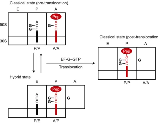
To develop the orthogonal ribosome–tRNA pair, the interaction between ribosome and tRNA needs to be engineered. During the elongation cycle of bacterial translation, where amino acids were sequentially added to the growing peptide chain, CCA-end of tRNA interacts with 23S rRNA of 30S subunit. The elongation cycle is divided to three steps127,128 (Figure 18). First step is accommodation of aa-tRNA into the A-site of ribosome. aa-tRNA is delivered to the A-site by EF-Tu•GTP in the A/T state where the anticodon forms base pairs with codon in the A-site of 30S and the acceptor end is bound to EF-Tu (the first letter represents the binding site on the 30S and the second letter the site on the 50S). Then GTP is hydrolyzed to GDP to release EF-Tu•GDP and the accommodation of the acceptor end of the aa-tRNA into the A-site of the 50S (A/A state, the classical state). Second is peptide bond formation between aa-tRNA in A/A state and peptidyl-tRNA in P/P state, which is catalyzed in the peptidyl transferase center (PTC). After the peptide bond formation, peptidyl-tRNA in A/A state is translocated to P/P state and deacylated tRNA in P/P state is translocated to E/E state facilitated by EF-G with associated GTP hydrolysis, which is the third step.
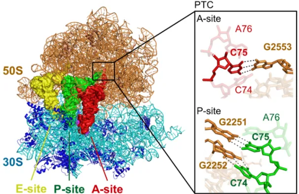
In the PTC of classical state of E. coli ribosome complex, critical Watson-Crick base pairs occur between the universally conserved 3′ end of tRNAs (C74 and C75) and 23S rRNA G2251 and G2252 at the P site as well as G2553 at the A site, which were revealed by biochemical experiments and crystal structural analyses62,129-136 (Figure 17
and 19a). Using an analogue of aa-tRNA fragment (C75 mutant puromycin derivatives of the form NPm) as an A-site substrate (Figure 19b and c), peptidyl transfer activity of
54
wild-type and G2553N mutant ribosome 50S subunits was measured by Kim and Green64.
They reported that the wild-type ribosome preferred the wild-type substrate (CPm) by about 2- to 5-fold relative to the other substrates (APm, GPm and UPm) (Figure 19b). Furthermore, G2553C ribosome preferred the compensatory mutated substrate (GPm) by at least 20-fold relative to the other three substrates (Figure 19c). These results indicated that G2253C ribosome-GPm pair has weak orthogonality to wild-type ribosome-CPm pair in peptidyl transfer reaction.
The first step of translocation is movement of acceptor end of peptidyl-tRNA in A/A state into A/P state and acceptor end of deacylated tRNA in P/P state into P/E state, which is called as hybrid state127 (Figure 18 and 20). This first step is promoted by EF-G•GTP but it can occur in the absence of GTP137. Then EF-G promotes translocation of peptidyl-tRNA from the A/P state to the P/P state and deacylated tRNA from the P/E state to the E/E state to be released from the ribosome. To my knowledge, there is no direct evidence of the interaction between 23S rRNA and CCA-end of tRNAs during the hybrid state. However, Dorner et. al. indicated the potential pairs between C74/C75 of peptidyl-tRNA in the A/P state and G2252/G2251 of 23S rRNA in the P-site63 (Figure 20). In this report, they measured EF-G translocation rate with the pairs of wild-type and mutant ribosomes and tRNAs (Table 7 and 8). When C74G tRNA is in A-site of wild-type ribosome, translocation rate reduced (Table 7). When potential pairing between tRNA in the A/P state and 23S rRNA in the P-site was restored (the pair of C74G tRNA and G2252C ribosome), translocation rate was restored (Table 7). Similar result was observed about the interaction between C75 of tRNA in the A/P state and G2251 of 23S rRNA (Table 7). These results indicates that the pair of G2251C or G2252C ribosome and C75G or C74G tRNA have weak orthogonality to the pair of wild-type ribosome and tRNA in translocation.
On the other hand, disruption of the interaction in the A/A state (the pairs of wild-type tRNA and G2253C/U/A ribosomes) increased the rate of translocation (Table 8) and restoring the base pair in the A/A state (the pair of C75G tRNA and G2253C ribosome) diminished the translocation rate. These results indicate that ribosome and tRNAs favored the stable state forming base pairs.
These studies described above showed that engineering the base pairs between G2251/G2252/G2553 of ribosome and C74/C75 of tRNAs in PTC had potential to develop the orthogonal ribosome–tRNA pairs. However, it was unknown whether a mutation (or mutations) of 23S rRNA and CCA-3′ end of tRNAs in the PTC affect the whole translation reaction to produce polypeptides using aa-tRNAs as substrates in an
55
mRNA-dependent manner, including other steps.
In this chapter 3, I measured the translation activity of the PTC-mutated ribosome–tRNA pairs using an E. coli custom-made in vitro translation system that was integrated with the flexizyme technology, referred to as the FIT (Flexible In vitro Translation) system126. In natural, tRNAs are aminoacylated by protein aminoacyl-tRNA synthetases (ARSs) and ARSs specifically recognize the sequence of cognate tRNAs. Because most ARSs also interact with the universally conserved CCA-3′ end of tRNAs4,5, ARSs are not suitable for preparation of aminoacyl-tRNA (aa-tRNA) bearing mutation in the CCA-3′ end6-8. In order to overcome this problem, flexizymes (eFx and dFx) bearing compensatory mutated 3′ ends were used because flexizymes recognize CCA-3′ end of substrate RNA via base pairs as described in chapter 1. Finally, the pair of mutant ribosome and tRNAs which acted orthogonally and used only the cognate genetic code consisting of ncAAs65.
56
Figure 17 | Crystal structure of classical state of ribosome-tRNAs-mRNA complex. The interaction between 23S rRNA and CCA-end of tRNAs at the peptidyl transferase center (PTC) is shown in black rectangle. Black dashed lines indicate hydrogen bonds. This structure was reported in previous paper62
57
Figure 18 | Schematic illustration of elongation cycle of bacterial translation. The ribosomes are described as black rectangles and divided into 30S and 50S subunits and partitioned into the A-, P- and E-sites. The biding states of the tRNAs as previously defined127 are indicated at the bottom of
each sites. “Pep” means peptide and “AA” means amino acid. Bold lines indicate tRNAs.
58
Figure 19 | Peptidyl transfer reaction using G2553N mutant ribosome and C75 mutant puromycin derivatives64. The ribosomes are described as
wheat ellipse and divided into 50S and 30S subunits. (a) Schematic illustration of natural peptidyl transfer between peptidyl-tRNA at P-site and aa-tRNA at A-site. (b) Schematic illustration of peptidyl transfer between N-acetyl-phenylalanyl-tRNA (AcPhe-tRNA) at P-site and C75 puromycin
derivatives shown as CPm at A-site using reconstituted wild-type 50S subunit. (c) Schematic illustration of peptidyl transfer between N-acetyl-phenylalanyl-tRNA (AcPhe-tRNA) at P-site and C75G puromycin derivatives shown as
GPm at A-site using reconstituted G2553C mutant 50S subunit.
59
Figure 20 | Schematic illustration of classical and hybrid state of tRNA-ribosome complex and translocation induced by EF-G–GTP. The ribosomes are described as black rectangles and divided into 30S and 50S subunits and partitioned into the A-, P- and E-sites. The biding states of the tRNAs as previously defined127 are indicated at the bottom of each sites. “Pep”
in red circle means peptidyl-tRNA. Bold lines indicate tRNAs and bold Gs indicate G2251, G2252 and G2553 of 23S rRNA interacting with CCA-end of tRNAs. This figure was adapted from previous report63,138.
60
Table 7 | EF-G translocation of WT, G2252C and G2251C mutant ribosomes. This table was adapted from previous report63.
Translocation rate (s-1) Translocation rate (s-1)
P-site tRNA A-site tRNA WT G2252C P-site tRNA A-site tRNA WT G2251C WT WT 3.8 1.5 WT WT 3.8 2.1 WT C74G 1.5 5.0 WT C75G 1.4 9.3
Table 8 | EF-G translocation with WT and G2553 mutant ribosomes. ND indicates “not determined”. This table was adapted from previous report63.
Translocation rate (s-1) P-site
tRNA
A-site
tRNA WT G2553C G2553U G2553A
WT WT 3.8 10.7 12.2 12.4
61
3.2. Results and discussions
The study in this chapter 3 was published as a paper in the scientific journal and the figures in this chapter are reproduced or adapted from the published paper65.
3.2.1. Aminoacylation of CCA-mutated tRNAs
To confirm that CCA-3′ end mutated tRNAs (denoted as tRNAs-NNA) can be aminoacylated by compensatory mutated flexizymes, I arbitrarily chose lysine and tyrosine as amino acids substrates (Figure 21), and tRNAAsnE2
CUU and tRNAAsnE2GUA
(Figure 22) bearing corresponding anticodons. Three mutant tRNAs derived from
tRNAAsnE2
CUU and tRNAAsnE2GUA bearing single or double C to G mutation (or mutations)
(C74G, C75G or C74G/C75G) and the compensatory single or double G to C mutated flexizymes derived from dFx and eFx (Figure 5) were prepared by in vitro transcription. The aminoacylation efficiency were measured by acid-PAGE, which indicates the cognate mutant dFxs and eFxs can aminoacylate the tRNAs-NNA in the presence of Lys-DBE and Tyr-CME (Figure 23). On the other hand, wild-type flexizymes could not aminoacylate non-cognate tRNA mutants (Figure 23). In the case of C75U tRNA mutant, aminoacylation efficiency of mutant flexizymes bearing GAU-3′ end, which form two Watson-Crick base pairs with tRNAs, was lower than that of wild-type bearing GGU-3′ end, which form one Watson-Crick base pair and one G•U wobble base pair (Figure 24). This result indicates that G•U wobble base pair is more suitable for aminoacylation by flexizymes than A-U Watson-Crick base pairs. Mutant flexizymes could also charge several other amino acids (Figure 21) onto the corresponding mutant tRNAs for further experiments (Figure 25).
62
Figure 21 | Amino acid substrates used in the chapter 3. The abbreviations of Fph, Aly, and Anv represent N-(5-FAM)-L-phenylalanine, L-acetyllysine,
and L-azidonorvaline, respectively. Carboxyl groups were activated using
63
Figure 22 | Secondary structures of transcribed (t.s.) tRNAs compared with native tRNAs of E. coli. The figure presents the secondary structures of tRNAfMet
CAU (a), tRNAAsnE2NNN (b), tRNAGluE2NNN (c), tRNALysCUU (d),
tRNAAsp
GUC (e), and tRNATyrGUA (f). s4U; 4-thiouridine, D; dihydrouridine,
Cm; 2′-O-methylcytidine, m7G; 7-methylguanosine, ψ; pseudouridine, Q; queuosine, ct6A: cyclic N6-threonylcarbamoyladenosine, mnm5s2U;
5-methylaminomethyl-2-thiouridine, m2A; 2-methyladenosine, acp3U;
3-(3-amino-3-carboxypropyl)uridine, T; ribothymidine, gluQ; glutamyl-queuosine, ms2i6A; 2-methylthio-N6-isopentenyladenosine. The sequences are based on
the Modomics database139 (http://modomics.genesilico.pl/).
a b
c d
64
Figure 23 | A model study of aminoacylation of tRNAs-NNA derived from
tRNAAsnE2. tRNAs were acylated by dFx in the presence of Lys-DBE (a), and
eFx in the presence of Tyr-CME (b). Acid-PAGE separated the bands of aa-tRNA (upper) and uncharged aa-tRNA (lower). The data were generated from a sample of the end product of aminoacylation reaction. Note that the mobility of flexizymes was different depending on the 3′-terminal mutations.
a
65
Figure 24 | Aminoacylation of tRNA-CUA derived from tRNAAsnE2.
tRNAs were acylated by dFx in the presence of Lys-DBE (a), and eFx in the presence of Tyr-CME (b). Acid-PAGE separated the bands of aa-tRNA (upper) and uncharged tRNA (lower). The data were generated from a sample of the end product of aminoacylation reaction.
66
Figure 25 | Efficiency of flexizymes-catalyzed aminoacylation with canonical (Tyr, Lys, and Asp) and non-canonical (Fph, Aly, and Anv) amino acids. The tRNAs-CCA, -GCA, -CGA, -GGA and -CUA were acylated by the compensatory flexizymes, dFx/eFxGGU, GCU, CGU, -CCU and -AGU. The data represent the average of three independent reactions. The error bars represent the standard deviation. The data were generated from a sample of the end product of aminoacylation reaction.
67
3.2.2. Preparation of tagged ribosomes
Mutant ribosomes bearing a mutation (or mutations) at PTC were prepared using an MS2-tag affinity purification method140. Professor Rachel Green in Johns Hopkins University kindly gifted me a plasmid p278 MS2 (AmpR) encoding 23S rRNA inserted with MS2-tag sequence in helix 98 of 23S rRNA (Figure 26a), pcI857 (KanR) encoding a temperature-sensitive mutant of the lambda repressor protein cI and a plasmid pMAL-c2g (AmpR) encoding His6-tagged MS2 coat protein fused with maltose binding protein
(His6-MS2-MBP).
The plasmids p278 MS2 bearing mutations at PTC were constructed by site-directed mutagenesis. First, the plasmid pMAL-c2g was used to transform E. coli and His6-MS2-MBP was expressed to be purified by FPLC using Ni-NTA affinity column.
Next, the plasmids pcI857 and p278 MS2 were used to transform E. coli. Because the
mutant ribosomes may be toxic to E. coli, this transformed E. coli was pre-cultured at 30°C where mutant ribosomes were not expressed and the temperature was increased to 42°C to express the mutant ribosomes. His6-MS2-MBP was immobilized onto the MBP
column by FPLC and the expressed tagged-ribosomes (wild-type, G2251C, G2252C, G2553C, G2251C/G2553C, G2251C/G2252C/G2553C and G2251A/G2553A) were purified by MS2 affinity purification using FPLC (Figure 26b). Contamination of wild-type untagged-ribosome into the purified tagged-ribosomes were quantified by primer extension method140. This result showed the negligible contamination rate less than 3%
68
Figure 26 | Schematic illustration of the insertion sites for the MS2 tag and purification of tagged ribosome. (a) Helix 98 in 23S rRNA which is the insertion sites for the MS2 tag was indicated as green circle. The image of structure was produced from the previous report 62 (PDB ID: 2WDK/2WDL).
(b) Representative results of FPLC purification of MS2-tagged ribosome.
69
Figure 27 | Primer extension analysis of rRNAs extracted from MS2-tagged ribosomes purified by FPLC. (a) A primer that is complementary to the bases of 2254–2273 in 23S rRNA was used for the detection of mutations of 2251 and 2252. (b) A primer that is complementary to the bases of 2556– 2575 in 23S rRNA was used for the detection of mutation of 2553. MS2-taggd ribosomes bearing mutations produced different ddGTP or ddTTP stops from those produced by WT ribosome. The abundance of tagged ribosomes relative to untagged wild-type in each population was shown below the gel. The calculated values of abundance were derived from a single experimental data set.
70
3.2.3. Translation activity of wild-type/mutant ribosome-tRNA pairs
An assay system to detect peptides translated by tagged-ribosomes and aa-tRNAs-NNA was established. Because E. coli ARSs do not uniformly charge amino acids onto tRNAs-NNA6-8, a conventional methods using radioisotope-labeling amino acids is unsuitable. Instead, fluorescently-labeled amino acid (N-(5-FAM)-L-phenylalanine, Fph, Figure 21) was aminoacylated onto initiator tRNAfMetCAU and incorporated at N-terminus
of peptide by FIT system126 to reassign the initiation codon from fMet to Fph. Lysine and tyrosine were charged onto tRNAAsnE2CUU-CCA and tRNAAsnE2GUA-CCA by cognate dFx
and eFx, respectively. As a model template, a DNA template encoding the heptapeptide-1 (Fph-Lys-Tyr-Lys-Lys-Tyr-Lys) was prepared and the expression of heptapeptide-heptapeptide-1 in the FIT system, whose components were described in the section of materials and methods, was confirmed. Tricine-SDS-PAGE analysis of the translated product enabled to visualize a fluorescent band (Figure 28a) and quantify an expressed peptide by comparison with a band generated by a known concentration of Fph (Figure 28b), and the peak corresponding to heptatpeptide-1 was confirmed by MALDI-TOF-MS (Figure 28c). The production of heptapeptide-1 plateaued at 30 min, with a final concentration of approximately 1.0 µM. We also examined expression of heptapeptide-1 using
Lys-tRNAGluE2CUU-CCA and Tyr-tRNAGluE2GUA-CCA under the same condition, and the
amount of expressed peptide increased to 3.5 µM (Figure 29a and b). The increase of expression rate could be attributed to the higher binding affinity of aa-tRNAsGluE2-CCA compared to aa-tRNAAsnE2-CCA (Figure 30). Therefore, we decided to use tRNAGluE2 for further translation experiments.
Next, I investigated whether tRNAs-NNA can be used by the wild-type ribosome for mRNA-dependent translation. Four mutant aa-tRNAs bearing GCA-3′, CGA-3′ and GGA-3′ were prepared by cognate dFxs and eFxs (Figure 25) and tested these expressions. Surprisingly tRNA-GCA could be used by wild-type ribosome to express heptapeptide-1 even only one base pair forms at P-site and the final concentration of the peptide was 2.7 µM (Figure 29). On the other hand, tRNAs-CGA and -GGA were inactive with wild-type ribosome (Figure 29). These results indicate that tRNAs-CGA and tRNAs-GGA but not tRNAs-GCA are orthogonal to the wild-type tRNAs-CCA (Figure 31).
Then, I measured the translation activity of mutant ribosomes and tRNAs. As predicted from the results with the pair of wild-type ribosome and non-cognate tRNAs-GCA, the G2252C ribosome was also active with both cognate tRNAs-GCA and non-cognate tRNAs-CCA, but not with tRNAs-CGA and tRNAs-GGA (Figure 29a). The expression rate of cognate pair of G2252C ribosome and tRNAs-GCA was almost the
71
same as that of wild-type pair, and that of non-cognate pair of G2252C ribosome and tRNAs-CCA was also the same as that of non-cognate pair of wild-type ribosome and tRNAs-GCA (Figure 29b). These both cognate and non-cognate pairs yielded heptapeptide-1 as major products (Figure 29c). These results indicated that mispair between G2252 and G74 or C2252 and C74 could be tolerated during the translation reaction.
The double mutant ribosome G2251C/G2553C ribosome was sufficiently active with only cognate tRNAs-CGA to yield heptapeptide-1 (Figure 29). Time-course analysis indicated that expression plateaued at a final concentration of 1.7 µM. This suggests that the cognate pair of G2251C/G2553C ribosome and tRNAs-CGA functions orthogonally to the wild-type pair (Figure 31). However, this mutant pair has a slower translation rate than the wild-type pair, resulting in a lower yield of peptide due to the hydrolysis of the aa-tRNAs. Faint bands observed when using tRNAs-CCA and tRNAs-GCA could be ascribed to a peptide originating from the background translation by little amount (< 3%) of wild-type ribosome contamination to the respective mutant ribosome (Figure 27). When tRNAs-GGA was used, a very faint band was detected because mispair between G2252 and G74 was tolerated as described above. However, these bands were too faint to reliably quantify the intensity for the time-course experiment below and determine the product identity by MALDI-TOF analysis.
The triple mutant ribosome (G2251C/G2252C/G2553C) was active with only cognate tRNAs-GGA to yield heptapeptide-1 like the double mutant ribosome (Figure 29). However, the translation rate dramatically decreased and the final concentration of heptapeptide-1 was 0.4 µM, which was reflected as a tiny peak of heptapaptide-1 in MALDI-TOF-MS spectrum (Figure 29c).
I also tested the translational activity of other ribosome mutants (G2251A/G2553A, G2251C and G2553C) and tRNA mutant (tRNA-CUA) (Figure 32). Like G2251C/G2553 ribosome and G2251C/G2252C/G2553C ribosome, G2251A/G2553A ribosome was active with only cognate tRNA-CUA, but the band was too faint to reliably quantify the intensity for the time-course experiment and determine the product identity by MALDI-TOF analysis. A very faint band observed when using tRNAs-CCA could be due to the contamination of wild-type ribosome (Figure 27). These results indicate that A-U pair between 23S rRNA and tRNA is not appropriate for translation. In addition, wild-type ribosome was inactive with tRNAs-CUA, which suggests that G•U wobble pair between ribosome and tRNA at the PTC is not tolerated. In the case of G2251C and G2553C ribosomes, translation activity was not restored when
72
using tRNA-CCA or tRNA-CGA. This result and the fact that the pair of G2251C/G2553C ribosome and tRNA-CGA had comparable translation activity to wild-type pair, combination of two base pairs, G2553-C75 at the A-site and G2251-C75 at the P-site, is essential for translation.
These decrease of translation rate of cognate pair of mutant ribosome and tRNAs indicates that, despite the fact that PT activity and translocation activity of mutant ribosomes bearing mutations at PTC was restored by the compensatory mutations into CCA end of tRNAs63,64, these compensatory mutations did not fully restore the whole translation activity. This further suggests that these bases have crucial roles in not only the PT reaction and translocation but also other steps of translation, potentially involving interactions with other rRNAs or protein factors. It was confirmed that the binding affinity of aa-tRNAs-NNA to form ternary complexes (EF-TU•GTP•aa-tRNA) were almost the same as that of wild-type, which was coincident with the previous report130,141. In the accommodation step, C75 of tRNA pack between EF-Tu residue 219 and the flipped base of A55 of 16S rRNA, which is 99.5% conserved in all species of known sequence142. It is possible that these interaction is critical for efficient translation reaction. Nonetheless, further studies are necessary to elucidate why compensatory mutations at CCA-end could not fully restore the translation activity of mutant ribosomes.
73
Figure 28 | Translation activity of pairs of wild-type ribosome–tRNA using tRNAsAsnE2 as elongator tRNAs. (a) Tricine-SDS-PAGE analysis of
heptapeptide-1 (FphLysTyrLysLysTyrLys) synthesized using the wild-type pairs of ribosome–tRNAs. tRNAfMet
CAU bearing the CCA-3′ end were used as
initiator tRNAs, and tRNAsAsnE2
NNN (NNN denotes CUU and GUA
designating Lys and Tyr, respectively) bearing the CCA-3′ end were used as elongator tRNAs. This image showed the result of the end of the translation reaction. Two bands were observed for Fph (indicated by Fph1 and Fph2), presumably due to the presence of Fph, Fph-CME and Fph-tRNAfMet
CAU. The
bands above and below of Fph1 showed truncated peptides generated by peptidyl-tRNA drop-off143 (b) Time-course analysis of heptapeptide-1
production. The data represent the average of three independent reactions and error bars represent the standard deviation of the individual measurements. (c) MALDI-TOF MS analysis of heptapeptide-1. C and O denote calculated and observed mass values, respectively. † indicates a potassium adduct of heptapeptide-1. Other minor peaks were also present in the non-templated translation product likely originating from the translation system. The data were generated from a sample of the end product of translation reaction.
a b
74
Figure 29 | Translational activity of pairs of ribosome and tRNA mutants. (a) Tricine-SDS-PAGE analysis of heptapeptide-1 (Fph-Lys-Tyr-Lys-Lys-Tyr-Lys) synthesized using the designated pairs of ribosome–tRNA mutants. tRNAsfMet
CAU bearing the respective NNA-3′ end were used as initiator
tRNAs, and tRNAsGluE2
NNN (NNN denotes CUU and GUA designating Lys
and Tyr, respectively) bearing the respective NNA-3′ end were used as elongator tRNAs. This image showed the result of the end of the translation reaction. (b) Time-course analysis of heptapeptide-1 production by active pairs of ribosomes and tRNAs. I investigated four cognate pairs of the wild-type ribosome–tRNAs-CCA (cyan solid line), G2252C-ribosome–tRNAs-GCA (yellow solid), G2251C/G2553C-ribosome–tRNAs-CGA (red solid), and G2251C/G2252C/G2553C-ribosome–tRNAs-GGA (purple solid), and two non-cognate pairs of the wild-type ribosome–tRNAs-GCA (black dashed) and G2252C-ribosome–tRNAs-CCA (brown dashed). The data represent the average of three independent reactions. The error bars represent the standard deviation. (c) MALDI-TOF MS analysis of heptapeptide-1 synthesized using cognate and non-cognate ribosome–tRNA pairs. Lane numbers are those of the tricine-SDS-PAGE gel described in Figure 29. C and
a b
75
O denote calculated and observed mass values, respectively. † indicates a potassium adduct of heptapeptide-1. ‡ indicates the molecular mass value consistent with FphLysTyrLysLysTyr, which was presumably produced by peptidyl-tRNA drop-off143. The identity of major peak in lane 16 is unknown;
however, since this peaks as well as other minor peaks were also present in the sample of G2251C/G225C/G2553C-ribosome alone, it could be originated from a contaminant of the G2251C/G225C/G2553C-ribosome preparation. The data were generated from a sample of the end product of translation reaction.
76
Figure 30 | Gel-shift assay for ternary complex of elongation factor Tu (EF-Tu), guanosine triphosphate (GTP) and aa-tRNAs-CCA, GCA, CGA and GGA. Tyrosine was charged onto tRNAAsnE2GUA and tRNAGluE2GUA, and
lysine was charged onto tRNAAsnE2CUU and tRNAGluE2CUU. Band intensities of
the ternary complexes were normalized relative to EF-Tu, GTP and aa-tRNAs-CCA complexes. The values of the relative intensity of the ternary complex (y-axis) were derived from a single experimental set.
77
Figure 31 | Illustration of compatibility and orthogonality of the ribosome–tRNAs mutant pairs. Line thickness indicates the compatibility of translational activity between each ribosome–tRNA pair.
78
Figure 32 | Translational activity of pairs of ribosome–tRNA mutants bearing CCA, GCA, CGA, GGA and CUA-3′ ends. tRNAsfMet were used
as initiator tRNAs and tRNAsGluE2 as elongator tRNAs. (a) The entire
image of the tricine-SDS-PAGE analysis of a heptapeptide-1 (FphLysTyrLysLysTyrLys) synthesized using pairs of ribosome–tRNA mutants. This image showed the result of the end of the translation reaction. The bands above and below of Fph1 showed truncated peptides generated by peptidyl-tRNA drop-off143, which were also observed for the wild-type
control of lane 1. The faint bands in lanes 3, 8, 9 and 13 could be ascribed to a peptide originating from the background translation by little amount (< 3%) of wild-type ribosome contamination to the respective mutant ribosome (Figure 27). (b) Time-course analysis of heptapeptide-1 production by active pairs of ribosomes and tRNAs. Lane numbers are those of the tricine-SDS-PAGE gel described in Figure 32a, in which the solid line in cyan indicates the reaction of a cognate pairs of the wild-type ribosome–tRNAs-CCA (the identical result shown in Figure 29b) while the dashed line in yellow shows that of a non-cognate pair G2253C-ribosome–tRNAs-CCA. The data represent the mean value of each sample (n = 3) and error bars show the standard deviations obtained from independent measurements. (c) MALDI-TOF MS analysis of heptapeptide-1 synthesized using a non-cognate pair G2253C-ribosome–tRNAs-CCA. Lane numbers are those of the Tricine-SDS-PAGE gel described in Figure 32a. C and O denote calculated and observed mass values, respectively. ‡ indicates the molecular mass value consistent with FphLysTyrLysLysTyr, which could be one of the bands corresponding to the truncated peptides appeared in the tricine-SDS-PAGE. The data shown in Figure 32a and c were generated from a sample of the end product of translation reaction.
a b
79
3.2.4. Simultaneous expression of two different peptides from single
mRNA template
Because the pair of G2251C/G2553C ribosome and tRNAs-CGA had comparable translation activity and orthogonality to the wild-type ribosome and tRNA-CCA pair, I examined whether two distinct peptides could be expressed from a single mRNA template under artificially programmed genetic code in one pot. Four amino acids were assigned to the respective genetic codes as follows; For the wild-type ribosome– tRNAs pair, a genetic code (referred to as WT-code) assigned Fph (AUG, initiation), lysine (AAG), tyrosine (UAC) and aspartic acid (GAC), and for the G2251C/G2553C ribosome–tRNAs-CGA pair, an orthogonal genetic code (referred to as OR-code) assigned Fph (AUG, initiation), lysine (AAG), L-azidonorvaline (Anv; UAC) and L -acetyllysine (Aly; GAC) (Figure 33). The tRNAs-CCA and tRNAs-CGA were aminoacylated with respective amino acids using wild-type or compensatory mutated flexizymes. The DNA template encoding heptapeptide-2 according to the WT-code and heptapeptide-3 according to the OR-code was added to the FIT system, which comprised the combination of the wild-type ribosomes. G2251C/G2553C ribosomes and/or the above aa-tRNAs (Figure 34a).
In the presence of wild-type ribosome and wild-type aa-tRNAs-CCA, heptapeptide-2 was expressed according to WT-code (Figure 34 b-d). When aa-tRNAs-CGA was added into wild-type pair of ribosome and aa-tRNAs-CCA, only heptapeptide-2 was observed in tiricine-SDS-PAGE and MALDI-TOF-MS spectrum without any hybrid products (for example, Fph-Lys-Anv-Asp-Lys-Tyr-Aly, [M+H]+ = 1313.51)
generated from potential cross-reading (or readings) of codons in non-cognate genetic code (Figure 34 b and c). This indicates that aa-tRNAs-CGA was not used as translation substrates by wild-type ribosome. Similarly, the mutant pair of G2251C/G2553C ribosome and aa-tRNAs-CGA or this pair with the wild-type aa-tRNAs-CCA yielded only heptapeptide-2 (Figure 34 b-d). The decrease of peptide expression was observed in the presence of one ribosome with both cognate and non-cognate aa-tRNAs (Figure 34 c and d). This decreased expression of peptide could not be restored by increasing EF-Tu concentrations from 10 µM to 20 µM, which rules out EF-Tu sequestration by the non-cognate aa-tRNAs as an explanation for this effect (Figure 35). From this result and the fact that the affinity of tRNAs-CCA to EF-Tu was almost the same as that of tRNAs-CGA (Figure 30), it is possible that ternary complex including non-cognate aa-tRNAs compete with that including cognate aa-aa-tRNAs in accommodation step. Nonetheless, further studies are necessary to restore this decrease of expression rate.
80
In the presence of both wild-type and mutant pair of ribosome and aa-tRNAs, the desired both heptapeptide-2 and -3 were expressed from a single DNA template in one pot according to the WT- and OR-codes (Figure 34). In the absence of DNA template, no peptide was observed (Figure 34c), which is consistent with the observation that translation proceeded in an mRNA-dependent manner. Moreover, the same experiment using different DNA template and aa-tRNAs produced two different heptapeptides from one DNA template (Figure 36 and 37).
In these experiments, two distinct peptides can be expressed from one DNA template without any hybrid products. These results indicate that these coexisting translational machineries acted orthogonally and used only the cognate genetic codes.
Figure 33 | Two genetic codes programmed for simultaneous expression of two distinct peptides from a single mRNA sequence. (a) WT-code. This code comprises the wild-type ribosome–tRNAs-CCA pair. (b) OR-code. This code comprises the G2251C/G2553C-ribosome–tRNAs-CGA pair.
81
Figure 34 | Simultaneous expression of two distinct peptides from a single mRNA sequence under two artificially programmed genetic codes. (a) Schematic illustration of simultaneous expression. The wild-type ribosome– tRNAs-CCA and G2251C/G2553C-ribosome–tRNA-CGA pairs generated heptapeptide-2 and heptapeptide-3, respectively, according to the WT- and OR-codes. (b) MALDI-TOF MS analysis of translational products. Calculated (C) and observed mass (O) values are shown in the right panel of the spectra. † and ‡ denote a potassium adduct of heptapeptide-2 and heptapeptide-3, respectively. ¶ denotes heptapeptide-3 whose azide group was reduced to a primary amino group presumably by thiols present in the FIT system144. Since other minor unidentified peaks were present in the
non-templated translation product, they are likely present in the translation system. The data shown in Figure 34b and c were generated from a sample of the end product of translation reaction. (c) Tricine-SDS-PAGE analysis of the respective heptapeptides. (d) Time-course analysis of simultaneous
a b
c
82
expression of heptapeptides. Heptapeptide-2 expression is seen in lane 1 (black dashed line), lane 2 (purple) and lane 5 (cyan, simultaneous expression); heptapeptide-3 expression is seen in lane 3 (brown dashed line) lane 4 (yellow) and lane 5 (red, simultaneous expression). Lane numbers are those described in Figure 34c. The data represent the average of three independent reactions and error bars represent the standard deviation.
83
Figure 35 | Time-course analysis of heptapeptide production with an elevated concentration of EF-Tu. The concentration of EF-Tu was increased to 20 µM from 10 µM which was the condition in the experiments in Figure
84
34. (a) Production of heptapeptide-2 when wildype ribosome coexisted with cognate aa-tRNAs-CCA and noncognate aa-tRNA-CGA with 20 µM EF-Tu/Ts were shown as green dashed line. (b) Production of heptapeptide-3 when orthogonal ribosome coexisted with its noncognate and cognate aa-tRNAs with 20 µM EF-Tu/Ts were shown as pink dashed line. (c) Production of heptapeptide-2 and -3 when both ribosomes with all aa-tRNAs coexisted with 20 µM EF-Tu/Ts were shown as blue and orange dashed lines. The other lines represent the same result as those shown in Figure 34d. Lane numbers are those described in Figure 34c. The data represent the average of three independent reactions, and the error bars represent the standard deviation of the individual measurements.
85
Figure 36 | Simultaneous expression of two heptapeptides-4 and -5 from a single mRNA template according to the two artificially programmed genetic codes. (a) Peptide translation from a single mRNA sequence in a single reaction mixture. The pair of wild-type ribosome–tRNAs-CCA and G2251C/G2553C-ribosome–tRNA-CGA generated Fph-Lys-Asp-Lys-Lys-Asp-Lys 4) and Fph-Lys-Aly-Lys-Lys-Aly-Lys (heptapeptide-5), respectively, according to their respective genetic codes. (b) Tricine-SDS-PAGE analysis of the respective heptapeptides, 4 and 5. Other faint based are truncated peptides generated by peptidyl-tRNA drop-off143. (c) MALDI-TOF
MS of the peptides isolated by gel filtration. Calculated (C) and observed mass (O) values are shown in the right panel of the spectra. † indicates the potassium adduct of heptapeptide-4, ‡ indicates the potassium adduct of heptapeptide-5, *species also present in the “no template” translation product and were likely derived from the translation mixture. The data shown in Figure 36b and c were generated from a sample of the end product of translation reaction.
a b
86
Figure 37 | Simultaneous expression of two heptapeptides-1 and -6 from a single mRNA template according to the two artificially programmed genetic codes. (a) Peptide translation from a single mRNA sequence in a single reaction mixture. The pair of wild-type ribosome–tRNAs-CCA and G2251C/G2553C-ribosome–tRNA-CGA generated Fph-Lys-Asp-Lys-Lys-Asp-Lys (heptapeptide-1) and Fph-Lys-Anv-Lys-Lys-Anv-Lys (heptapeptide-6), respectively, according to their respective genetic codes. (b) Tricine-SDS-PAGE analysis. Heptapeptides-1 and -6 could were observed at the same position. Other faint based are truncated peptides generated by peptidyl-tRNA drop-off143. (c) MALDI-TOF MS of the peptides isolated by
gel filtration. Calculated (C) and observed mass (O) values are shown in the right panel of the spectra. † indicates the potassium adduct of heptapeptide-1, ‡ indicates the potassium adduct of 2, ¶ indicates heptapeptide-6 whose azide group was reduced to a primary amino group presumably by thiols present in the FIT system144, *species also present in the “no template”
translation product and were likely derived from the translation mixture. The data shown in Figure 37b and c were generated from a sample of the end product of translation reaction.
a b
87
3.3. Conclusion
In this chapter, I developed the method to prepare aa-tRNAs-NNA using compensatory mutated flexizymes. Then, I measured the translation activity of PTC-mutated ribosome–tRNA pair and developed the orthogonal ribosome–tRNAs pair.
First, CCA-3′ end mutated tRNAs were aminoacylated with various amino acids by compensatory mutated flexizymes. In addition to C74N/C75N mutants, A76N mutant tRNA also can be aminoacylated by prototype of flexizymes “pre-24”40,58, which
indicates that also compensatory mutated flexizymes (eFx, dFx and aFx) can aminoacylate A76N mutant tRNAs. From other groups, an efficient method to prepare homogeneous aa-tRNAs using flexizyme was recently reported145. These results indicate
that wild-type and compensatory mutated flexizymes have great potential to charge various amino acids onto any RNA substrates bearing NNN-3′ end.
Next, translation activity of several mutant ribosome–tRNAs pairs were measured. To my knowledge, this is the first report about the whole translation activity of CCA-3′ end mutated tRNAs and compensatory mutated ribosome although peptidyl transfer64 and translocation63 of those were already reported. Compensatory mutation of 23S rRNA in the PTC restored the whole translation activity, which is consistent with the results of peptidyl transfer64 and translocation63. However, compensatory mutations did not fully restore the activity of some mutant ribosome–tRNAs pairs. To fully restore the translation activity, more engineering of translation machinery including other positions of rRNAs and translation factors will be required.
Finally, orthogonal mutant ribosome–tRNAs functioning independently to wild-type ribosome–tRNAs according to artificially designed genetic code. Combination of wild-type and orthogonal ribosome–tRNAs pair can produce two different peptides from one mRNA. Unfortunately, the efficiency of translation was decreased when both cognate and non-cognate tRNA were present with ribosomes. To increase the translation efficiency, the more detailed interaction of CCA-3′ end with ribosomes and translation factors should be investigated and more engineering of translation machinery will be required.
The study in this chapter 3 established a novel method for genetic code reprogramming, and also demonstrated the importance of interactions between the rRNA and the tRNAs in translation. These results open an entrance to a new opportunity of in
vitro synthetic biology involving the engineering of the genetic codes and translation