α
-Fetoprotein Messenger RNA in the Blood Predicts Poor Prognosis of
the Patients with Hepatocellular Carcinoma
Manri Kawakami, Masahiko Koda*, Yoshikazu Murawaki*, Hironaka Kawasaki* and Shiro Ikawa
Department of Clinical Laboratory Medicine and *Second Department of Internal Medicine, Faculty of Medicine, Tottori University, Yonago 683-0826, Japan
α-Fetoprotein (AFP) messenger RNA (mRNA) in the peripheral blood of patients with hepatocellular carcinoma (HCC) may indicate hematogenous spread of HCC. This study examined the presence of AFP mRNA in the blood of 148 patients, in terms of clinical parameters, tumor metastasis and survival rate. For the prospective study, 109 patients with HCC were followed in the period between March 1996 and March 1999. AFP mRNA in the blood was examined by means of nested reverse transcription poly-merase chain reaction. AFP mRNA was detected in the blood in 23 (15.5%) of 148 patients with HCC. AFP mRNA in the blood was significantly correlated with protein induced by vitamin K absence or antagonist II level, higher AFP level (200 IU/mL or more) and extrahepatic metastases, but not with tumor size, number of tumor nodules or tumor-nodule-metastasis stage. This prospective study confirmed that intra- and extra-hepatic metastases developed more frequently in the 22 AFP mRNA-positive patients than in the 87 AFP mRNA-negative patients (P < 0.01). The cumulative survival rate was significantly lower in the former than in the latter (P < 0.01). In conclusion, AFP mRNA in the blood is closely related to hematogenous spread and might be a good predictor of metastasis and poorer survival rate in HCC patients.
Key words: α-fetoprotein messenger RNA; hepatocellular carcinoma; metastasis; tumor marker
Abbreviations: AFP, α-fetoprotein; bp, base pair; CT, computed tomography; HCC, hepatocellular carci-noma; mRNA, messenger RNA; PCR, polymerase chain reaction; PIVKA-II, protein induced by vitamin K absence or antagonist II; RT-PCR, reverse transcription-PCR; TNM, tumor-nodule-metastasis
Hepatocellular carcinoma (HCC) is a common malignancy throughout the world, especially in Asian countries. Despite advances in diagnos-tic tools and therapeudiagnos-tic options, intra- or extra-hepatic tumor metastases are frequently found after surgical and medical treatments such as hepatic resection, transcatheter arterial emboli-zation , percutaneous ethanol injection and liver transplantation (Yokoyama et al., 1990; Behghiti et al., 1991; Okuda, 1992; Nagasue et al., 1993; Adachi et al., 1995; Zhou, et al, 1996). HCC me-tastases include intrahepatic meme-tastases and/or de novo tumors (Chen et al., 1989; Heu et al., 1991; Sheu et al., 1993; Nakano et al., 1994; Takenaka et al., 1994; Tarao et al., 1994; Toyosaka et al., 1996; Kumada et al., 1997; Kubo et al., 1998). Therefore, it is important to detect
micrometastases in order to choose effective therapeutic approaches.
α-Fetoprotein (AFP), a serum protein pro-duced in large amounts during fetal life, rapidly reduces from late fetal life and is essentially scarce in normal adults. The synthesis of AFP is often associated with the development of HCC and yolk sac tumors (Gitlin and Boesman, 1966; Adinolfi et al., 1975). The detection of se-rum AFP provides a useful marker for diagnosis and prognosis of these tumors (Adinolfi et al., 1975). However, the serum AFP level does not always correspond to the clinical stage of HCC (Okuda et al., 1975; Nomura et al., 1989; Inoue et al., 1994; Suehiro et al., 1994; Nakagawa et al., 1999). Recent molecular biological tech-niques have provided a method for detecting
malignant cells in the peripheral blood by amplification of messenger RNA (mRNA) of various genes specific to a particular cell type from peripheral blood mononuclear cells (Smith et al., 1991; Mattano et al., 1992; Moreno et al., 1992; Tada et al., 1993; Brandt et al., 1996; Krüger et al., 1996; Nomoto et al., 1996; Ishikawa et al., 1998). AFP mRNA has been demonstrated to be one of the candidate molecules for detecting HCC cells in the blood (Matsumura et al., 1994, 1995, 1999; Komeda et al., 1995; Jiang et al., 1997; Lemoine et al., 1997; Louha et al., 1997; Nambu et al., 1997; Omichi-Funaki et al., 1997, 1998; Wong et al., 1997; ; Luo et al., 1999; Okuda et al., 1999). With this background, this study examined AFP mRNA in patients with HCC using nested reverse transcription polymerase chain reaction (RT-PCR), in terms of clinical parameters, tumor metastasis and survival rate.
Subjects and Methods Patients and cell line
All patients were admitted to Tottori University Hospital between March 1996 and March 1999. Samples were obtained from the 148 patients with HCC. Informed consent was obtained from each patient or from family members if the patient could not make a decision. Table 1 sum-marized the patients’ profiles. In 116 of 148 pa-tients, HCC was histologically classified ac-cording to the General Rules for the Clinical and Pathological Study of Primary Liver Cancer in Japan (Liver Cancer Study Group of Japan, 1989) by tumor biopsies under the guidance of ultra-sonography. The remaining patients were diag-nosed by several imaging modalities such as ultrasonography, computed tomography (CT), magnetic resonance imaging or angiography. On the basis of these findings, the tumors were classified according to the tumor-nodule-metastasis (TNM) classification of the General Rules for the Clinical and Pathological Study of Primary Liver Cancer in Japan (Liver Cancer Study Group of Japan, 1989) when the sample was obtained. Extrahepatic metastases were
Table 1. Profiles of HCC patients
Number of patients 148
Age (year) 66 ± 9
Gender (male/female) 94/54 Etiology of liver disease
HBV-related 33
HCV-related 96
HBV- and HCV-related 4
Alcoholic 5
Cryptogenic 10
Underlying liver disease
Chronic hepatitis 19 Cirrhosis Child-Pugh grade A 67 Child-Pugh grade B 36 Child-Pugh grade C 26 TNM stage* I 10 II 30 III 27 IVA 54 IVB 27 Tumor differentiation† Well 40 (34.5%) Moderate 65 (56.0%) Poor 11 ( 9.5%) Serum AFP (ng/mL) Median 159 Range 1 – 332,500
Serum PIVKA-II (mAU/mL)‡
Median 80
Range 4 – 1,270,000
AFP, α-fetoprotein; HBV, hepatitis B virus; HCC, hepatocellular carcinoma; HCV, hepatitis C virus; PIVKA-II, protein induced by vitamin K absence or antagonist II; TNM, tumor-nodule-metastasis. * Clinical staging of the tumor was assessed
accord-ing to the TNM system of the General Rules for the Clinical and Pathological Study of Primary Liver Cancer in Japan on the basis of imaging studies.
† Tumor biopsies were performed in 116 lesions and classified according to the General Rules for the Clinical and Pathological Study of Primary Liver Cancer in Japan.
‡ Serum PIVKA-II levels were measured in 126 pa-tients.
detected by means of interviews, physical ex-aminations and imaging techniques such as chest X-ray, scintigraphy or CT. The mean dia-meters of the main tumors were 1.7 ± 0.4 cm in stage I, 2.5 ± 1.3 cm in stage II, 3.7 ± 1.6 cm in stage III, 3.3 ± 1.8 cm in stage IVA and 4.6 ± 4.1 cm in stage IVB. No HCC patients underwent treatment in the 2 months before the blood samples were obtained.
Samples were also obtained from healthy volunteers, who agreed to donate blood. A human hepatic carcinoma cell line, HepG2 cells known to produce AFP, was kindly supplied by Prof. Kenzo Sato of the Dept. of Molecular Bio-chemistry, School of Life Sciences, Faculty of Medicine, Tottori University. For the prospec-tive study, 109 patients (72 males and 37 fe-males) with a median age of 66 ± 9 years and a range of 34–80 years were enrolled. Four (3.7%) of 109 patients underwent hepatic re-section, 71 (65.1%) were treated medically by transcatheter arterial embolization and/or percutaneous ethanol injection and 33 (30.3%) remained untreated because of advanced liver dysfunction, advanced liver cancer or the deci-sion of each patient. These patients were exa-mined every 1 or 2 months at our outpatient clinic for the detection of metastasis by the measurement of tumor markers, AFP and pro-tein induced by vitamin K absence or antagonist II (PIVKA-II), every 1 or 2 months, ultrasonog-raphy every 3 months and CT every 6 months. The patients who developed a diffuse, infiltra-tive HCC were not treated. The patients were followed up for a mean period of 16.2 ± 11.2 months.
Extraction of RNA
Five milliliters of heparinized blood were col-lected from each patient. RNA was extracted from blood nuclear cells and HepG2 cells. Pe-ripheral mononuclear cells that may have con-tained tumor cells were separated by discontin-uous gradient centrifugation using a Ficoll-P a q u e ( A m e r s h a m - Ficoll-P h a r m a c i a - B i o t e c h , Buckinghamshire, United Kingdom). Total RNA from mononuclear cells was extracted with an RN easy Mini Kit (QIAGEN, Hilden,
Germany), which was based on the acid-guanidium phenol-chloroform method and stor-ed at –80°C until use.
Synthesis of complementary DNA (cDNA)
One microgram of RNA was heated at 65°C for 10 min and cooled rapidly on ice, diluted to 13-mL buffer with 45-mmol/L Tris (pH 8.3), 68-mmol/L KCl, 9-68-mmol/L MgCl2, 15-mmol/L
1,4-dithiorethreitol, 0.2-mg random hexadeoxynu-cleotides, RNA guard (porcine), 0.8-µg/mL RNase/DNase-free bovine serum albumin, 1.8-mmol/L each dNTP, and Moloney Murine Leu-kemia Virus reverse transcriptase (First-Strand cDNA synthesis kit, Amersham-Pharmacia-Biotech). cDNA was synthesized by means of incubation at 37°C for 60 min. It was then heated at 94 °C for 5 min for inactivation of re-verse transcriptase, cooled rapidly and stored at –20°C until use for the 1st PCR.
Sequence of primers used in nested PCR
The sequences of primers used in the experi-ment were as follows. The primers for the AFP gene were i) 5'-ACA GCA GCC ACT TGT TGC CAA-3' (nucleotides 1413 to 1433) and ii) 5'-CTC TTC AGC AAA GCA GAC TTC-3' (nucleotides 1809 to 1829) for the outer prim-ers; and iii) 5'-GCT GAC ATT ATT ATC GGA CAC-3' (nucleotides 1473 to 1493) and iv) 5'-AGC CTC AAG TTG TTC CTC TGT-3' (nu-cleotides 1734 to 1754) for the inner primers.
The PCR amplification of the β-globin gene was done to confirm the successful extraction of mRNA from blood nuclear cells and HepG2 cells. The primer sequences for the β-globin gene were v) 5'-TGG TCT CCT TAA ACC TGT CTT G-3' and vi) 5'-ACA CAA CTG TGT TCA CTA GC-3' for the outer primers; and vii) 5'-GTC TCC TTA AAC CTG TCT TG-3' and viii) 5'- ACA ACT GTG TTC ACT AGC-3' for the inner primers. The sense and antisense primers described above were selected from different exons to distinguish amplification of RNA from contamination of DNA (Saiki et al., 1988).
240 bp 167 bp AFP β-Globin Health y volunteer
HCC patient with AFP mRNAHepG2 cells
Nested RT-PCR
One microliter of cDNA solution was mixed with 9 µL of the PCR reaction mixture contain-ing 20-mmol/L Tris-HCl (pH 8.0), 100-mmol/L KCl, 0.mmol/L EDTA, 0.5% Tween 20, 1-mmol/L 1,4-dithiothreitol, 50% glycerol, 15-mmol/L Mg2+, 1 mmol of each primer (Nos. i, ii, v
and vi) and 0.25-unit Taq polymerase (Gene Taq; Wako Pure Chemical Ind., Osaka, Japan). The reaction mixture was overlaid with mineral oil (Perkin Elmer, Foster City, CA) and heated at 95°C for 5 min. It was subjected to a total of 35 cycles of heat at 94°C for 30 s, 54°C for 30 s and 72°C for 1 min with a thermal cycler (Gene Amp PCR System 9600-R, Perkin-Elmer). The reac-tion terminated by heating at 72°C for 7 min and cooling at 4°C. One microliter of the am-plified product was then used in the 2nd PCR.
One microliter of the amplified sample was mixed with the same buffer as in the 1st PCR for the 2nd PCR, except that the primers were inner primers (Nos. iii, iv, vii and viii). The am-plification program was also the same as for the 1st PCR. Three microliters of the above sec-ondary amplified products were electrophores-ed on a 1.8% agarose gel containing ethidium bromide and photographed on a UV transil-luminator (ATTO densitograph, ATTO Corpo-ration, Tokyo, Japan). The amplified products of AFP and β-globin were 240 and 167 base pair (bp), respectively.
Assay for AFP and PIVKA-II
Serum AFP and PIVKA-II concentrations were measured with a chemiluminescence assay (Wako Pure Chemical) and an electrochemilu-minescence assay (Eizai Pharmaceutical Co., Tokyo), respectively.
Statistics
Statistical analysis was performed with the Mann-Whitney U test. The χ2 test was used for
statistical analysis between group frequencies. The cumulative overall and metastasis-free survival rates were calculated by the
Kaplan-Meier method. Survival curves were compared with a generalized Wilcoxon test. A P value less than 0.05 was considered significant.
Results
A specific band for the amplified AFP gene (240 bp) was noted in the samples obtained from HepG2 cells and an HCC patient with AFP mRNA, but not in the sample from a healthy volunteer (Fig. 1).
AFP mRNA in the peripheral blood was detected in 23 (15.5%) of 148 patients with HCC. The frequency of positive patients in each TNM stage was as follows: 1 of 10 patients (10.0%) in stage I, 4 of 30 patients (13.3%) in stage II, 4 of 27 patients (14.8%) in stage III, 7 of 54 patients (13.0%) in stage IVA and 7 of 27 patients (25.9%) in stage IVB (Table 2). Al-though the frequency tended to be higher in stage IVB, there was no statistical significance. HCC patients with AFP mRNA in the peripheral blood were analyzed in relation to tu-mor size, number of tutu-mor nodules, the pres-ence of portal vein thrombosis, portocaval shunt, intrahepatic metastasis, extrahepatic
Fig. 1. Nested reverse transcription-polymerase
chain reaction of α-fetoprotein (AFP) messenger RNA (mRNA). bp, base pair; HCC, hepatocellular carcinoma.
metastasis and serum tumor markers (AFP and PIVKA-II) (Tables 2 and 3). The largest tumor was described in cases with two or more tumors in the liver. Tumor diameter was not signifi-cantly different between the HCC patients with AFP mRNA in the blood and those without AFP mRNA. The incidence of AFP mRNA in the blood was not correlated with tumor size.
The number of tumor nodules in the liver was not significantly different between the HCC patients with AFP mRNA in the blood and those without AFP mRNA. The incidence of AFP mRNA in the blood was not correlated with the number of tumor nodules (Table 2).
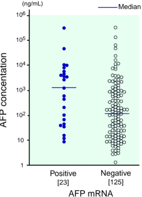
Serum AFP concentration was higher in the HCC patients with AFP mRNA (median: 1209 ng/mL, range: 9–319,900 ng/mL) than in the patients without AFP mRNA (median: 125 ng/ mL, range: 1–332,500 ng/mL), the value being not statistically significant (Fig. 2, Table 3). When the patients were divided into 2 groups according to serum AFP concentration, the
inci-Table 2. AFP mRNA in the blood of 148 HCC patients
Number AFP mRNA in blood P value
of Positive Negative Positive rate
patients [23] [125] (%) Tumor diameter ≥ 5 cm 26 5 21 19.2 NS < 5 cm 122 18 104 14.8 Number of tumors ≥ 3 90 17 73 18.9 NS < 3 58 6 52 10.3 AFP ≥ 200 ng/mL 64 15 49 23.4 < 0.05 < 200 ng/mL 84 8 76 9.5 PIVKA-II* ≥ 40 mAU/mL 72 10 62 13.9 NS < 40 mAU/mL 54 8 46 14.8 TNM stage† I 10 1 9 10.0 II 30 4 26 13.3 NS III 27 4 23 14.8 IVA 54 7 47 13.0 IVB 27 7 20 25.9 NS
Portal thrombosis Present 27 7 20 25.9
Absent 121 16 105 13.2
Portocaval shunt Present 37 3 34 8.1 NS
Absent 111 20 91 18.0
Extrahepatic metastasis Present 27 8 19 29.6 < 0.05
Absent 121 15 106 12.4
[ ], number of patients.
AFP, α-fetoprotein; HCC, hepatocellular carcinoma; mRNA, messenger RNA; NS, not significant; PIVKA-II, protein induced by vitamin K absence or antagonist II; TNM, tumor-nodule-metastasis.
* Serum PIVKA-II levels were measured in 126 patients.
† Clinical staging of the tumor was assessed according to TNM system of the General Rules for Clinical and Pathological Study of Primary Liver Cancer in Japan on the basis of imaging studies.
Median Negative [125] Positive [23] (ng/mL) AFP mRNA AFP concentation 106 105 104 103 102 10 1
Fig. 2. Serum α-fetoprotein (AFP) concentrations in 23 patients with AFP messenger RNA (mRNA) in the blood (median: 1209 ng/mL, range; 9–319,900 ng/mL) and 125 patients without AFP mRNA (median: 125 ng/ mL, range; 1–332,500 ng/mL). [ ], number of patients.
dence of AFP mRNA in the blood was signifi-cantly higher in the patients with 200 ng/mL or more (23.4%) than those with less than 200 ng/ mL (9.5%; P < 0.05; Table 2). PIVKA-II concen-tration in the serum was significantly higher in HCC patients with AFP mRNA in the blood (76,443 ± 298,178 mAU/mL) than in the patients without AFP mRNA (12,209 ± 50,415 mAU/mL; P < 0.05). However, when the patients were di-vided into 2 groups according to PIVKA-II con-centration, the incidence of AFP mRNA was not different between the patients with PIVKA-II concentrations of 40 mAU/mL or more and those with less than 40 mAU/mL (Table 2).
The presence of portal thrombosis was found in 7 (30.4%) of the 23 patients with AFP mRNA in the blood and in 20 (16.0%) of the
Table 3. Profiles of 148 patients with or without AFP mRNA
AFP mRNA in blood P value
Positive Negative
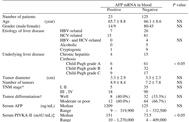
Number of patients 23 125
Age (year) 65.7 ± 8.8 66.1 ± 8.6 NS
Gender (male/female) 14/9 80/45 NS
Etiology of liver disease HBV-related 7 26
HCV-related 15 81
HBV- and HCV-related 0 4 NS
Alcoholic 0 5
Cryptogenic 1 9
Underlying liver disease Chronic hepatitis 4 15 Cirrhosis Child-Pugh grade A 6 61 < 0.05 Child-Pugh grade B 4 32 Child-Pugh grade C 9 17 Tumor diameter (cm) 3.3 ± 2.9 3.3 ± 2.3 NS Number of tumors 8.9 ± 8.4 7.2 ± 7.8 NS TNM stage* I, II 5 35 NS III , IV 18 90
Tumor differentiation† Well 8 (40.0%) 32 (33.3%) NS
Moderate or poor 12 (60.0%) 64 (66.7%)
Serum AFP (ng/mL) Median 1209 125 NS
Range 9 – 319,900 1 – 332,500
Serum PIVKA-II (mAU/mL)‡ Median 151 73.5 < 0.05
Range 10 – 1,270,000 4 – 409,000
AFP, α-fetoprotein; HBV, hepatitis B virus; HCV, hepatitis C virus; mRNA, messenger RNA; NS, not signifi-cant; PIVKA-II, protein induced by vitamin K absence or antagonist II; TNM, tumor-nodule-metastasis. * Clinical staging of the tumor was assessed according to the TNM system of the General Rules for the
Clinical and Pathological Study of Primary Liver Cancer in Japan on the basis of imaging studies. † Tumor biopsies were performed in 116 lesions (20 cases in the AFP mRNA-positive group and 96 cases in
the AFP mRNA-negative group) and classified according to the General Rules for the Clinical and Pathological Study of Primary Liver Cancer in Japan.
‡ Serum PIVKA-II was measured in 126 patients (18 in the AFP mRNA-positive group and 108 in the AFP mRNA-negative group).
125 patients without AFP mRNA (Table 2). The presence of a portocaval shunt was found in 3 (13.0%) of the 23 patients with AFP mRNA in the blood and in 34 (27.2%) of the 125 patients without AFP mRNA (Table 2). Thus, AFP mRNA in the blood did not correlate with portal thrombosis or a portocaval shunt.
Extrahepatic metastases were noted in 8 (34.8%) of the 23 patients with AFP mRNA in the blood and 19 (15.2%) of the 125 patients without AFP mRNA, the value being signifi-cantly higher in the former than in the latter (P < 0.05; χ2 test). When the patients were classified
according to the presence or absence of extra-hepatic metastases, AFP mRNA in the blood was detected in 8 (29.6%) of the 27 patients with extrahepatic metastases and in 15 (12.4%)
of the 121 patients without extrahepatic metas-tases, the value being statistically significant (P < 0.05; Table 2).
Aspartate aminotransferase and alanine aminotransferase concentrations in the serum were significantly higher in the 23 HCC patients with AFP mRNA in the blood (aspartate amino-transferase; 203 ± 297 IU/L, alanine amino-transferase; 113 ± 134 IU/L) than in the 125 pa-tients without AFP mRNA (104 ± 116, P < 0.01,72 ± 61 IU/L, P < 0.05).
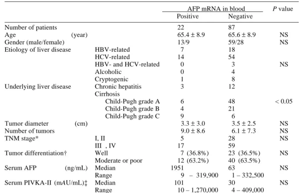
Table 4 summarizes the profiles of 109 pa-tients undergoing follow-up studies which disclosed the cause of death in 51 patients (46.8%); 35 died from HCC progression, 9 from hepatic failure, 3 from gastrointestinal
Table 4. Profiles of the 109 HCC patients with or without AFP mRNA
AFP mRNA in blood P value
Positive Negative
Number of patients 22 87
Age (year) 65.4 ± 8.9 65.6 ± 8.9 NS
Gender (male/female) 13/9 59/28 NS
Etiology of liver disease HBV-related 7 18
HCV-related 14 54
HBV- and HCV-related 0 3 NS
Alcoholic 0 4
Cryptogenic 1 8
Underlying liver disease Chronic hepatitis 3 12 Cirrhosis Child-Pugh grade A 6 48 < 0.05 Child-Pugh grade B 4 21 Child-Pugh grade C 9 6 Tumor diameter (cm) 3.3 ± 3.0 3.5 ± 2.5 NS Number of tumors 9.0 ± 8.6 6.1 ± 7.3 NS TNM stage* I, II 5 28 NS III , IV 17 59
Tumor differentiation† Well 7 (36.8%) 23 (36.5%) NS
Moderate or poor 12 (63.2%) 40 (63.5%)
Serum AFP (ng/mL) Median 1951 63 NS
Range 9 – 319,900 1 – 332,500
Serum PIVKA-II (mAU/mL)‡ Median 101 30 NS
Range 10 – 1,270,000 4 – 409,000
AFP, α-fetoprotein; HBV, hepatitis B virus; HCC, hepatocellular carcinoma; HCV, hepatitis C virus; mRNA, messenger RNA; NS, not significant; PIVKA-II, protein induced by vitamin K absence or antagonist II; TNM, tumor-nodule-metastasis.
* Clinical staging of the tumor was assessed according to the TNM system of the General Rules for the Clinical and Pathological Study of Primary Liver Cancer in Japan on the basis of imaging studies. † Tumor biopsies were performed in 82 lesions (19 cases in the AFP mRNA-positive group and 63 cases in
the AFP mRNA-negative group) and classified according to the General Rules for the Clinical and Pathological Study of Primary Liver Cancer in Japan.
‡ Serum PIVKA-II was measured in 94 patients (17 in the AFP mRNA-positive group and 77 in the AFP mRNA-negative group).
bleeding and 4 from other diseases. AFP mRNA was noted in 8, 4, 0 and 0 patients, respectively. Extrahepatic metastasis was observed in 13 (11.9 %) patients (7 in the AFP mRNA-positive and 6 in the AFP mRNA-negative group). Tumor metastases were found in the lung in 6 patients, the bone in 3, the adrenal glands in 4, the spleen in 1 and the pharynx in 1.
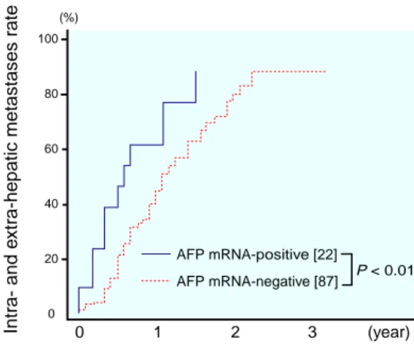
Next, the following parameters were ana-lyzed: the cumulative rate of the extrahepatic metastasis, the cumulative rate of intra- and extra-hepatic metastases and the cumulative survival rate for each AFP mRNA status. The cumulative rate of extrahepatic metastasis was similar between the 2 groups (data not shown). However, the cumulative rate of intra- and
extra-hepatic metastases was significantly higher in the 22 AFP mRNA-positive patients than that in the 87 AFP mRNA-negative pa-tients (P < 0.01; Fig. 3). The cumulative surviv-al rate was significantly lower in the 22 AFP mRNA-positive group than in the 87 AFP mRNA-negative group (P < 0.01; Fig. 4).
AFP mRNA-positive [22] AFP mRNA-negative [87] (%) Intr a- and e xtr a-hepatic metastases r ate 100 80 60 40 20 0 0 1 2 3 (year) P < 0.01 AFP mRNA-positive [22] AFP mRNA-negative [87] (%) Culumativ e sur viv al r ate 100 80 60 40 20 0 0 1 2 3 (year) P < 0.01
Fig. 4. Cumulative survival curve calculated by the
Kaplan-Meier method for 22 patients with positive
α-fetoprotein (AFP) messenger RNA (mRNA) and 87 patients with negative AFP mRNA. The difference is statistically significant (P < 0.01). [ ], number of patients.
Fig. 3. Rates of intra- and extra-hepatic metastases
calculated by the Kaplan-Meier method for 22 pa-tients with positive α-fetoprotein (AFP) messenger RNA (mRNA) and 87 patients with negative AFP mRNA. The difference is statistically significant (P < 0.01). [ ], number of patients.
Discussion
In this study, AFP mRNA in the blood was detected in the 23 (15.5%) of the total 148 HCC patients. Previous studies showed the positive rate of AFP mRNA in the peripheral blood to be from 25 to 95% of HCC patients (Matsumura et al., 1994, 1999; Komeda et al., 1995; Jiang et al., 1997; Lemoine et al., 1997; Louha et al., 1997; Nambu et al., 1997; Luo et al., 1999; Okuda et al., 1999). The lower frequency in the present study might be partly due to the selection of patients, in whom extrahepatic metastases were noted only in 27 patients. Furthermore, there were more patients with AFP non-producing HCC in the present study than in the previous reports (Matsumura et al., 1994; Jiang et al., 1997). The next possible reason is the difference in the timing of the examination of blood samples. Louha et al. (1997) reported that AFP mRNA producing cells in the blood were detected after locoregional therapy such as transcatheter arterial embolization or percutaneous ethanol injection. Matsumura et al. (1994) and Okuda et al. (1999) also showed that tumor cells do not always circulate in the blood, but may appear intermittently. We analyzed the blood sample before therapy or at least 2 months after ther-apy.
Next, we investigated the relationship be-tween AFP mRNA in the blood and the clinical parameters of HCC. The AFP mRNA in the blood did not correlate with tumor size, number of tumor nodules, presence of portal thrombosis and portocaval shunt, or TNM stage. Clinical significance of AFP mRNA in the blood is con-troversial even now (Matsumura et al., 1994, 1999; Komeda et al., 1995; Lemoine et al., 1997; Louha et al., 1997; Nambu et al., 1997; Luo et al., 1999). What is interesting is that this study confirmed a significant correlation between se-rum aspartate aminotransferase or alanine ami-notransferase concentration and AFP mRNA in the blood. The present study also demonstrated that the HCC patients with high levels of serum AFP showed higher detectable rates of AFP mRNA in the blood than those with low levels of serum AFP. The frequency of AFP mRNA
in the blood, however, was not so much higher in the HCC patients with a high AFP level (23.4%). This result suggests that the serum AFP level does not necessarily reflect the pres-ence of circulating cells with AFP mRNA. In other words, the serum AFP level is not a useful test to indicate the presence of circulating HCC cells. Nambu et al. (1997) and Luo et al. (1999) showed that the PIVKA-II value may be a good indicator of AFP mRNA in the blood. The posi-tive rate of AFP mRNA in the blood was not different between PIVKA-II-positive patients (40 mAU/mL and more) and -negative patients (less than 40 mAU/mL) in this study, although HCC patients with AFP mRNA-positive in the blood had higher PIVKA-II values than those with lower PIVKA-II. It is conceivable that the positivity of AFP mRNA did not correlate with the serum PIVKA-II value, because PIVKA-II-positive and AFP-negative HCCs are commonly experienced and the nested RT-PCR in the pres-ent study does not detect AFP non-producing cells.
In agreement with some previous research (Matsumura et al., 1994, 1999; Komeda et al., 1995; Louha et al., 1997), we found that the positivity of AFP mRNA in the blood was asso-ciated with extrahepatic metastasis. The detec-tion of AFP mRNA was clearly more frequent in the patients with extrahepatic metastasis than those without. This suggests that circulating AFP producing cells might be responsible for extrahepatic metastasis.
The present study compared the survival rate and intra- and extra-hepatic metastases, in which there were no differences in serum AFP levels, PIVKA-II levels or TNM stages. Matsumura et al. (1999) reported that the incidence of extra-hepatic metastasis was significantly higher in the AFP mRNA-positive group than that in the AFP mRNA-negative group. However, the extrahepatic metastasis was not significantly different between the 2 groups in the present study. The cumulative rate of intra- and extra-hepatic metastases was significantly higher in the patients positive with AFP mRNA in the blood than in the patients without. This result may indicate that tumor cells in the systemic circulation are responsible not only for
extra-hepatic metastasis but also intraextra-hepatic metas-tasis, which have been considered to be caused mainly by the spreading of cancer cells via the portal vein (Toyosaka et al., 1996). Funaki et al. (1997) reported that cancer cells released from the primary tumor to the systemic circula-tion might participate in intrahepatic metas-tasis. Since the liver provides an essentially fa-vorable environment for HCC cells, the inci-dence of intrahepatic metastasis through the systemic circulation might be more frequent than previously considered. Therefore, it is reasonable to assume that HCC patients who undergo liver transplantation do not always have good prognoses and often experience me-tastases in the transplanted livers (Zhou et al., 1996).
In the present follow-up study, we found that the cumulative survival rate was signifi-cantly poorer in the AFP mRNA-positive group than that in the AFP mRNA-negative group. Matsumura et al. (1999) found that overall sur-vival was similar between patients in AFP mRNA-positive and -negative groups, but the survival rates in continuous AFP mRNA-posi-tive patients was poorer than that in continuous AFP mRNA-negative patients. This result shows that the presence of AFP mRNA cells in the blood might be a predictive factor in HCC patients. Since the present AFP mRNA-posi-tive group had more severe underlying liver dis-ease than the AFP mRNA-negative group, this might affect the survival rate. However, it is indisputable that the high frequency of intra-and extra-hepatic metastases in the AFP mRNA-positive group was responsible for their poor prognoses.
In conclusion, AFP mRNA in the blood of HCC patients related to PIVKA-II levels and was significantly detectable in patients with an AFP level of 200 IU/mL or more, or with extra-hepatic metastases. Thus, the detection of AFP mRNA in the blood might be a predictor for tumor metastasis and survival of HCC patients.
References
1 Adachi E, Maeda T, Matsumata T, Shirabe K, Kinukawa N, Sugimachi K, et al. Risk factors for intrahepatic recurrence in human small hepato-cellular carcinoma. Gastroenterology 1995;108: 768–775.
2 Adinolfi A, Adinolfi M, Lessof MH. Alpha-feto-protein during development and in disease. J Med Genet 1975;12:135–151.
3 Behghiti J, Panis Y, Farges O, Benhamou JP, Fekete F. Intrahepatic recurrence after resection of hepatocellular carcinoma complicating cirrho-sis. Ann Surg 1991;214:114–117.
4 Brandt B, Junker R, Griwatz C, Heidl S, Brinkmann O, Semjonow A, et al. Isolation of prostate-derived single cells and cell clusters from human peripheral blood. Cancer Res 1996; 56:4556–4561.
5 Chen P-J, Chen D-S, Lai M-Y, Chang M-H, Huang G-T, Yang P-M, et al. Clonal origin of recurrent hepatocellular carcinomas. Gastro-enterology 1989;96:527–529.
6 Gitlin D, Boesman M. Serum α-fetoprotein, albumin, and γG-Globulin in the human con-ceptus. J Clin Invest 1966;45:1826–1838. 7 Heu H-C, Chiou T-J, Chen J-Y, Lee C-S, Lee
P-H, Peng S-Y. Clonality and clonal evaluation of hepatocellular carcinoma with multiple nodules. Hepatology 1991;13:923–928.
8 Inoue S, Nakao A, Harada A, Nonami T, Takagi H. Clinical significance of abnormal prothrom-bin (DCP) in relation to postoperative survival and prognosis in patients with hepatocellular carcinoma. Am J Gastroenterol 1994;89:2222– 2226.
9 Ishikawa T, Kashiwagi H, Iwakami Y, Hirai M, Kawamura T, Aiyoshi Y, et al. Expression of α -fetoprotein and prostate-specific antigen genes in several tissues and detection of mRNA in normal circulating blood by reverse transcriptase-poly-merase chain reaction. Jpn J Clin Oncol 1998;28: 723–728.
10 Jiang SY, Shyu R-Y, Huang M-F, Tang H-S, Young H-S, Roffler SR, et al. Detection of alphafeto-protein-expressing cells in the blood of patients with hepatoma and hepatitis. Br J Cancer 1997; 75:928–933.
11 Komeda T, Fukuda Y, Sando T, Kita R, Furukawa M, Nishida N, et al. Sensitive detection of circulating hepatocellular carcinoma cells in peripheral venous blood. Cancer 1995;75:2214– 2219.
12 Krüger W, Krzizanowski C, Holweg M, Stockschläder M, Kröger N, Jung R, et al. Reverse transcriptase/polymerase chain reaction detection of cytokeratin-19 mRNA in bone marrow and blood of breast cancer patients. J Cancer Res Clin Oncol
1996;122:679–686.
13 Kubo S, Nishiguchi S, Hirohashi K, Shuto T, Kuroki T, Minamitani S, et al. Clinicopatho-logical criteria for multicentricity of hepato-cellular carcinoma and risk factors for such carcinogenesis. Jpn J Cancer Res 1998;89:419– 426.
14 Kumada T, Nakano S, Takeda I, Sugiyama K, Osada T, Kiriyama S, et al. Pattens of recurrence after initial treatment in patients with small hepatocellular carcinoma. Hepatology 1997;25: 87–92.
15 Lemoine A, Le Bricon T, Salvucci M, Azoulay D, Pham P, Raccuia J, et al. Prospective evalua-tion of circulating hepatocytes buy alpha-fetopro-tein mRNA in humans during liver surgery. Ann Surg 1997;226:43–50.
16 Louha M, Poussin K, Ganne N, Zylberberg H, Nalpas B, Nicolet J, et al. Spontaneous and iatro-genic spreading of liver-derived cells into periph-eral blood of patients with primary liver cancer. Hepatology 1997;26:998–1005.
17 Luo W, Yatsuhashi H, Hamada R, Matsumoto T, Inoue O, Koga M, et al. Analysis of α-fetoprotein mRNA in peripheral blood: detection and semi-quantitation by reverse transcription polymerase chain reaction. Hepatol Res 1999;14:1–12. 18 Matsumura M, Niwa Y, Hikida Y, Okano K,
Kato N, Shiina S, et al. Sensitive assay for de-tection of hepatocellular carcinoma associated gene transcription (alpha-fetoprotein mRNA) in blood. Biochem Biophys Res Commun 1995; 207:813–818.
19 Matsumura M, Niwa Y, Kato N, Komatsu Y, Shiina S, Kawabe T, et al. Detection of α -fetoprotein mRNA, an indicator of hematogenous spreading hepatocellular carcinoma, in the circulation: a possible predictor of metastatic hepatocellular carcinoma. Hepatology 1994;20: 1418–1425.
20 Matsumura M, Shiratori Y, Niwa Y, Tanaka T, Ogura K, Okudaira T, et al. Presence of α -feto-protein mRNA in blood correlates with outcome in patients with hepatocellular carcinoma. J Hepatol 1999;31:332–339.
21 Mattano LA Jr, Moss TJ, Emerson SG. Sensitive detection of rare circulating neuroblastoma cells by the transcriptase-polymerase chain reaction. Cancer Res 1992;52:4701–4705.
22 Moreno JG, Croce CM, Fischer R, Monne M, Vihko P, Mulholland SG, et al. Detection of hematogenous micrometastasis in patients with prostate cancer. Cancer Res 1992;52:6110–6112. 23 Nagasue N, Uchida M, Makino Y, Takemoto Y, Yamanoi A, Hayashi T, et al. Incidence and fac-tors associated with intrahepatic recurrence fol-lowing resection of hepatocellular carcinoma. Gastroenterology 1993;105:488–494.
Imamura M, Itoh T, et al. Clinicopathologic sig-nificance of protein induced vitamin K absence or antagonist II and a-fetoprotein in hepatocellular carcinoma. Intern J Oncol 1999;14:281–286. 25 Nakano S, Haratake J, Okamoto K, Takeda S.
In-vestigation of resected multinodular hepato-cellular carcinoma: assessment of unicentric or multicentric genesis from histological and prog-nostic viewpoint. Am J Gastroenterol 1994;89: 189–193.
26 Nambu S, Nishimori H, Maekawa M, Higuchi K, Watanabe A. Plasma PIVKAII values as an indicator of AFP mRNA in the circulation in pa-tients with advanced hepatocellular carcinoma. Hepatol Res 1997;8:28–36.
27 Nomoto S, Nakao A, Kasai Y, Harada A, Nonami T, Takagi H. Detection of ras gene mutations in perioperative peripheral blood with pancreatic adenocarcinoma. Jpn J Cancer Res 1996;87:793– 799.
28 Nomura F, Ohnishi K, Tanabe Y. Clinical fea-tures and prognosis of hepatocellular carcinoma with reference to serum alpha-fetoprotein levels. Cancer 1989;64:1700–1707.
29 Okuda K. Hepatocellular carcinoma: recent progress. Hepatology 1992;15:948–963. 30 Okuda K, Kotoda K, Obata H, Hayashi N,
Hisamitsu T, Tamiya M, et al. Clinical obser-vations during a relatively early stage of hepato-cellular carcinoma, with special reference to serum α-fetoprotein levels. Gastroenterology 1975;69:226–234.
31 Okuda N, Nakao A, Takeda S, Oshima K, Kanazumi N, Nonami T, et al. Clinical signifi-cance of α-fetoprotein mRNA during periopera-tive period in HCC. Hepatogastroenterology 1999;46:381–386.
32 Omichi-Funaki N, Tanaka J, Imamura M. Quan-titative analysis of alpha fetoprotein mRNA in circulating peripheral blood of patients with hepatocellular carcinoma and alpha fetoprotein producing gastric carcinoma. Life Sci 1998;62: 1973–1984.
33 Omichi-Funaki N, Tanaka J, Seto S, Kasamatsu T, Kaido T, Imamura M. Hematogenous spread-ing of hepatocellular carcinoma cells: possible participation in recurrence in the liver. Hepatol-ogy 1997;25:564–568.
34 Saiki RK, Gelfand DH, Stoffel S, Scharf SJ, Higuchi R, Horn GT, et al. Primer-directed enzy-matic amplification of DNA with a thermostable
DNA polymerase. Science 1988;239:487–491. 35 Sheu J-C, Huang G-T, Chou H-C, Lee P-H,
Wang J-T, Lee H-S, et al. Multiple hepatocellu-lar carcinomas at the early stage have different clonality. Gastroenterology 1993;105:1471– 1476.
36 Smith B, Selby P, Southgate J, Pittman K, Bradley C, Blair GE. Detection of melanoma cells in peripheral blood by means of reverse transcriptase and polymerase chain reaction. Lancet 1991;338:1227–1229.
37 Suehiro T, Sugimachi K, Matsumata T, Itasaka H, Taketomi A, Maeda T. Protein induced by Vitamin K absence or antagonist II as prognostic marker in hepatocellular carcinoma. Cancer 1994;73:2464–2471.
38 Tada M, Omata M, Kawai S, Saisho H, Ohto M, Saiki RK, et al. Detection of ras gene mutation in pancreatic juice and peripheral blood of patients with pancreatic adenocarcinoma. Cancer Res 1993;53:2572–2574.
39 Takenaka K, Adachi E, Nishizaki T, Hiroshige K, Ikeda T, Tsuneyoshi M, et al. Possible multi-centric occurrence of hepatocellular carcinoma: a clinicopathological study. Hepatology 1994; 19:889–894.
40 Tarao K, Hoshino H, Shimizu A, Ohkawa S, Nakamura Y, Harada M, et al. Role of increased DNA synthesis activity of hepatocytes in multi-centric hepatocarcinogenesis in residual liver of hepatectomized cirrhotic patients with hepato-cellular carcinoma. Jpn J Cancer Res 1994;85: 1040–1044.
41 Toyosaka A, Okamoto E, Mitsunobu M, Oriyama T, Nakao N, Miura K. Intrahepatic metastasis in hepatocellular carcinoma: evidence for spread via the portal vein as an efferent vessel. Am J Gastroenterol 1996;91:1610–1615.
42 Wong IH-N, Leung T, Ho S, Law WY, Chan M, Johnson PJ. Semiquantification of circulating hepatocellular carcinoma cells by reverse tran-scriptase polymerase chain reaction. Br J Cancer 1997;76:628–633.
43 Yokoyama I, Todo S, Iwatsuki S, Starzl TE. Liver transplantation in the treatment of primary liver cancer. Hepatogastroenterology 1990;37: 188–193.
44 Zhou X-D, Tang Y, Yang B-H, Yu Y-Q, Lin Z-Y, Lu J-Z, et al. Long-term results of surgery for small primary liver cancer in 514 adults. J Cancer Res Clin Oncol 1996;122:59–62.